
Abstract
Background: Hepatic stellate cells (HSCs) are considered to be of vital importance in the pathogenesis of liver fibrosis. In vitro studies have demonstrated antiproliferative effects of 5-hydroxytryptamine2 (5-HT2, serotonin) receptor antagonists upon HSCs. Terguride, recognized as a partial dopamine agonist, also has potent 5-HT2 receptor antagonist qualities and has been shown to prevent serotonin-induced organ changes and fibrosis in rats. Aim: In the current study, the carbon tetrachloride (CCL4) hepatic fibrosis rat model was used in combination with daily administration of terguride to evaluate potential preventive effects of a 5-HT2 receptor antagonist upon liver fibrosis. Materials and Methods: Forty rats (Sprague-Dawley) were included in the study. Twelve animals received terguride in combination with CCL4 and 12 animals were given only CCL4. The remaining 16 animals served as model controls. The extent of fibrosis was evaluated by liver weight, histologic analysis, biochemical analysis, and α-smooth muscle actin (α-SMA) immunohistochemistry. Results: There were no significant differences in liver weight, hydroxyproline content, serum alanine and aspartate transaminases, serum hyaluronic acid, or α-SMA immunostaining between rats treated with terguride in combination with CCL4 and rats given only CCL4. All parameters, however, were significantly lower (P < 0.01) in the model controls. Conclusion: Terguride, a potent 5-HT2 antagonist, did not prevent the development of liver fibrosis in rats given CCL4.
Introduction
The hepatic stellate cell (HSC) is recognized as playing a key role in the development of hepatic fibrogenesis (1, 2). In the normal liver, HSCs reside in the space of Disse, regulating sinusoidal blood flow and production of extracellular matrix (ECM). Furthermore, HSCs function as a retinoid storage site. Following liver injury, HSCs undergo activation, which is characterized by transformation into myofibroblast-like cells, expression of α-smooth muscle actin (α-SMA), increased production of ECM, and loss of retinoids (1, 2, 3). In addition, the normally quiescent HSCs reenter the cell cycle.
Serotonin has been associated with hepatic fibrosis, partly due to the increased plasma serotonin levels found in patients with liver cirrhoses (4), but more importantly, in vitro investigations have demonstrated both 5-HT receptors in HSCs and suppression of HSC proliferation in response to 5-HT2 receptor antagonists (5). In addition, serotonin increases and 5-HT2 antagonists suppress HSC expression of key fibrogenic factors (transforming growth factor-β, SMAD4) (6). Terguride (1.1 diethyl-3-(6-methyl-8α-ergo-linyl)urea), an ergoline used for treating hyperprolactinemia, is a partial dopamine agonist (7), but also characterized by 5-HT receptor properties. In contrast to the majority of ergoline agents, terguride antagonizes 5-HT2 receptors (5-HT2A,2B,2C) (7, 8). We have recently demonstrated that long-term serotonin administration induces organ changes and localized skin fibrosis in rats and that all of these effects are prevented by co-administration of terguride (9). An increased expression of 5-HT2B receptors has been found in CCL4-induced liver fibrosis in rats (5). In the present study, we used the CCL4 rat model to investigate whether terguride, as a 5-HT2 receptor antagonist, has the potential to modulate the development of hepatic fibrosis in vivo.
Materials and Methods
Animals
Forty female age-matched Sprague Dawley rats (Møllegaard, Denmark) were housed in steel cages (6–4 animals in each cage) rigged in two steel/glass chambers equipped with a separate ventilation system (airflow, 165 l/min) to protect the investigators from potential CCL4 exposure. The chamber temperature was 23 ± 1°C, with relative humidity ~70%. The rats were maintained on a 12-hr light/dark cycle and had free access to rat diet (B&K Universal Ltd., Hull, UK) and tap water. The study was approved by the Norwegian National Animal Research Authority. The animals were allocated into four experimental groups as presented in Table 1. The experiment had a schedule of 8 weeks and thus was designed to induce hepatic fibrosis. The model of CCL4-induced hepatic fibrosis has been described in detail (10). Briefly, the animals received 0.2 ml/100 g CCL4 intraperitoneally in a 1:1 solution with olive oil (Sigma Aldrich, St. Louis, MO) twice weekly, with constant intervals for 2 weeks. The dose was then reduced to 0.1 ml/100 g and administrated for 6 more weeks. Terguride (Ergonex Pharma GmbH, Appenzell, Switzerland) 2.5 mg/kg in 5% Cremophor (Sigma-Aldrich, Steinheim, Germany) was administrated twice daily by gavage (volume 1 ml) during the whole study. The control groups were given vehicles (olive oil and Cremophor). Prior to sacrifice, the animals were anesthetized with a 4-ml/kg body weight combination of fentanyl (0.02 mg/ml), midazolam (2.0 mg/ml), and haloperidol (1.25 mg/ml) and were sacrificed by exsanguination. The organs were immediately collected and weighed.
Histology
Liver samples from three separate locations (right, middle, and left parts of the liver) were formalin-fixed, paraffin-embedded, and cut into 4-μm sections. The sections were stained using hematoxylin-eosin-saffron and reviewed, in a blinded fashion, by a pathologist.
A semiquantitative scoring system was established to evaluate the extent of fibrosis. The degree of central vein fibrosis, portal field fibrosis, and hepatic steatosis was evaluated independently, using the scoring system shown in Table 2.
Immunohistochemistry
Immunostaining for α-SMA was performed as previously described (11). Briefly, the sections were incubated with monoclonal (mouse) anti-human SMA (Dako, clone 1A4, Glostrup, Denmark) for 2 hrs. The EnVision-HRP kit (Dako, K5007) was used as a secondary antibody, and the immunoreactions were visualized using 3-amino-9-ethylcarbazole (AEC, Vector Laboratories, Burlingame, CA).
Biochemical Analysis
Serum levels of aspartate transaminase (AST) and alanine transaminase (ALT) were determined using a dry chemical strip analyzer (Reflotron plus, Roche Diagnostics, Basel, Switzerland) according to the manufacturer’s instructions. Hydroxyproline levels were analyzed in frozen liver homogenates (50 mg each) from both the left and right parts of the liver (all aliquots were analyzed separately). The samples were subjected to amino acid hydrolysis at 110°C in 1 ml 6 N HCl for 16 hrs. The hydroxyproline content was then determined using a colormetric assay, as previously described (12). Data were presented as micrograms of hydroxyproline per gram of wet liver weight using hydroxyproline standards (Sigma Chemicals). The hydroxyproline data obtained from the left and right samples were pooled prior to the final statistical analysis.
Serum hyaluronic acid was measured using a commercially available enzyme-linked immunoabsorbent assay kit (Corgenix Ltd., Peterborough, UK), following the kit instructions.
Statistical Analysis
A two-tailed nonparametric test, Mann-Whitney U test, was used in all calculations. The significance level was set as α = 0.05. The null hypothesis was rejected when P ≤ 0.05.
Results
Animals
The experimental procedures were generally well tolerated; however, 4 animals died during the study: one rat in Group 2, two rats in Group 3, and one rat in Group 4. Two deaths were caused by aspiration pneumonia related to the gavage. One rat possibly died of peritonitis, and the last rat died shortly after an intraperitoneal injection, for unknown reasons. The median body weight at the end of the study was 259 g (range 233–295 g), and there were no significant differences among the experimental groups.
The liver weight data are presented in Table 3. There was no significant difference in liver weight between rats given CCL4 in combination with terguride (Group 1) and rats given only CCL4 (Group 2); however, there was a significant difference (P < 0.01) between CCL4 animals and model controls (Groups 1 and 2 vs Groups 3 and 4).
Spleen weights were also measured and found to be similar in all groups.
Histology
The histologic evaluation is summarized in Table 4. Established fibrosis was evident in animals given CCL4 in contrast to the model controls, where no changes were found. There was no significant difference in the extent of fibrotic changes between groups given CCL4 (Groups 1 and 2); however, there was a slightly less degree of hepatic steatosis in rats given terguride (Group 1).
Immunohistochemistry
In the fibrotic livers, α-SMA–positive cells were observed in perisinusoidal spaces and in fibrous septa. More intense α-SMA staining was evident in rats given CCL4 (Groups 1 and 2) compared to model controls (Groups 3 and 4). However, no disparities were apparent between rats given (Group 1) or not given (Group 2) terguride. The α-SMA–positive staining was focally and unevenly distributed, and a more quantitative approach was not reasonable.
Biochemical Analysis
The biochemical data are summarized in Figure 1. Terguride had no effect upon AST/ALT levels in the CCL4 model. In the model controls (Groups 3 and 4), the AST/ALT levels were significantly lower compared to those in the CCL4 animals.
Terguride (Group 1) did not influence serum hyaluronic acid levels in the CCL4 model. However, the model control animals (Groups 3 and 4) had significantly lower levels compared to the CCL4 animals (Groups 1 and 2).
In addition, terguride had no significant effects upon hydroxyproline levels in the CCL4 model. The model controls (Groups 3 and 4) had significantly lower levels compared to the CCL4 animals (Groups 1 and 2).
Discussion
An experimental model using injections of a toxic and highly reactive agent is not directly comparable to the usual mechanisms of hepatic injury causing fibrosis and cirrhosis. However, evidence supports a common pathway in hepatic fibrogenesis following the initial hepatocellular injury; particularly, the activation of hepatic stellate cells is regarded crucial in processing fibrotic events (13). Based on their findings regarding the antiproliferative effects on hepatic stellate cells, in vitro studies have suggested potential preventive effects of 5-HT2 receptor antagonists upon the development of liver fibrosis (5, 6). The current investigation, using a potent 5-HT2 antagonist, did not demonstrate any amelioration of fibrotic liver changes in the CCL4 rat model, which thereby calls into question any significant relationship between serotonin and liver fibrosis pathogenesis. The serotonin-producing small bowel carcinoids are associated with fibrosis in peritumoral areas, retroperitoneal tissues, and heart valves (14). However, despite extensive liver metastases and massive serotonin exposure, there is no known association with liver fibrosis in these patients. We have previously demonstrated increased liver weight in rats exposed to serotonin administration over a prolonged period of time; however, no evident fibrotic liver changes were found (9). Thus, serotonin acting as a direct mediator of liver fibrosis seems less likely. Although serotonin could serve as a cofactor in the fibrotic process following liver injury, our study utilizing the CCL4 rat model could not demonstrate any significant effect of 5-HT2 receptor antagonism upon liver fibrogenesis.
Experimental Groups
Semiquantitative Scoring of Hepatic Fibrosis
Liver Weight
Histology a
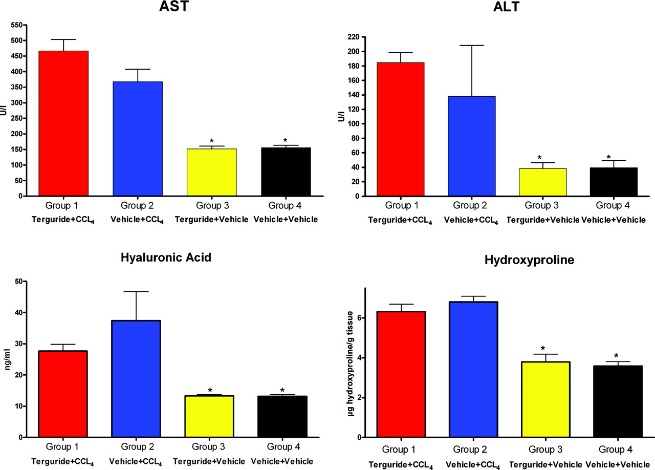
A summary of the biochemical data. *P < 0.001.
Footnotes
This work was supported by Ergonex Pharma GmbH and the Cancer Foundation of St. Olavs Hospital.