



Abstract
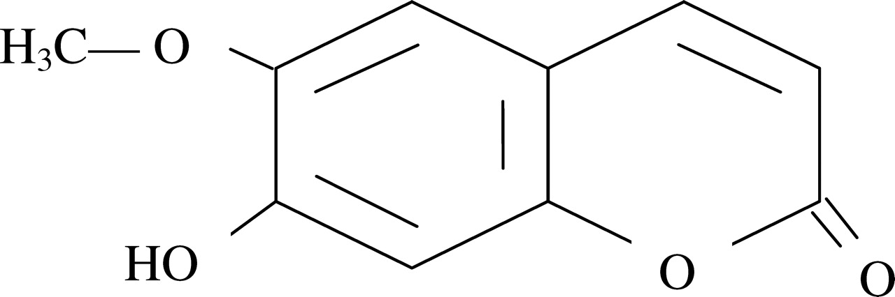
The chemical structure of the main fluorescenting compound in the ethanolic extract (mother tincture) of the American yellow jasmine, Gelsemium sempervirens, was determined by employing 1H nuclear magnetic resonance (NMR), 13C NMR, mass spectroscopy, high-performance liquid chromatography (HPLC), correlation spectroscopy (COSY), and Fourier transform infrared (FTIR) spectroscopy analyses. Spectrofluorometric analysis has been made of the mother tincture and its agitated serial dilutions (up to 12th potency) prepared according to a homeopathic procedure in which serial, agitated dilutions were made separately in glass and polypropylene containers. The succussions were made by employing three different modes: hand jerk, sonication, and vortexing. The chemical formula of scopoletin, the main fluorescent compound, was determined to be C10H8O4 having a molecular weight of 192.17. Significant differences were noted between the remedies prepared in the two types of containers. Further, a comparison between any two methods of agitation revealed significant differences in fluorometric data of remedies at certain potency levels. The biological (anticancer) action of the crude extract, the alkaloid scopoletin, and 2C potency of Gelsemium sp were tested in vitro on the HeLa cell line through fluorescence microscopy, the 3(4, 5-dimethylthiazol-2-yl)-2, 5-diphenyltetrazolium bromide (MTT) assay, and fluorescent activated cell sorting (FACS). The role of nanoparticles presumably derived from the containers, their orientation, and their interaction with the starting substance during the dynamization process initiated by different modes of agitation could possibly be attributed to the differences noted in the fluorometric data of potencies prepared in the two types of containers and among the three different means of succussion tested.
Introduction
Homeopathy is a controversial science. The homeopathic procedure of dilution and agitation to potentize the remedies is one of the causes of this controversy: when the dilutions are at 12C or higher (i.e., 1024 or more), the remedy cannot in theory contain even a single molecule of the original drug substance. Further, homeopathy tends to use drugs in microdoses; however, the effect of dilution and succussion has been claimed to be of enormous importance in the process of dynamization of potentized homeopathic remedies. As a matter of practice, homeopathic remedies are diluted and succussed in glass vials according to the original method adopted by Dr. Samuel Hahnemann, the proponent of homeopathy. However, the procedure of succussion varies. The hand-jerk method, the traditional practice used for dynamization of homeopathic remedies, has now made room for the semiautomatic potentizer, which ensures that each succussion is performed with the same number of strokes and with the same force on each stroke. Alternatively, sonication is also sometimes used as a method of potentization (1); the agitation is also done by vortexing. In some situations, homeopathic practitioners use polypropylene containers, particularly when using 50 millisemal potencies in ethanolic water. The initial potencies are generally made by grinding the starting drug material in mortar and pestle, after which the tiny powdery substances are dissolved in ethanolic water contained in polypropylene vials. The patients are advised to give 10 jerks to the container with the remedy before use each time and take a few drops from it as a single dose. Our inquisitiveness in this aspect led us to examine whether the physicochemical properties of different modes of preparations were the same or different for the various processes of preparation.
Thus, the hypotheses to be tested were (i) whether scopoletin, an alkaloid that has been separated and chemically characterized from the plant Gelsemium semerviriens (family Loganiaceae) and that is fluorescent upon exposure to UV light, showed any change in its fluorescence upon agitation in three different modes (traditional hand jerk [10 for each serial dilution], vortexing, and sonication), in two different containers (glass and polypropylene vials) and (ii) whether the quanta of fluorescence differs between the separated alkaloid and the crude ethanolic extract of G. sempervirens and shows differences in anticancer activity on human HeLa cells as revealed by 4′,6-diamidino-2-phenylindole (DAPI) staining, the 3(4, 5-dimethylthiazol-2-yl)-2, 5-diphenyltetrazolium bromide (MTT) assay, and fluorescence activated cell sorting (FACS) analysis.
The dried rhizomes and roots of the American yellow jasmine (G. sempervirens, family Loganiaceae) are indigenous to the southern United States. The ethanolic plant extract contains 45 alkaloids, many of which are toxic (2). Gelsemine is the principal alkaloid and is the one most elaborately studied. Other toxic compounds are gelsemicine, 1-methoxy- and 21-oxogelsemine, 14-hydroxy gelsemicine, gelsedine, 14-hydroxy gelsedine, and sempervirine. A thorough literature search revealed that this plant indeed contains a highly fluorescent compound, possibly scopoletin, which is responsible for emission of blue fluorescence upon exposure to UV light and is reported to have anticancer activity (3). Therefore, we isolated different alkaloid components of the extract by high-performance liquid chromatography (HPLC) and analyzed the chemical nature of the separated fluorescent alkaloid further with the aid of methods such as 1H nuclear magnetic resonance (NMR), 13C NMR, mass spectroscopy, Fourier transform infrared (FTIR), and correlation spectroscopy (COSY) to confirm that only this compound was undergoing fluorometric analysis.
Materials and Methods
The chemical properties of Gelsemium sp mother tincture was determined by 1H NMR, 13C NMR, COSY, FTIR, mass spectroscopy, and HPLC analyses. One milliliter of the mother tincture of G. sempervirens (Boiron Laboratory, lot TJ0071, 65% ethanol) was initially diluted with 9 ml of 65% ethanol to obtain the stock solutions (1× solution). One milliliter each of the stock solution was diluted with 99 ml of 65% ethanol and given succussions in a glass container in three different modes—hand jerk, sonication, and vortexing—to produce successive centesimal (C) potencies.
Polypropylene vials were also used in the same process of potentization.
Spectrofluorometric Studies.
Spectrofluorometric analysis of different dilutions of G. sempervirens was made in a spectrofluorometer (Luminescence Spectrometer, LS 50 B; Perkin Elmer, Waltham, Massachusetts). The study was performed with three replicates of each type of dilution.
Cell Cultures.
The HeLa cell line obtained from National Center for Cell Science, Pune, was grown at 37°C in an atmosphere of 5% carbon dioxide in Dulbecco’s modified Eagle’s medium (DMEM) supplemented with 10% fetal bovine serum and 1% antibiotic (PSN). For experimental studies, cells were grown to 80% to 90% confluence, harvested with ice cold phosphate-buffered saline (PBS), plated at the desired density, and allowed to re-equilibrate for 24 hrs before any treatment.
Drug and Placebo (65% Ethanol) Treatment.
The alkaloid scopoletin was isolated from the mother tincture of Gelsemium sp by the standard column chromatography method (3:1 petroleum ether: ethyl acetate; silica gel 60–120 mesh size). Next scopoletin was dissolved in pure ethanol. Different amounts of scopoletin (20 μg/105 cells, 40 μg/105 cells, 80 μg/105 cells, 100 μg/105 cells, 160 μg/105 cells, and 200 μg/105 cells) were poured into different wells.
Four microliters of the mother tincture of Gelsemium sp and 2C dilutions made with the mother tincture in two vials (glass and polypropylene) by three different modes of succussion (sonication, vortexing and hand jerks) were added to different wells containing 105 cells. A well was also provided with the ethanol placebo (4 μl). After 24 hrs, either DAPI or acridine orange/ethidium bromide (AO/EB) were added to each well, and the contents of the well were subjected to fluorescence microscopy.
Fluorescence Microscopy.
DAPI Staining.
The cell-permeable DNA dye DAPI was used at the rate of 10 μg/105 cells to visualize chromatin condensation and/or fragmentation typical of apoptotic cells (4).
AO/E B Double Staining.
Apoptotic morphology was studied by staining cells with a combination of the fluorescent DNA-binding dyes AO and EB. To distinguish the live apoptotic and necrotic cells, the conventional EB/AO staining procedure (20 μg/105 cells) was followed (5). The color and structure of different cell types were observed under a fluorescence microscope.
MTT Assay.
The MTT assay was used to determine energetic cell metabolism by measuring the activity of one of the oxidative enzymes (6). The dye is reduced in mitochondria by succinic dehydrogenase to an insoluble violet formazon product. HeLa cells (106/ml) were cultured for 24 hrs on 96-well microplates. The cells were incubated for 24 hrs with and without compounds during the test. MTT was added, and after 2 hrs, formazon crystals were solubilized with acidic isopropanol, and the absorbance of the solution was measured at 595 nm by using an enzyme-linked immunoabsorbent assay (ELISA) reader.
Cell Cycle Analysis.
Cellular DNA was stained with propidium iodide (PI) and quantified by flow cytometry according to Nicoletti’s procedure (7). Briefly, cells were collected after treatment, washed in PBS, fixed with 70% (v/v) cold aqueous ethanol (−20°C), and stored at 4°C for at least 12 hrs. The cells were washed in PBS; after cell centrifugation, cell pellets were stained with 10 μg/ml PI and treated with 5000 units of RNase. The cell suspension was incubated in the dark at room temperature for 30 mins. DNA content was determined by using a FACS Calibur flow cytometer (Becton Dickinson, Mountain View, CA). A total of 25,000 events was acquired, MOD-FIT LT software (Becton Dickinson) was used for cell cycle analysis of the cytostatic effect, and sub-G1 peak detection of the apoptotic effect was analyzed.
Statistical Methods.
The differences in data between the diluted drugs prepared in different ways and their respective controls were analyzed by using Student’s t test. Similarly the significance of differences between dilutions made in glass vials and those made in polypropylene containers were also analyzed by the t test. In the same way, significance of differences was also ascertained between data of any two methods of succussion: between hand jerk and vortexing, between hand jerk and sonication, between vortexing and sonication. Similar tests of the MTT assay were also conducted.
Results
With the help of 1H NMR, 13C NMR, COSY, FTIR, mass spectroscopy, and HPLC analyses, the structure of the principal alkaloid of Gelsemium sp was determined to be C10H8O4, as shown in Scheme 1.
Spectral Data.
Mass Spectra of purified compound MS: m/z = 193(M+). 1H NMR spectra of purified compound. 1H NMR(DMSO, 300 MHz), δH = 3.95(S, 3H, OCH3) 6.15(S, 1H, OH), 6.25(d, 1H, J = 9.4Hz, ArH) 6.84(S, 1H, ArH), 6.92(S, 1H, ArH), 7.58 (d, 1H,J = 9.4Hz, ArH). 13C NMR spectra of purified compound. 13C NMR (DMSO, 75 MHz): 56.8, 103.5, 110.3, 111.3, 112.4, 145.4, 146.1, 150.3, 151.9, 161.6 ppm.
The data obtained for dilutions made in glass and polypropylene containers and by different succussion procedures, are summarized in Table 1. The data are the mean of three replicates, which did not vary much (a representative example in Figure 1a shows the results for all three replicates), whereas only one or two are shown in other panels to avoid superimposition and for clarity (Fig. 1b–d and Fig. 2a–f; standard errors for each dilution are shown in Tables 1–9).
The control (unsuccussed 65% alcohol) and the succussed alcohol (“vehicle”—2C, 3C, 4C, and 5C) did not yield any fluorescence emission at 425 nm. When the Gelsemium sp mother tincture was diluted without succussion, fluorescence was weak or totally lost after the 2C dilution. However, dilutions up to 4C and, in some cases, up to 5C made with succussions in three different modes in glass and polypropylene containers exhibited fluorescence emission. The results of significance testing are shown in Tables 2 through 9. Analysis of data of different potencies of drug prepared by sonication (Table 1) revealed differences in their fluorescence data, which were highly significant for 2C, 3C, and 4C potencies (P < 0.001), and moderately significant for the 1C dilution (P < 0.05) prepared in glass and polypropylene containers. The differences for other dilutions were not significant. In case of succussion by vortexing, highly significant differences were noted, particularly at 2C, 3C, 4C (P < 0.001), between the drugs prepared in glass and polypropylene vials. Interestingly, the data from hand succussions using glass and polypropylene vials were highly significantly different for the 3C dilution and moderately to fairly significantly different for 1C, 2C, and 5C dilutions, whereas there were no significant differences for other dilutions. Thus, the data were significantly different for certain potency levels, when considered either between containers or between any two procedures of agitation. This finding would indicate that the fluorometric property of Gelsemium sp was affected to a different extent when the dilutions were made in glass and polypropylene vials. The differences were more pronounced at certain steps and between any two means of dilution. Further, no fluorescence was detected in the agitated dilutions beyond 5C through 12C. However, there was striking difference in fluorescence of the potentized remedies when they were compared with their ethanolic counterparts without any starting substance (Table 4) prepared in both glass vials (Table 4) and polypropylene vials (Table 5), between vortexing and hand succussion to produce differences for 1C through 5C prepared in glass containers (Table 6) but only for 2C and 3C prepared in polypropylene containers (Table 7), and between sonication and hand jerk in glass containers at 1C through 4C levels (Table 8) and in polypropylene containers at 2C and 3C levels (Table 9). Further, highly significant differences in fluorometric properties were found particularly at 2C and 3C levels (P < 0.001) for all methods of comparison. Data compared between mechanical vortexing and sonication showed few differences, but significant differences were found in dilutions in glass vials prepared by hand jerk rather than in those made in polypropylene containers.
Results of DAPI staining and AO/EB staining are provided in Figure 3, and the data of the MTT assay and FACS are summarized in Figures 4 and 5 and Tables 10 through 13. Green living cells showed normal morphology in the control (Fig. 3e and f), whereas EB intercalated with fragmented chromatin to produce orange fluorescence. In contrast, DAPI specifically stained the nuclei of the cells, particularly those studded with fragmented or condensed chromatin, indicating the cells were undergoing apoptosis. Thus, in general, more apoptotic cells were present in the drug-treated series than in the control. Further, critical analysis revealed that the number of apoptotic cells slightly varied in relation to the various types of dilutions made in different containers. The results of the cell viability assay (the MTT assay) showed that there was a linear decrease in the cell viability along with an increase in the concentration of scopoletin from 0 to 200 μg (Fig. 4a). In the case of the mother tincture of Gelsemium sp, there was a significant decrease in the cell viability (Fig. 4b), indicating scopoletin and the mother tincture share the anticancer property. Results of FACS (Fig. 5 and Table 10) indicated that both Gelsemium sp mother tincture and scopoletin (100 μg/ml and 200 μg/ml) arrested the cells more at the sub-G1 stage, that is, they did not allow a large number cells to enter the preparatory stage of cell division. A critical analysis of the data revealed that 2C dilutions of Gelsemium sp made by different modes of succussion in two types of containers also showed subtle differences; some of these tended to arrest the cells in sub-G1 but to a lesser extent than that of the mother tinctures of Gelsemium sp and scopoletin. The other stage of the cycle that showed the inhibitory effect of the different dilutions of Gelsemium sp was G2, which follows the synthetic phase. These results indicate that the cells were affected at various stages of the cell cycle and that there are subtle differences in action among the crude extract, scopoletin, and 2C dilutions prepared in glass and polypropylene vials by three methods of succussion. The actions of the sonication-made preparations differed considerably from those of hand-jerked preparations, but the action of the vortex-made preparations differed only slightly from that made by hand-jerk made ones. Similarly, comparison of the data from the MTT assays revealed a statistically significant difference between preparations made in glass and polypropylene vials.
Discussion
The 45 alkaloids derived from G. sempervirens are categorized into five structurally different groups; scopoletin is the one that has been identified as displaying fluorescence. Analysis of physicochemical properties confirmed that this compound has the chemical formula of C10H8O4 and a molecular weight of 192.17. Therefore, the results of our study are in conformity with those of an earlier one (2). In the present study we did not proceed beyond the dilution of 12C (BRAN, i.e., beyond Avogadro’s number) because there was no detectable differences in fluorescence between the vehicle and the drug after 5C. A critical analysis of the data revealed that there could be some differences in fluorescence, which served as a marker of change in any major physicochemical property of Gelsemium sp, while homeopathic procedures of dilution and succussion were under way. The results of the present study suggest that the homeopathic dilution up to 5C potency can be distinguished from the placebo (diluted without initial drug component) by its fluorescence property. Further, the result indicates that the container in which the homeopathic remedies were made is also important, because the fluorescence of the drugs prepared in glass and polypropylene containers to 2C and 3C dilutions differed significantly (P < 0.001). Although why such differences were encountered is not precisely known, one possible explanation could be that the differences in the occurrences and the nature of nanoparticles in the two types of containers could have contributed to the alteration of fluorescence. They also had subtle differences in their biological action (anticancer activity), as revealed by the fluorescence assays.
Milgrom et al. (8) reported that a small amount of silicon dioxide and ions dissolve from the glass walls into aqueous solution during succussion. The quantities dissolved are larger for soda glass and smaller for borosilicate glass, but there is always some (9). Demangeat et al. (10) found higher-than-expected silica in remedies prepared in glass vials and more silica in certain remedies than in succussed controls; Anick and Ives (9) thought the silica served as an active ingredient, as advocated in their “silica hypothesis for homeopathy.”
Glass vials have also been reported to shed nanoparticles (delamination), particularly during heating for depyrogenation (11). It has been shown that various proteins kept in glass vials can adsorb glass particles, potentially causing altered protein structures that also alter their ANS fluorescence emission intensity when they are compared with untreated proteins (11).
Water can take on many possible oligomeric and polymeric structures; that is, it can form complex networks of molecules in three-dimensional space, held together by various forces that include not only hydrogen bonds (relatively strong) but also van der Waals forces (much weaker; Ref. 12). Even if specific molecules or small molecular complexes leave their places in the network, other water structure complexes can take their place within the network structure itself, thereby maintaining the overall nanostructure within the solution, in part via configurational entropy or electromagnetic forces maintaining organizational stability of the network (13). In 1 min, the H-bond network of liquid water can undergo literally trillions of rearrangements (9). The principal ingredient in glass is silicon dioxide (SiO2). This compound can dissolve in water by combining with two H2O molecules to form a molecule of silicic acid, Si(OH)4, but this reaction depends on many factors such as temperature and salinity (14). Two molecules of silicic acid can link to form a dimer H6Si2O7 by expelling a single H2O and forming a Si-O-Si bond (also known as siloxene bond). The dimer may join with another monomer of Si(OH)4 to form a trimer and so on. Likewise, chains of polymerized Si(OH)4 can acquire various configurations by bending at different angles and create an infinite variety of silicate structures composed of polymeric species of silicon, hydrogen, and oxygen molecules (9); some of them can bind with either anionic form (with Si-H) and cationic form (with Si-OH) of dyes or elements (15). For cationic compounds, the molecules interact with the O site, but for anionic ones, the interaction is preferentially with the H sites. Therefore, scopoletin may be attached to this anionic form of silicates through ion-dipole interactions to form various configurations of scopoletin-silicate structure in the glass vials, depending on the modes of succussion; the mode of succussion made differences in the compound’s fluorescence. Alternatively scopoletin molecule can also join with water and leave its “molecular imprint” within water clathrates. However, the exact nature of interaction of the complex cannot be specified until more specific experiments (e.g., other relevant spectroscopic analyses) are carried out. Theoretically, this interaction of silicon-scopoletin is possible in view of the occurrence of the 7-hydroxyl group in the structure of scopoletin, which can interact with silicic acid and water. This interaction may explain how the “molecular imprinting” of the original drug substance can be made. Incidentally, nanocomposites of porous silicon with rhodamine and zinc oxide have been reported to differ in their photoluminescence property as compared to individual luminescence shown by the two elements (15). The porous silicon structure was obtained from boron-doped silicon substrates by electrochemical etching in HF/C2H5OH/H2O solution.
Literature on nanoparticles of polypropylene is scanty. However, Jin et al. (16) studied the structure of polypropylene crystallized in confined nanolayers and observed that crystallization was affected when the film thickness was on a microscale or nanoscale. Discoid lamellae frequently take a preferred crystallographic orientation with respect to the surface. Crystallization in lamellae generally occurs with hydrogen-bonded planes; therefore, it is possible that scopoletin interacted in an unknown manner with nanocrystals of polypropylene that possibly affected its fluorescence.
Although amber glass vials are ideal for preserving the medicinal properties of homeopathic remedies, plastic such as polypropylene is used by some because of its lesser cost. However, polypropylene, a propene polymer, has the disadvantage of being highly permeable to oxygen, which can have a deleterious effect on some remedies (17). Polypropylene can also release nanoparticles as a result of friction with ethyl alcohol during the process of homeopathic potentization or dynamization. Because the fluorescence of the material in a glass container differed to some extent from that produced in a polypropylene container and by modes of stirring, there could be some role of nanoparticles in interacting with the starting material in the formation of the “molecular imprint” or specific “signal” that possibly initiates the remedial biological action. The orientation of the nanoparticles partly derived from the containers and partly pre-existing as contaminants and the interaction between the products of the starting material and the nanomaterials possibly make the differences in fluorescence. The use of three different forms of succussion showing capabilities of modulation of fluorescence indicates that a uniform succussion procedure that produces the optimum result should be searched and recommended for adoption by different drug manufacturers. However, to achieve this goal, issues related to the biological activity of the remedies produced by different means of succussion should be critically investigated further in in vitro and in vivo systems.
Fluorescence Emission Spectra of Gelsemium sp Mother Tincture and Successive Homeopathic Dilutions Made by Sonication
Fluorescence Emission Spectra of Gelsemium sp Mother Tincture and Successive Homeopathic Dilutions Made by Vortexing
Fluorescence Emission Spectra of Gelsemium sp Mother Tincture and Successive Homeopathic Dilutions Made by Hand Jerk
Comparison of Fluorescence Emission Spectra of Gelsemium sp Mother Tincture and Successive Homeopathic Dilutions Made in Glass Containers: Sonication Compared with Vortexing
Comparison of Fluorescence Emission Spectra of Gelsemium sp Mother Tincture and Successive Homeopathic Dilutions Made in Polypropylene Containers: Sonication Compared with Vortexing
Comparison of Fluorescence Emission Spectra of Gelsemium sp Mother Tincture and Successive Homeopathic Dilutions Made in Glass Containers: Vortexing Compared with Hand Jerk
Comparison of Fluorescence Emission Spectra of Gelsemium sp Mother Tincture and Successive Homeopathic Dilutions Made in Polypropylene Containers: Vortexing Compared with Hand Jerk
Comparison of Fluorescence Emission Spectra of Gelsemium sp Mother Tincture and Successive Homeopathic Dilutions Made in Glass Containers: Sonication Compared with Hand Jerk
Comparison of Fluorescence Emission Spectra of Gelsemium sp Mother Tincture and Successive Homeopathic Dilutions Made in Polypropylene Containers: Sonication Compared with Hand Jerk
Cell Cycle Distribution of HeLa Cells
Percentage of Viable Cells Determined by Using the MTT Assay
Cell Viability Compared Between Two Modes of Succussion in Glass Containers
Cell Viability Compared Between Two Modes of Succussion in Polypropylene Containers

Fluorescence emission spectra of Gelsemium sp mother tincture in different dilutions made by (a) vortexing in a polypropylene container, (b) vortexing in a glass container, (c) hand jerk in a glass container, and (d) hand jerk in a polypropylene container.

Fluorescence emission spectra of Gelsemium sp mother tincture in different dilutions made by sonication in (a–c) glass containers and (d–f) polypropylene containers. (p = 1x, q = 1c, r = 2c, s = 3c, t = 4c, u = 5c)

(a and b) Morphology of HeLa cells treated without (control) or with Gelsemium sp (200 μg/ml) (upper panel). (c and d) Cells were stained with DAPI (middle panel). (e and f) AO/EB staining (lower panel). Photographs demonstrate brightly stained apoptotic nuclei in Gelsemium sp–treated cells.

(a) Percentages of cell viability after the addition of different concentrations of scopoletin. (b) Percentages of cell viability after the addition of placebo and Gelsemium sp mother tincture.

Cell cycle distribution at G1, S, and G2/M phases of (a) control cells, (b) control cell and ethanol, (c) cells treated with Gelsemium sp mother tincture, (d) cells treated with scopoletin (200 μg), (e) cells treated with material sonicated in a glass vial, (f) cells treated with material sonicated in a polypropylene vial, (g) cells treated with material vortexed in a glass vial, (h) cells treated with material vortexed in a polypropylene vial, (i) cells treated with material hand-jerked in a glass vial, and (j) cells treated with material hand-jerked in a polypropylene vial.
Footnotes
This work was supported by a grant provided to A.R.K.-B. by the Boiron Laboratory, Lyon, France.