
Abstract
Traditional medical extracts are commonly used as complex mixtures, which may contain naturally occurring contact sensitizers. In this investigation, the mice local lymph node assay (LLNA) was performed to evaluate the dermal sensitization potential of Myrrh, Borneolum, Olibanum, Moschus and Cassia Bark, which are widely used in topical traditional medication. In the radioactive LLNA, the stimulation index (SI) values were calculated for each medical extract. Myrrh, Borneolum, Olibanum and Moschus induced dose-dependent cell proliferation and SI was more than 3. Cassia Bark showed no positive response over the range of test concentrations. In the flow cytometry analysis, the total number of CD3+, CD4+, and CD8+ cells in local lymph nodes was increased in Moschus-, Olibanum-, Myrrh- and Borneolum-treated mice. The ratio of the B220+/CD3+ (B/T cell ratio) and the percentage of I-Ak+ cells that was also positive for the CD69 marker (I-Ak+/ CD69+) were increased in the Moschus-, Olibanum- and Myrrh-treated mice. However, no ofbvious change was observed in Borneolum-treated mice. Cassia Bark did not induce changes in the lymphocyte subpopulations. These results indicate that Moschus, Olibanum and Myrrh can be regarded as sensitizers, and Borneolum regarded as an irritant. Cassia Bark is neither a sensitizer nor an irritant. The combination of radioactive and flow cytometric LLNA can be used for the prediction of sensitizing potential of medical extracts which lead to allergic contact dermatitis in humans.
Introduction
Allergic contact dermatitis (ACD) is the most common manifestation of immunotoxicity in humans (1). In China, the ACD caused by topical traditional Chinese medication (TCM), which is commonly used for the treatment of skin eruptions and musculoskeletal pain, remains an important health problem. TCM often employs herbal plant extracts as the main ingredients, most of which contain terpenes and essential oils like limonene, alpha terpinene and sesqui terpenoids (2, 3). These ingredients have been implicated in causing allergic reactions (4).
The extracts from Moschus, Borneolum, Olibanum, Myrrh and Cassia Bark are most widely used in topical medication, such as in the treatment of bone injury, musculoskeletal and rheumatic pains as well as insect bites and itchy skin lesions. These traditional medical extracts (TMEs) are mainly composed of sesquiterpene hydrocarbons, furanosesquiterpenes and/or aromatic oil (5–7). Clinical cases have shown that these extracts can cause adverse effects on skin, and the common symptoms include red rash, eczema or blisters and burning skin, implying a skin sensitizing potential of these extracts (8–10). However, there is no laboratory evidence available supporting this assumption.
It is known that the local lymph node assay (LLNA) is an alternative to guinea pig models for the assessment of the contact sensitization potential (11–13). The contact sensitization of an allergen is characterized by lymphocyte proliferation in the lymph nodes (14). The assay provides information on a substance’s ability to induce sensitization, and importantly, delivers quantitative data that enable dose response assessment. Previous studies show that some essential oils such as basil, clove leaf and palmarosa can result in a dose-dependent local lymph node cell (LNC) proliferation (15, 16). Therefore, LLNA could be useful for detecting the allergy potential of TMEs containing these components. Although a great majority of non-sensitizing irritants are negative in the LLNA, chemically induced cellular proliferation has been observed with some irritants (17). Therefore, it is necessary to establish a new method to distinguish sensitizer from irritant.
An increased understanding of events in the draining lymph nodes (DLN) during induction of contact sensitization has led to the development of additional endpoints for the LLNA which improve the predictability, selectivity, and sensitivity of the assay. It has been demonstrated that the DLN of mice exposed topically to contact allergens, not the irritants, could display an increased frequency of B lymphocytes measured as a function of B220+ cells (18–20). Moreover, during the induction phase of contact hypersensitivity, the activation of antigen-presenting cells can also be an indicator of sensitization, such as the total nodal percentage of I-Ak+ cells that were also positive for the CD69 marker (I-Ak+/CD69+). Therefore, the flow cytometry (FC) analysis for these lymphocyte subpopulations has been introduced to improve the accuracy of LLNA and to discriminate sensitizer and irritant (21, 22).
In this study we used both radioactive and FC-LLNA to investigate the sensitizing and allergenic potential of the five TCMs described above. The results suggest that Olibanum, Myrrh and Moschus have the skin sensitizing potential, whereas Borneolum is an irritant. Cassia Bark is neither a sensitizer nor an irritant. The combination of radioactive and FC-LLNA can be used for the prediction of sensitizing potential of medical extracts which lead to allergic contact dermatitis in humans.
Materials and Methods
Test Substances.
The test substances Myrrh, Borneolum, Olibanum, Moschus and Cassia Bark were purchased from the National Institute for the Control of Pharmaceutical and Biological Products (NICPBP) (Beijing, China). Dimethyl sulfoxide (DMSO), acetone and olive oil were purchased from Sigma (St. Louis, MO). [3H] methyl thymidine ([3H]-TdR) was from the China Institute of Atomic Energy (Beijing, China). Allophycocyanin (APC)-labelled anti-mouse CD3, phycoerythrin-Cy5 (PE-Cy5)-labelled anti-mouse B220 (CD45R), fluorescein isothiocyanate (FITC)-labelled anti-mouse CD69 and phycoerythrin (PE)-labelled anti-mouse I-Ak were purchased from eBio-science Corporation (San Diego, USA).
Animals.
Specific pathogen-free female CBA mice (8–12 weeks old) were kept in cages in groups of five at room temperature (21 ± 3°C), and given food and water ad libitum. The mice were acclimatized for at least 5 days prior to the start of the test. Animal treatments were in accordance with the guidelines established by the Association for Assessment and Accreditation of Laboratory Animal Care (AAALAC).
Radioactive Murine LLNA.
Groups of four mice were treated by topical application with 25 μL of the test material (treated) or vehicle (untreated) on the dorsum of both ears. Treatments were performed daily for three consecutive days (days 1, 2, and 3). There was no treatment on days 4 and 5. Ear thickness was measured 3 times on days 1, 3 and 6 using a digital gauge. On day 6 following the first application, all mice were injected intravenously by the tail vein with 250 μl of phosphate buffered saline (PBS) containing 20 μCi of [3H]-TdR. Five hours later the draining lymph node of each ear was excised and pooled in PBS for each animal. Both bilateral draining lymph nodes were collected. The lymph node cell (LNC) was prepared in PBS by gentle mechanical separation through 200-mesh stainless steel gauze. LNC was washed twice with an excess of PBS and the precipitated with 5% trichloroacetic acid at 4°C for approximately 18 hours. The pellets were resuspended in 1 ml TCA and transferred to 10 ml of scintillation fluid. Incorporation of [3H]-TdR was measured by β-scintillation-counting as disintegrations per minute (dpm) for each mouse and expressed as dpm/mouse (13).
Flow Cytometric Analysis.
In contrast to the radioactive LLNA, the non-radioactive one was performed with FC analysis. The FC-LLNA is very similar to the radioactive murine LLNA but adapted for flow cytometric evaluation on day 6 (21, 22). Specifically, the mice were treated as described above from day 1 to day 5, but they were sacrificed on day 6 without injecting [3H]-TdR and ear swelling was measured. The auricular draining lymph nodes were excised and pooled. Single cell suspensions were prepared by mechanical disaggregation through 200-mesh stainless steel gauze, washed twice, and then resuspended at a concentration of 107 cells/ml in RPMI 1640 medium supplemented with 10% fetal bovine serum, 25 mM HEPES, 100 U/ml penicillin and 100 μg/ml streptomycin. Cell counting was performed with a Z2 cell counter (Beckman Coulter, Villepinte, France). Immunophenotype analysis of the nodal cells was conducted with the marker combinations using flow cytometry (FACS420, BD Biosciences, USA).
Statistical Evaluation.
For each concentration of test material, a stimulation index (SI) relative to the vehicle-treated control was calculated. Results for each treatment group were expressed as the mean SI according to an equation: SI = (mean test dpm/node)/(mean vehicle dpm/ node). If one or more concentrations of the test chemical resulted in an SI of 3 or greater, the chemical was considered to have a significant potential to cause contact sensitization (11–13).
Data are expressed as the mean ± standard deviation. The data were analyzed by a one-way analysis of the variance followed by Dunnett’s method as a post-hoc test. Differences were considered significant when P < 0.05. Statistical analysis was performed using Sigma Stat version 2.0 (Sigma Stat, USA).
Results
Thickness of Mouse Ears.
None of the animals studied showed any abnormal clinical signs. Moschus at the 5% and 10% concentrations (w/v) increased the ear thickness by 25% and 38%, respectively, and showed statistical significance compared to the vehicle control group. Borneolum at the 25% concentration increased the ear thickness by 19% compared to the control. The thickness remained unchanged by treatment of Myrrh, Olibanum and Cassia Bark (Table 1).
Lymph Node Weights and Cell Proliferation Measured by [3H]-TdR.
The weights of auricular lymph nodes increased following the treatment of these test materials (Table 2). Borneolum at 10% and 25% concentrations led to statistically significant increases in lymph node weights (55% and 89%, respectively) compared to vehicle treated animals. The 10% and 25% Borneolum also induced positive responses of cell proliferation, showing the SI 3.6 and 7.1, respectively. Topical exposure to Myrrh increased the lymph node weight by 75% at the 10% concentration and by 111% at the 25% concentration (P < 0.05), and the SI was 3.1 and 6.2, respectively. The three concentrations of Olibanum and Moschus induced higher proliferative responses compared to the vehicle treatment, resulting in a dose-dependent induction in LNC proliferation. The maximal SI achieved in response to Moschus was 9.6, and Olibanum was 7.4; moreover, the weights of lymph nodes treated by these TMEs also showed an increase with increasing concentrations. Therefore, each of them can be regarded as a potential skin sensitizer. Cassia Bark caused no change in lymph nodes weight, and SI values at three test concentrations were less than 3. Therefore, Cassia Bark can be considered as non-sensitizing under the conditions of the test.
Flow Cytometric Analysis of Lymphocyte in Auricular Lymph Node.
Some irritant substances give false positive responses in radioactive LLNA (17). In order to confirm the potential sensitization of the medical extracts tested above, the FC-LLNA was performed to distinguish the irritant and sensitizer. Flow cytometry analysis showed Cassia Bark had no significant effect on the proportion of CD3+, B220+ and I-Ak+/CD69+ as compared with the vehicle treatment group (Table 3). Borneolum at the concentrations of 5%, 10% and 25% did not induce significant change in CD3+ and B220+ cells as well as CD69+ on the surface of I-Ak+ lymph node cells (I-Ak+/ CD69+) (Fig. 1, 2). It also produced no change in the B/T cell ratio. In the 2.5%, 5%, and 10% Moschus groups, the proportion of B220+ cells was 7.5%, 8.6%, and 10.3%, respectively, which were increased significantly compared to 5.3% in the control, whereas the proportion of CD69+/ I-Ak+ significantly elevated by 39%, 78% and 88%, respectively, at these three concentrations. In the 5%, 10%, and 25% Myrrh groups, the percentage of B220+ cells was 5.8%, 6.1%, and 7.5%, respectively, whereas the I-Ak+/ CD69+cells showed an increase at the concentration of 10% and 25%. Olibanum at the three concentrations increased the B220+ cells by 11%, 26% and 38%, respectively. The percentages of I-Ak+/CD69+ were 1.9%, 2.1%, 2.1%, respectively.
Discussion
Many traditional medical extracts used as topical medicines and transdermal drugs are known to produce contact allergy in humans, but have not been extensively tested in preclinical models. It remains one of the most important and troublesome areas of TME safety evaluation. In recent years, TME related-allergic contact dermatitis seems to be an increasing issue and now represents approximately 10% of the patch test population in China. Myrrh, Borneolum, Olibanum, Moschus and Cassia Bark have been widely used in topical traditional medication for hundreds of years. However, there is still no information available on the utility of the LLNA to assess their potential dermal risk.
As the substitute for guinea pig tests (GPT) for skin sensitization, the advantages of the radioactive LLNA over GPT include improvements in animal welfare, a more scientific approach to hazard identification, and the inclusion of a dose-response element in the endpoint, which enables an estimation of potency. Therefore, it presents an objective and quantitative measure and high sensitivity in predicting allergic potential. In our experimental treatments with five medical materials over three consecutive days, the absolute numbers of T-lymphocytes consistently increased in the DLN as measured by [3H]-TdR incorporation. Moschus, Olibanum, Myrrh and Borneolum not only increased the lymph node weight, but also significantly increased SI values of greater than 3 in one or more of the concentrations. Therefore, each of them can be regarded as a potential skin sensitizer. Haptens in these TMEs may penetrate the skin barrier and bind covalently to a skin protein and form a macromolecular immunogen. These antigens, together with co-stimulatory molecules are taken up and presented by Langerhans cells to T cells and induce antigen-specific T-cell proliferation and the formation of memory T cells (20, 23). Thus, during the induction period, [3H]-TdR can be incorporated in LNC to measure the cell proliferation. The components existing in Moschus, Olibanum, Myrrh and Borneolum like sesquiterpene hydrocarbons and/or aromatic oil may contribute to this effect, since they possess the lipophilic feature and can penetrate the skin.
Although the results indicated that the four TMEs had the potential of skin sensitization, a series of experiments showed that some irritants could also induce a positive response in LLNA. Previous studies have shown that sodium lauryl sulfate could lead to LNC proliferation, but it is just an irritant (24, 25). Croton oil and nonanoic acid, which induced changes in ear thickness, lymph node weight, and cell counts, are also irritants (21, 26). It is not known whether the increases in T and B lymphocytes in the DLN during induction are due strictly to proliferation, or whether the changes in lymphocyte trafficking are involved as well. The endpoint of radioactive LLNA has been thought to be non-specific and non-antigen directed.
Flow cytometry is one of methods which could distinguish allergen- from irritant-induced proliferation by enumerating changes in the lymphocyte populations. In flow cytometric analysis, both the irritants and allergens can increase the total numbers of T cells (both CD4+and CD8+), but only allergens preferentially increase the percentage of B220+ cell. As the ‘very early activation antigen,’ research has shown that the most prominent differences between irritant and allergic reactions are in the expression of CD69+ on I-Ak+lymph node cells (27). Although allergen treatment dramatically up regulates CD69+on I-Ak+lymph node cells, irritant treatment fails to induce significant changes in CD69 expression. Therefore, the percentage of B220+ and I-Ak+/ CD69+ cells is considered to be a consistent and robust endpoint to discriminate the allergen from irritant.
In this study, we found that both the Moschus and Olibanum not only increased lymph node weight and cell proliferation in a dose-response manner, but also induced a relative increase in B-cells as well as I-Ak+/CD69+ cells. Thus, Moschus and Olibanum are considered to be allergens, which is consistent with the results of radioactive LLNA. Myrrh was tested positive in the endpoints of lymph node weight and cell proliferation at the higher concentrations. This response was accompanied by the changes in the proportion of T-cells and B-cells, but without significant increase in I-Ak+/CD69+ cells in phenotypic analysis. These results indicate that Myrrh is a weak sensitizer, which is consistent with those clinical case reports regarding contact allergy induced by Myrrh (28, 29). Interestingly, we found that Borneolum markedly increased the lymph node weight and cell proliferation in LLNA. However, the cellularity including ratio of B/T, and I-Ak+/CD69+ did not consistently change in flow cytometric evaluation. These findings show that Borneolum is not a sensitizer, but an irritant. Previous research observed an increase in the number of B220+ cells in the lymph nodes treated with the allergens, dinitrochlorobenzene and toluene diisocyanate, but no increase was observed in the case of the irritant, sodium lauryl sulfate (30). A study showed that a sensitizer, oxazolone, increased dramatically the percentage of I-Ak+/ CD69+ (31). These results also support our findings that combination of radioactive LLNA with FC-LLNA is useful to test sensitizing potential of TMEs.
We also investigated the ear thickness of mice after treatment with medical extracts. However, no significant dose-dependent response in each of the test materials was found. Previous research showed that the ear thickness assay could only identify strong contact sensitizers due to its low sensitivity, indicating that it is not reliable for detecting weak to moderate allergens (32, 33). Therefore, the reason why ear thickness remained unchanged in this study may be that these medical extracts have a weak potential as skin sensitizers. Moreover, the ear thickness is not a sensitive parameter predicting the skin sensitization. Further investigations need to be performed to validate predictability of this endpoint for weak skin sensitizers. Collectively, our study demonstrates that Moschus, Olibanum and Myrrh possess the dermal sensitizing potential that can result in ACD as clinical cases report. Borneolum is an irritant, although it can cause the same symptoms as a sensitizer induces in clinical topical application.
TMEs are widely used for treating skin and musculoskeletal diseases, especially beneficial for the chronic ones, and nearly half of topical traditional medication contains one of these five TMEs tested. The findings obtained from this study would be very useful for risk assessment when developing new topical pharmaceutical preparations with these TMEs. Corresponding measures could be taken to relieve the side effects based on their different mechanism of dermal toxicity.
The findings reported here indicate that combination of radioactive LLNA and FC-LLNA are of value in investigating the sensitizing capacity of a mixture or crude medicine which is applied in topical administration. It provides a more systematic, rigorous dermatotoxicity study of therapeutic extracts in preclinical safety evaluation.
Change of Ear Thickness Following Treatment of Mice with Five Medical Extracts a
Changes of Weight and Cell Proliferation in Auricular Lymph Node Following Treatment with Medical Extracts a
Flow Cytometric Analysis of Cellular Changes in Auricular Lymph Node Cells a
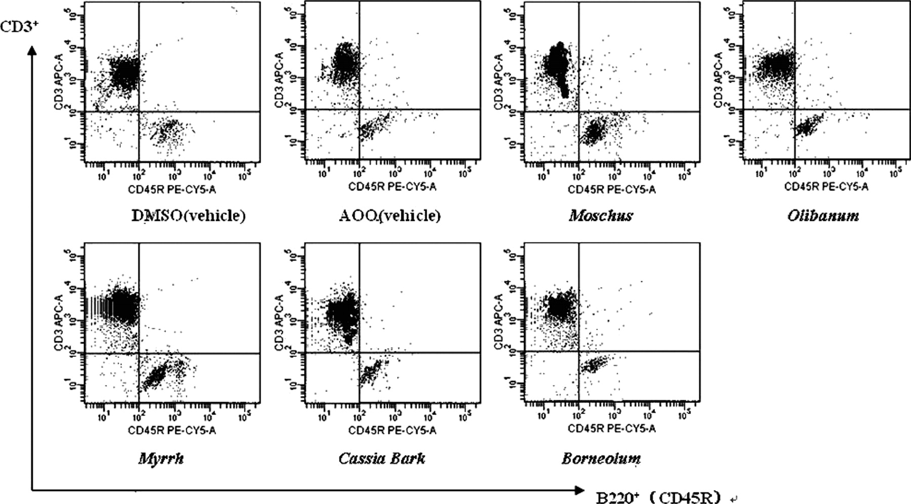
Lymphocyte composition of auricular lymph node cells treated by TMEs. Lymphocytes were isolated from CBA mice. Single cell suspensions were stained with APC-labelled anti-mouse CD3 and PE-Cy5-labelled anti-mouse B220 (CD45R), followed by flow cytometry measurement. Plots shown are from the representative experiments of each high dose TME group and vehicle groups. The vehicle for Moschus, Olibanum, Myrrh and Cassia Bark was dimethyl sulfoxide (DMSO), and the vehicle for Borneolum was acetone-olive oil (AOO). Moschus, Olibanum and Myrrh increased significantly the percentage of B220+ cell comparing with vehicle group, but Cassia Bark and Borneolum had no effect on the percentage of B220+ cell.
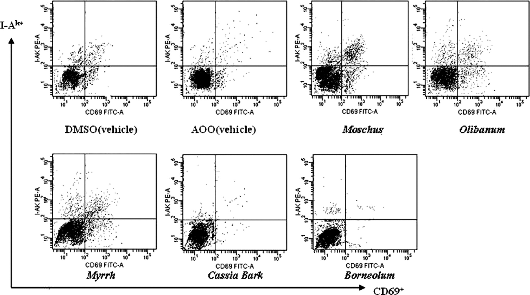
Flow cytometric analysis of the expression of I-Ak and CD69 in auricular lymph node cells treated by five TMEs. Single cell suspensions were stained with FITC-labelled anti-mouse CD69 and PE-labelled anti-mouse I-Ak, followed by flow cytometry measurement. Plots shown are from the representative experiments of each high dose TME group and vehicle group. The vehicle for Moschus, Olibanum, Myrrh and Cassia Bark was dimethyl sulfoxide (DMSO), and the vehicle for Borneolum was acetone-olive oil (AOO). Moschus and Myrrh showed higher expression of I-Ak+/CD69+ than vehicle group.
Footnotes
This study was supported by National Scientific & Technological Research Program of China “Key Technology Research for Drug Safety Evaluation” (2006BAI14B06), by Innovative Drug Development Program of China “Technical Platform for Drug Safety Evaluation,” and by a National 863 project (2006AA03Z356).