
Abstract
This study reports the in vitro biocompatibility of a composite biomaterial composed of 46S6 bioactive glass in association with chitosan (CH) by using 3D osteoblast culture of SaOS2. The 46S6 and CH composite (46S6–CH) forms small hydroxyapatite crystals on its surface after only three days immersion in the simulated body fluid. For 2D osteoblast culture, a significant increase in cell proliferation was observed after three days of contact with 46S6 or 46S6–CH-immersed media. After six days, 46S6–CH led to a significant increase in cell proliferation (128%) compared with pure 46S6 (113%) and pure CH (122%). For 3D osteoblast culture, after six days of culture, there was an increase in gene expression of markers of the early osteoblastic differentiation (RUNX2, ALP, COL1A1). Geometric structures corresponding to small apatite clusters were observed by SEM on the surface of the spheroids cultivated with 46S6 or 46S6–CH-immersed media.
We showed different cellular responses depending on the 2D and 3D cell culture model. The induction of osteoblast differentiation in the 3D cell culture explained the differences of cell proliferation in contact with 46S6, CH or 46S6–CH-immersed media. This study confirmed that the 3D cell culture model is a very promising tool for in vitro biological evaluation of bone substitutes’ properties.
Introduction
Among synthetic biomaterials, bioactive glasses are particularly studied for several applications in bone tissue engineering [1]. They have the ability to bind directly to the bone by the formation of a carbonated hydroxyapatite (HCA) layer close to the mineral phase of bone. This HCA layer provides an ideal environment for colonization, proliferation and differentiation of osteoblasts [2].
“Composite” or “hybrid” biomaterials mimicking both the organic and inorganic phases of bone may promote osseointegration by combining the biological and mechanical properties of each constituent [3]. It was in this context that the association of 46S6 bioactive glass to chitosan (46S6–CH) was synthesized and the physico-chemical characteristics of 46S6–CH have been previously evaluated by our laboratory [4].
Chitosan (CH) is a natural polysaccharide derived from chitin by deacetylation [5,6]. The cationic nature of chitosan is primarily responsible for electrostatic interactions with compounds of the extracellular matrix like anionic glycosaminoglycans, proteoglycans and other negatively charged molecules [7]. Chitosan is also known to be biocompatible, biodegradable, non-antigenic, bacteriostatic and fungistatic [8,9]. It also has angiogenic properties, immunomodulatory effects and it plays an important role in the phenomena of bone repair [10].
46S6 bioactive glass is a four-component melt-derived glass with a wt% composition: 46% SiO2, 24% Na2O, 24% CaO, and 6% P2O5 [4]. This bioactive material forms a layer of hydroxyapatite after immersion in simulated body fluid (SBF) [11].
The goal of this study was to use, for the first time, a 3D osteoblast culture to evaluate the effects of 46S6–CH on cell proliferation and differentiation.
Material and methods
Elaboration of 46S6 bioactive glass
Bioactive glass was prepared by the melting method according to the specific heating regime as previously mentioned [12]. The melted bioactive glass was poured into pre-heated brass moulds and annealed for four hours at the glass transition temperature (about 536°C) to relieve the residual thermal stresses. Finally, after cooling to room temperature, the bulk glasses were ground to a fine powder and sieved, to obtain a bioactive glass with particle size less than 40 µm.
Preparation of 46S6 bioactive glass–chitosan composite (46S6–CH)
The 46S6–CH composite containing 17 wt% of chitosan polymer was prepared following several steps. Chitosan polymer (with degree of deacetylation 85%) was dissolved in a 1 wt% acetic acid aqueous solution. At the same time, the bioactive glass 46S6 suspension was prepared by ultrasonication of the glass microparticles in 2 ml of 1 wt% acetic acid for 30 min. Then, the prepared 46S6 suspension was gradually suspended into chitosan solution and stirred at 1200 rpm for two hours. The mixture of 46S6–CH was immersed in 10% NaOH solution, in order to neutralize the residuals of acetic acid. Finally, composite particles were frozen by liquid nitrogen for 30 min and transferred to a freeze-dryer [12].
Physico-chemical characterizations before and after soaking in simulated body fluid (SBF): In vitro assays in SBF solution
The in vitro study of 46S6 glass and 46S6–CH composite was carried out by soaking 100 mg of material samples in 200 ml of simulated body fluid with pH and mineral composition similar to those of human blood plasma. The SBF solution was prepared according to Kokubo’s technique. The powder samples were immersed in SBF solution and maintained at body temperature (37°C) under controlled agitation of 50 rpm for 1, 3, 7, and 15 days (dynamic in vitro degradation).
The physicochemical characterizations were performed on the outer layer formed on the surface of the bioactive glasses. This layer was removed by scraping. X-ray diffraction measurements were realized on a Bruker D8 Advance diffractometer. The powder samples were mixed homogeneously with cyclohexane (volatile solvent) and were placed on the surfaces of plastic tablets. Then, these tablets were introduced into a diffractometer. The X-Ray Diffraction (XRD) data were acquired in the range of 10–70° (
Bidimensional (2D) and three-dimensional (3D) cell culture of SaOS2 in basic culture medium (BCM)
SaOS2 is a human osteosarcoma cell line. For propagation, cells were cultured with DMEM medium, supplemented with 10% foetal calf serum, 15 mM HEPES buffer, 2 mM L-Glutamine, 100 UI · ml−1 penicillin and 100 µg · ml−1 streptomycin at 37°C in a humidified incubator. After reaching approximately 80% confluency, cells were harvested and subcultured.
For 2D cell culture, SaOS2 cells were plated in 96-well microplates with 200 µl of BCM (initial concentration: 2.5 × 10⁴ cells · ml−1 for the three day assays and 1 × 10⁴ cells · ml−1 for the six day assays). Overnight the monolayer cultures were incubated with dissolution products. The specific seeding densities were chosen in order to obtain 80–90% confluence at the end of incubation period.
For 3D cell culture, 96-well microplates were firstly coated with 50 µl of a 1.5% agarose gel. Secondly, 1 × 10⁴ cells · ml−1 SaOS2 cells were deposited in coated microplates with 200 µl of BCM. The microplates were incubated for four days to allow spheroids formation.
BCM was used as control for all the experiments with 46S6, CH or 46S6–CH-immersed media.
Preparation and contact with 46S6, CH or 46S6–CH-immersed media (Table 1)
46S6–CH-immersed medium was obtained by soaking 0.2% (w/v) of 46S6–CH in 100 ml of DMEM supplemented with antibiotics. Pure 46S6 or CH-immersed media were obtained with respect to the wt% ratio in the association (83% 46S6, 17% CH). The concentration of particles was respectively 0.16% (w/v) for 46S6 and 0.04% (w/v) for CH. After 24 h of incubation at 37°C in a humidified incubator of 5% (v/v) CO2, media were filtered through a 0.2 µm sterile filter. Filtrates were supplemented with FCS, L-Glutamine and HEPES buffer. Media were renewed on day 3 (D3).
Description of samples
Description of samples
Viable cells were determined by the MTT assay as described [13]. 50 µl of a solution of MTT (1 mg/ml) was added to the media during three hours. The media were then removed and 100 µl of DMSO was added to each well to dissolve the formazan crystals. After ten minutes agitation, the optical densities (OD) of the solutions were read at 550 nm with a spectrophotometer. The assay was performed on D3 and D6. All the experiments were conducted in triplicate.
Acid phosphatase (APH) assay
The APH was applied to determine cell viability without prior dissociation of spheroids [14]. Samples of 3D cultures were obtained by pooling four spheroids from the same culture conditions into microtubes. After centrifugation (2000 rpm; 3 min), spheroids were washed in phosphate buffer saline (PBS), and tested under 100 µl of PBS. Then 100 µl of an assay buffer (Immunopure p-nitrophenyl phosphate 20 mg/10 ml, sodium acetate 0.1M, Triton ×100, 0.1%) was added to each tube. Following incubation (37°C, 90 min), 10 µl of 1N NaOH was dispensed into each tube and OD were measured at 405 nm. The APH assay signal allows the establishment of regression lines from a standard range of SaOS2 cell suspensions to determine the number of viable cells at D3 and D6. All the experiments were conducted in triplicate.
Cell differentiation: Reverse transcription real time quantitative PCR
After being washed twice in PBS, the cells were lysed in 1 ml TRIzol per flask. Total RNA was isolated according to the manufacturers’ instructions. RNA was reverse transcripted into complementary DNA by the use of the Protoscript® M-MuLV First Strand cDNA Synthesis Kit. Quantitative real-time PCR was performed in a PCRq 7900 HT in the presence of 1× Power SYBR Green PCR Master Mix and 300–900 nM of each of forward and reverse primers. Table 2 shows primer sequences of Collagen 1 (COL1A1), RUNX2, osteopontin (OPN), bone sialoprotein (IBSP), alkaline phosphatase (ALP), osteocalcin (OCN) and hypoxanthine-guanine phosphoribosyltransferase (HPRT). The efficiency of the amplification for each target gene was assessed with the use of serial dilutions of the cDNA template. Samples were analysed in triplicate. Data analysis was carried out using SDS v2.3 software.
Sequences of primers used for real-time RT-PCR
Sequences of primers used for real-time RT-PCR
Spheroids from the same culture conditions were pooled into microtubes, rinsed twice in PBS and fixed for 24 h in 2.5% glutaraldehyde in PBS. Fixed samples were washed twice in PBS and dehydrated twice in graded alcohol (80°, 95°, 100°). After critical point drying (CPD 010, Balzers Union) according to the standard procedure using liquid carbon dioxide, the samples were sputtered with gold palladium and were examined via a scanning electron microscopy. All studies were performed in triplicate.
Statistical analysis
The data are expressed as the mean ± SD. Differences between groups were analysed by one way analysis of variance (ANOVA) followed by Fisher’s Protected Least Significant Difference (PLSD) with values of
Results
Physico-chemical characterizations before and after soaking in SBF: XRD characterization
The XRD diagrams of the 46S6 glass surface are shown in Fig. 1, before and after soaking in SBF. The XRD diagram of hydroxyapatite is presented as a reference to evaluate the bioactivity of the material as a function of immersion time [JCPDF 09-432 card]. The XRD diagram of the surface of initial glass did not show any evidence of a crystalline phase which confirmed the amorphous structure of bioactive glass.
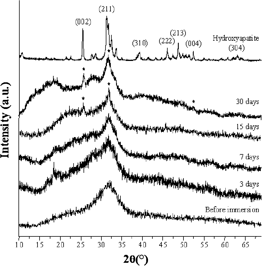
X-ray pattern of pure glass 46S6 before and after soaking in SBF for different durations.
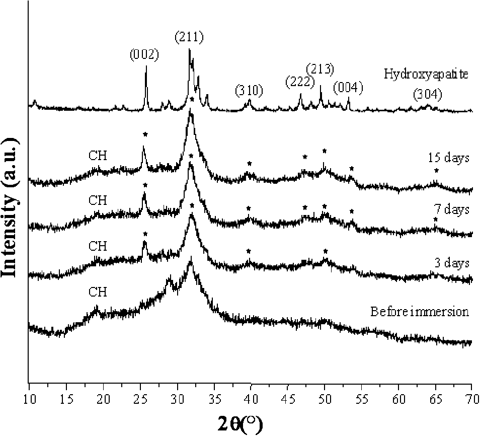
X-ray pattern of composite glass–chitosan 46S6–CH before and after soaking in SBF for different durations.
The XRD diagrams of the surface of 46S6–CH composite are shown in Fig. 2, before and after different durations of immersion in SBF solution. The hydroxyapatite XRD diagram served as reference [JCPDF 09-432 card]. The X-ray diffractogram of the initial 46S6–CH composite exhibited one characteristic peak of chitosan, at about 20° (
Figures 3 and 4 show the ratios (%) of optical densities (OD) measured from cell cultures cultivated with 46S6, CH or 46S6–CH-immersed media, compared with the OD measured from cell cultures cultivated with basic culture medium (BCM) after three days (D3) and six days (D6) of contact.
MTT assay
For 2D cell cultures (Fig. 3) at D3, there was a significant increase in cell proliferation for media containing 46S6 (
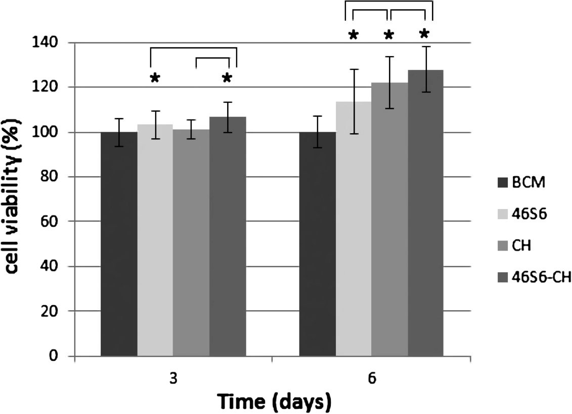
Cell viability of SaOS2 (2D cell culture) after three days and six days of contact with basic culture medium (BCM), 46S6, CH or 46S6–CH-immersed media. Data are represented as percentage of cell viability relative to BCM at day 3 (D3) and day 6 (D6) respectively. * Statistically significant difference from BCM (
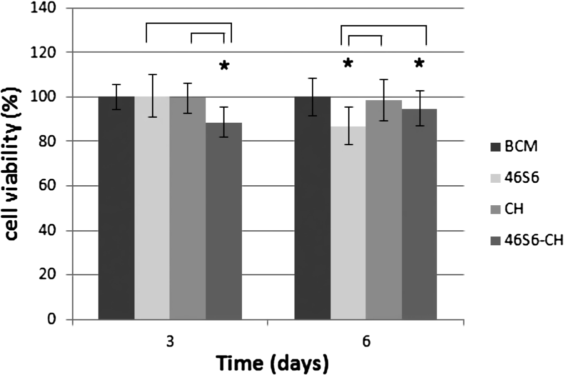
Cell viability of SaOS2 (3D cell culture) after three days and six days of contact with BCM, 46S6, CH or 46S6–CH-immersed media. Data are represented as percentage of cell viability relative to BCM at D3 and D6 respectively. * Statistically significant difference from BCM (
At D6, cell proliferation was significantly higher for all the culture media (
For 3D cell cultures (Fig. 4) at D3, there was no difference between 46S6, CH and BCM. For 46S6–CH, there was a significant decrease of cell proliferation by 11% (
Cell differentiation: Reverse transcription real time quantitative PCR
Regarding the 2D cultures, there was no significant variation in gene expression of the markers of osteogenesis for any experimental conditions (data not shown).
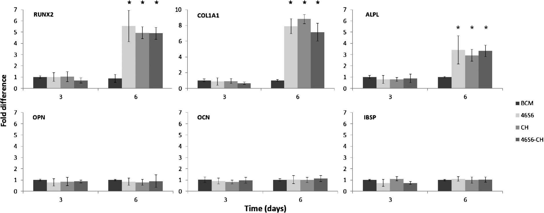
Gene expression profile of SaOS2 (3D cell culture) after three days and six days of contact with BCM, 46S6, CH or 46S6–CH-immersed media. Data are mRNA levels of the gene of interest and are represented as x-fold differences relative to BCM at D3 and D6 respectively. Data have been normalised to the expression of the housekeeping gene HPRT. *
For the 3D cultures, there was no significant variation in gene expression of the markers of osteogenesis after three days of contact. At D6 there was an increase in gene expression of markers of the early osteoblastic differentiation (RUNX2, ALP, COL1A1) with ratios greater than three for 46S6, CH, 46S6–CH-immersed media compared with BCM (Fig. 5). On the other hand, markers of the terminal osteoblastic differentiation (OCN, IBSP, OPN) did not vary.
At D3, all spheroids showed all the same aspect regardless of the culture media. At low magnification, they presented a regular spherical shape (Fig. 6) and the cells were perfectly nested within each other, forming a compact surface. With 46S6–CH, there were many signs of cellular stress with cells dropping out of the cell mass (Fig. 6).
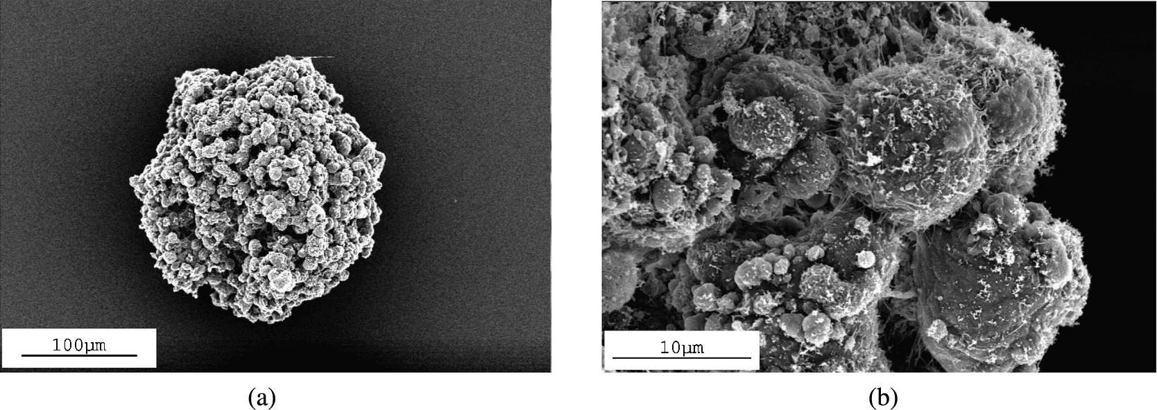
SEM image of spheroids of SaOS2 at D3. (a) They all show a regular spherical shape regardless of the culture media, (b) except for spheroids cultivated with 46S6–CH, cells form a compact surface but many cells drop out of the cell mass, corresponding to signs of cellular stress.
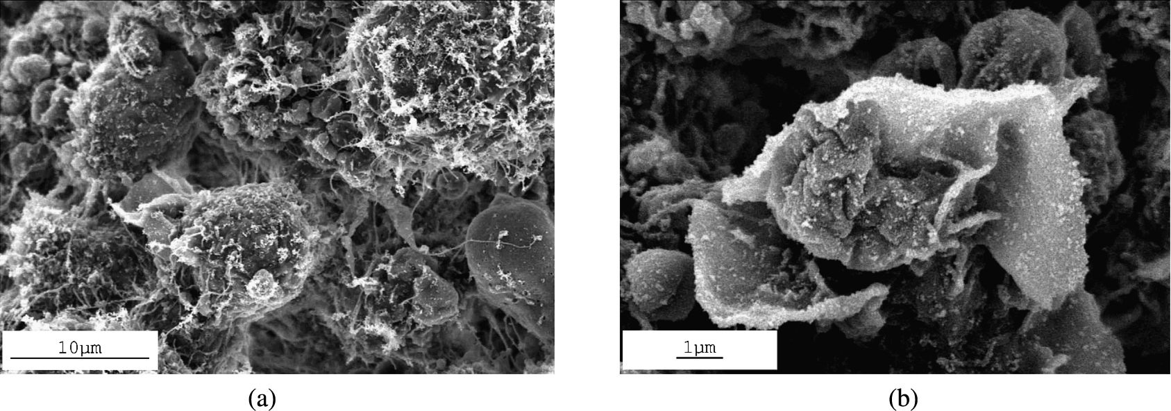
SEM image of spheroid of SaOS2 at D6. (a) Vesicles appear on the cell surface but many intercellular junctions remain. (b) For spheroids cultivated with 46S6-immersed media, geometric structures like flower petals appear.
At D6, vesicles appeared on the cell surface, even though there were many intercellular junctions (Fig. 7). It is also noteworthy that geometric structures like flower petals appeared on the surface of the spheroid cultivated with media containing pure 46S6 or 46S6–CH (Fig. 7).
Our study reports the biological effects of for 46S6, CH or 46S6–CH-immersed media on three-dimensional models of osteoblast culture. The bioactivity and chemical reactivity of 46S6 have been previously tested in combination with additive elements such as magnesium, zinc or strontium [11,15,16]. While 46S6 showed promising results both in terms of chemical and biological properties, the brittleness of bioglasses and the need for hybrid biomaterials with controlled resorption kinetics led to the development of new biomaterials mimicking both organic and inorganic phase for bone tissue engineering [17]. In this way, the association of bioactive glass with chitosan was elaborated [18]. The hypothesis is that such a composite material could be used to create robust bioactive coatings or a bioactive scaffold for bone tissue engineering applications [9].
X-ray diffraction highlights the interactions between glass matrix and chitosan. Chitosan’s structure corresponds to a type II orthorhombic crystallization [12,19] while pure glass presents an amorphous system. After three days of soaking in SBF solution, the characteristic peaks of hydroxyapatite clearly emerged in the X-ray pattern of the 46S6–CH composite. This illustrates the rapid rate of formation of a hydroxyapatite layer on the surface of the 46S6–CH composite in comparison with pure 46S6 glass. As the immersion time was prolonged, the intensities of the characteristic peaks of hydroxyapatite phase became more pronounced. This confirms the good combination of biocomposite [12]. After 7 and 15 days of immersion in SBF, the diffractogram of 46S6–CH composite presented all of the characteristic peaks of hydroxyapatite crystals. It is recognized that the hydroxyapatite peaks formed on the surface of biocomposite are more visible than those found on bioactive glass [20]. That highlights the good crystallization of the apatite layer on the surface of biocomposite.
Ionic dissolution products of bioactive glasses have been shown to change the intracellular ionic concentrations and therefore to mediate cell metabolism [21,22]. Many studies have demonstrated that silicon and other released products promote the proliferation of different cell types [23–26] and that these products play an important role for the osteogenic effects [27–29]. The release of silica could have a dual role, indirectly by promoting the adhesion of osteoblasts to the hydroxyapatite layer [30] and directly by stimulating osteogenesis through the Erk pathway and Runx2 expression in pre-osteoblastic cells [31] or through the expression of key genes Col1α1, Col1α2 and Runx2 [29]. The mechanisms by which dissolution products can modify cell physiology are not fully understood. The kinetics of release, the pH, the synergy between different chemical entities, and the role of trace elements are hypotheses being proposed. In this study, the association 46S6–CH led to a faster degradation and in fact it could allow ions such as silicon, calcium and phosphorous, which are required to form bone-like apatite, to be provided in a gradual manner. Further studies should provide better understanding of the significance of each ionic component in the 46S6–CH formulation toward the osteoblastic stimulation process.
On 2D cell cultures after three days of contact with 46S6–CH, we observed a statistically significant increase in cell viability. This is consistent with the study of Jun et al. who observed that the proliferation rate of osteoblastic cells cultivated on a bioactive coating was higher than that of the control substrate [8].
After six days of exposure, cell proliferation was significantly increased for the association 46S6–CH. This result clearly shows the positive effect of the association in terms of cell proliferation. Our results are corroborated by those of Luz et al. [32]. Lee et al. also found that after seven days of contact, the proliferation rate was significantly higher on the hybrid membrane [9]. But Mota et al. [33] did not find any significant differences in terms of proliferation.
In this study no cytotoxic effects of 46S6, CH or 46S6–CH-immersed media on 2D cell cultures were noted. A positive effect was observed for the association 46S6-chitosan on cell proliferation at D3 and D6, but the study of markers of osteogenesis did not show significant changes in gene expression. These results are not in agreement with various studies that report a positive effect on the cell cycle at D3 [34] and induction of differentiation at D6 [29,35]. Primocultures of human bone cells or rats were used in these previous works, which probably explains the discordance with our results obtained with the cell line SaOS2.
Three-dimensional cultures like spheroids have attracted great interest as a tool to screen therapeutic agent effects in cancer research that cannot be tested with conventional 2D cell-culture. This is because 3D cultures more closely mimic the cell-cell interactions and the topography found in vivo [36]. Spheroids models filled the gap between monolayer cultures and animal models [14].
Taken together, data on cell proliferation and gene expression suggest that the deficit in cell numbers observed with 46S6–CH in 3D cell-culture at D3 results from a relative toxicity of 46S6, CH or 46S6–CH-immersed media. Indeed, of all the culture media tested in 3D cell-culture, the study of gene expression markers of osteogenesis failed to show induction of gene expression of markers of osteoblastic differentiation at D3. This fact is corroborated by signs of cell suffering observed in the SEM. Cells come off the spheroid through rupture of the intercellular junctions. These differences between 2D and 3D cell-cultures have already been observed in our laboratory with other bioactive glasses [37]. Moreover, such a differential effect of external molecules on 3D cultures compared with 2D cultures has already been reported [38]. Cell viability in spheroids requires intimate interactions between cells, intercellular junctions and concentration gradients. We propose the hypothesis that, in 3D cell-culture, intercellular structures are disrupted by contact with the association 46S6/chitosan. This could induce either cell death or a rupture of the concentration gradient, resulting in an overall deficit of proliferation.
Regarding the 3D cell cultures at D6, the deficit in the number of cells compared with the 2D cell cultures is not due to a toxic effect but to an effect of induction of cell differentiation. Indeed, the study of gene expression markers of osteogenesis showed an induction of gene expression of early markers of osteoblast differentiation (RUNX2, ALP, COL1A1). It has previously been shown that gene expression of collagen is modified after 24 h of contact [29] and that the expression of most other markers is modulated in the first 96 h [39]. Some studies have reported up-regulation up to five-fold of genes involved in osteoblast metabolism, proliferation and cell–cell and matrix–cell adhesion when human osteoblastic cells were cultured with Bioglass®-immersed media [22,40,41]. Related studies by Jell et al. [27] showed six- and three-fold up-regulation of osteogenic markers, namely bone sialoprotein (IBSP) and ALP, in osteoblasts treated with medium containing bioactive glass dissolution products, resulting in enhanced cell differentiation. In our study, we did not detect any induction of gene expression of late markers of osteoblast differentiation (OPN, OCN, IBSP). This is probably due to the physiological difference between our 3D cell culture model and primocultures and to the physico-chemical properties of 46S6.
A study of Tsigkou et al. [35] investigated the effect of 45S5 Bioglass® dissolution products on 2D culture of human fetal osteoblasts. They demonstrated that the dissolution products create an extracellular matrix deposition and mineralization. In our study, structures like flower petals were observed by SEM on spheroid cultivated with pure 46S6 or 46S6–CH-immersed media. These formations correspond probably to the small apatite clusters described by Luz et al. [18], which represent the beginning of mineralization of the extracellular matrix.
Conclusion
This study identifies, for the first time, the biological properties of 46S6–CH-immersed media by using a three-dimensional osteoblast culture. The bioactivity of the association 46S6–CH has been shown because of the rapid rate of formation of a hydroxyapatite layer compared with pure 46S6. The model spheroids allowed the different cellular responses compared with 2D classical cell culture to be highlighted. The effects of dissolution products on cell proliferation were not the same, but depended on the cell culture model. This difference is due to the osteoblast differentiation induction in the 3D model. Our work demonstrates the usefulness of a 3D model to evaluate more precisely the in vitro biological properties of a bone substitute.
Footnotes
Acknowledgements
The authors would like to thank Jean Lecerf, Joseph Le Lannic, Hélène Solhi and Céline Allaire for their valuable assistance. This work was supported by the Fondation Langlois.