
Abstract
BACKGROUND:
Enzyme-linked immunosorbent assays (ELISA) are considered the gold standard in the demonstration of various immunological reactions with an application in the detection of infectious diseases such as during outbreaks or in patient care.
OBJECTIVE:
This study aimed to produce an ELISA-based diagnostic with an increased sensitivity of detection compared to the standard 96-well method in the immunologic diagnosis of infectious diseases.
METHODS:
A ‘3DStack’ was developed using readily available, low cost fabrication technologies namely nanoimprinting and press stamping with an increased surface area of 4 to 6 times more compared to 96-well plates. This was achieved by stacking multiple nanoimprinted polymer sheets. The flow of analytes between the sheets was enhanced by rotating the 3DStack and confirmed by Finite-Element (FE) simulation. An Immunoglobulin G (IgG) ELISA for the detection of antibodies in human serum raised against Rubella virus was performed for validation.
RESULTS:
An improved sensitivity of up to 1.9 folds higher was observed using the 3DStack compared to the standard method.
CONCLUSIONS:
The increased surface area of the 3DStack developed using nanoimprinting and press stamping technologies, and the flow pattern between sheets generated by rotating the 3DStack were potential contributors to a more sensitive ELISA-based diagnostic device.
Keywords
Introduction
Immunoassays such as Enzyme-linked Immunosorbent Assay (ELISA) are important methods for the detection of various proteins in immunologic reactions with an application in numerous disciplines including the diagnosis of infectious diseases [1–6]. ELISAs have high specificity and sensitivity and are usually carried out in 96-well microtiter plates which allow multiple assays to be carried out concomitantly [1–4]. Nonetheless, it is time-consuming (e.g., many hours) to complete the assay due to its small diffusion coefficient often requiring large volumes of reagents [2–9]. Achieving a larger surface area of the present 96-well would lead to an increase in its diffusion coefficient requiring more time and volume of reagents to complete the assays [10–12].
The reaction kinetics in high surface-to-volume ratio diagnostics is rapid due to the shorter diffusion distance for analytes to reach the reaction zones. The reaction kinetics during an assay is influenced by the flow of analytes (e.g., convection, diffusion and binding) and the structural characteristics of the ELISA vessel [11–14]. Improvements in the reaction kinetics by addressing the flow of analytes have been made using various propulsion mechanisms assisted by the use of hydraulic, pneumatic, electrochemical and centrifugal pumps [15,16]. Other advancements to improve the efficiency of the assay have also been addressed through advancements in microelectromechanical (MEMS) and micro total-analysis (µ-TAS) systems by producing high surface-to-volume ratio devices [10,17,18]. These include compact-disc (CD) – [15,19], Lab-On-a-Chip (LOC) – [2,20], and inkjet-driven µ-TAS [9,21–23] – based devices that incorporate the use of carbon-nano tubes [2,20,24], microbeads [9,25], centrifugal force [15] and the use of various polymers [8,9,15,19,26–28]. These enhancements have been useful in ensuring high-throughput and improving the sensitivity of the ELISA compared to the standard method [11,15,29]. Although many of these advancements have been validated, they are often complex to perform, require skilled instrumentation and fall short in terms of the number of samples that can assayed concomitantly compared to the standard methods [1,6].
Numerous conventional and more complex microfabrication technologies have been used in the development of various LOC, microfluidic and Point of Care (POC) diagnostics due to its availability, low-cost and high achievable accuracy [18,30]. The application of these technologies in biology and medicine has produced high-throughput, high surface-to-volume ratio devices that are composed of readily available thermoplastics. These biomedical devices are portable, easy to use and disposable, all of which are important consideration in the development of newer devices for immunoassays [18,30–34].
This study aimed at developing a high sensitivity ELISA vessel by increasing the surface area for reaction compared to the standard method using readily available, low cost fabrication technologies namely nano-imprinting and press stamping. The validation of this vessel called 3DStack was carried out through the immunologic diagnosis of infectious diseases without modifying the current practices for the standard 96-well ELISA.
Materials and methods
Specification and design rationale
The 3DStack prototype used in this study aimed at producing a higher surface area for reaction compared to 96-well plates by stacking multiple polymer sheets held together by a central support core composed of a cylindrically rolled stainless steel mesh and a magnetic base. The 3DStack was designed composed of 10 circular shaped polyethylene terephthalate (PET) sheets of 5.0 mm in diameter. The diameter of each sheet and the total height of the 3DStack did not exceed that of the diameter and the height of the 96-well plate, respectively. Each sheet in turn was designed to have a central opening of 2.0 mm in diameter to allow assembly on to the central support core. The diameter of each sheet and the total height of the 3DStack were important parameters to allow the prototype to sit in 96-well plates. In addition, the magnetic base of the central support core was to allow the 3DStack to be rotated using a magnetic stirrer to enhance the flow between sheets.
Parameters used for the development of 3DStack prototypes
Parameters used for the development of 3DStack prototypes
Surface area of 96-well: 151 mm2.
To further increase the surface area of the 3DStack, the upper surface of each sheet was imprinted with a micrometer-scale pattern (µ-pillars). Two different µ-pillars shapes were used in the imprinting of the sheets; square (S) and circular (C). The total surface area of 3DStack(S) and 3DStack(C) was 4 and 6 times more than the 96-well, respectively [surface area: 604 mm2 [3DStack(S)] and 924 mm2 [3DStack(C)] vs. 151 mm2 (96-well)]. The 3DStack(S) was designed without the use of spacers between sheets while the 3DStack(C) employed the use of spacers of 100 µm in height. The total combined height of 3DStack(S) and 3DStack(C) were 1.3 mm and 2.2 mm, respectively. The surface dimensions of the µ-pillars used in this study were [height, diameter and distance between pillars (µm)]: (S) 9.5, 45.0 and 48.0; (C) 1.8, 2.0 and 3.1, respectively. The different parameters used in the development of the 3DStack prototypes are summarized in Table 1. A schematic diagram of the 3DStack prototype is shown in Fig. 1.
The roller ultraviolet (UV) nano-imprinting machine (Soken Chemical & Engineering, Tokyo, Japan) consisted of an imprint forward roller with the appropriate µ-pillar surface mold and a support plate. The µ-pillar mold was mechanically laminated onto a UV curable acrylic resin-PET film composite at a rolling speed of 5 mm/min. UV (375 nm) curing was then performed on the resulting composite. The PET film was detached from the mold at a rolling speed of 2 mm/min and UV (365 nm). Each film used for the nano-imprinting process was 130 µm in thickness and
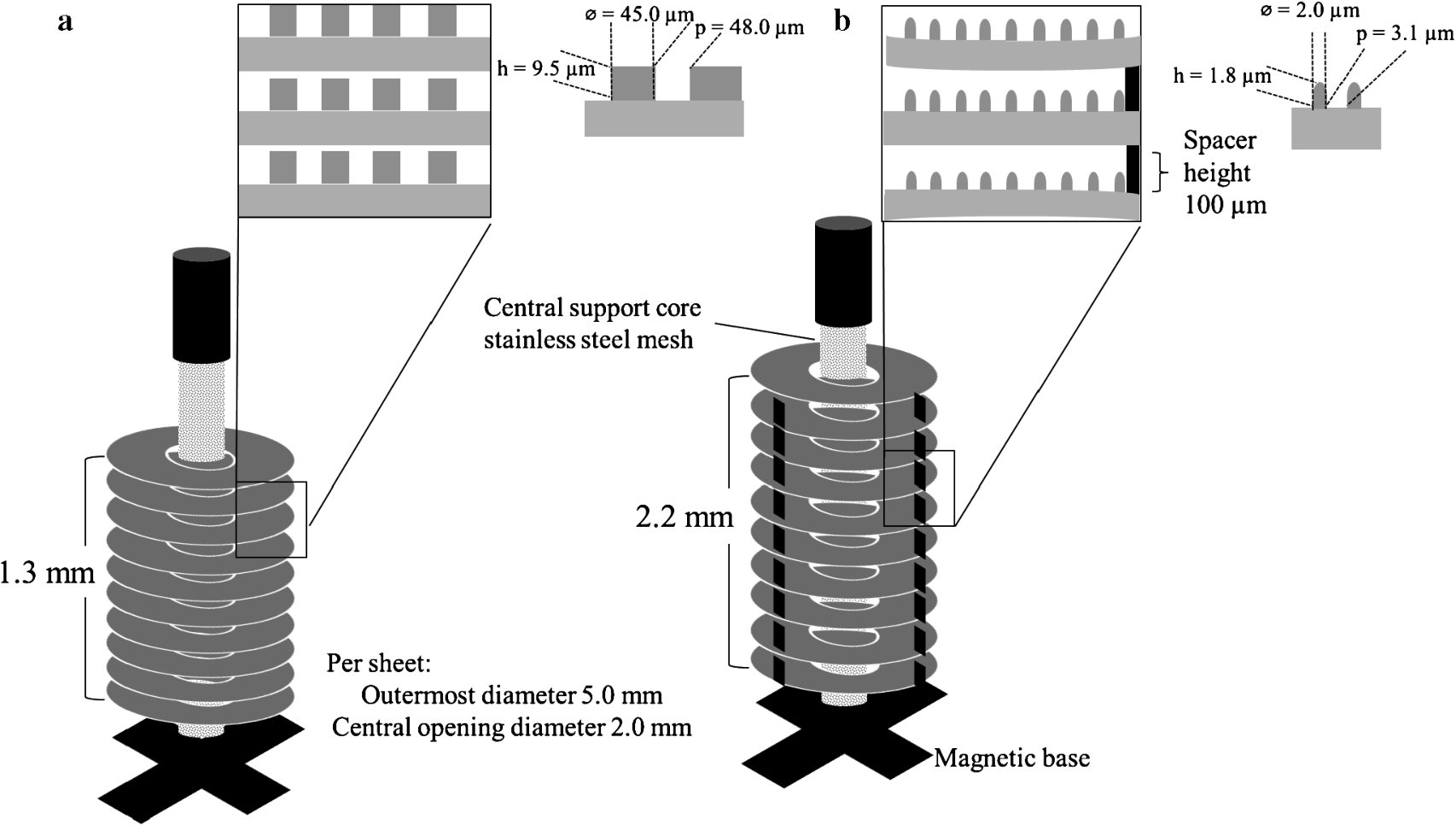
Design: Schematic diagram of the assembly to the central support core of (a) 3DStack(S) and (b) 3DStack(C) (both, 10 sheets). Insert shows µ-pillars patterns and dimensions on each sheet (h, height; ∅, diameter and p, pitch or distance between µ-pillars).
Numerical values of the parameters used for the FE simulation of 3DStack(S) and 3DStack(C)
The µ-pillar height and width of the 3DStack(C) were ignored in the FE simulation since the space between the sheets were due to the height of the spacer (i.e., 100 µm).
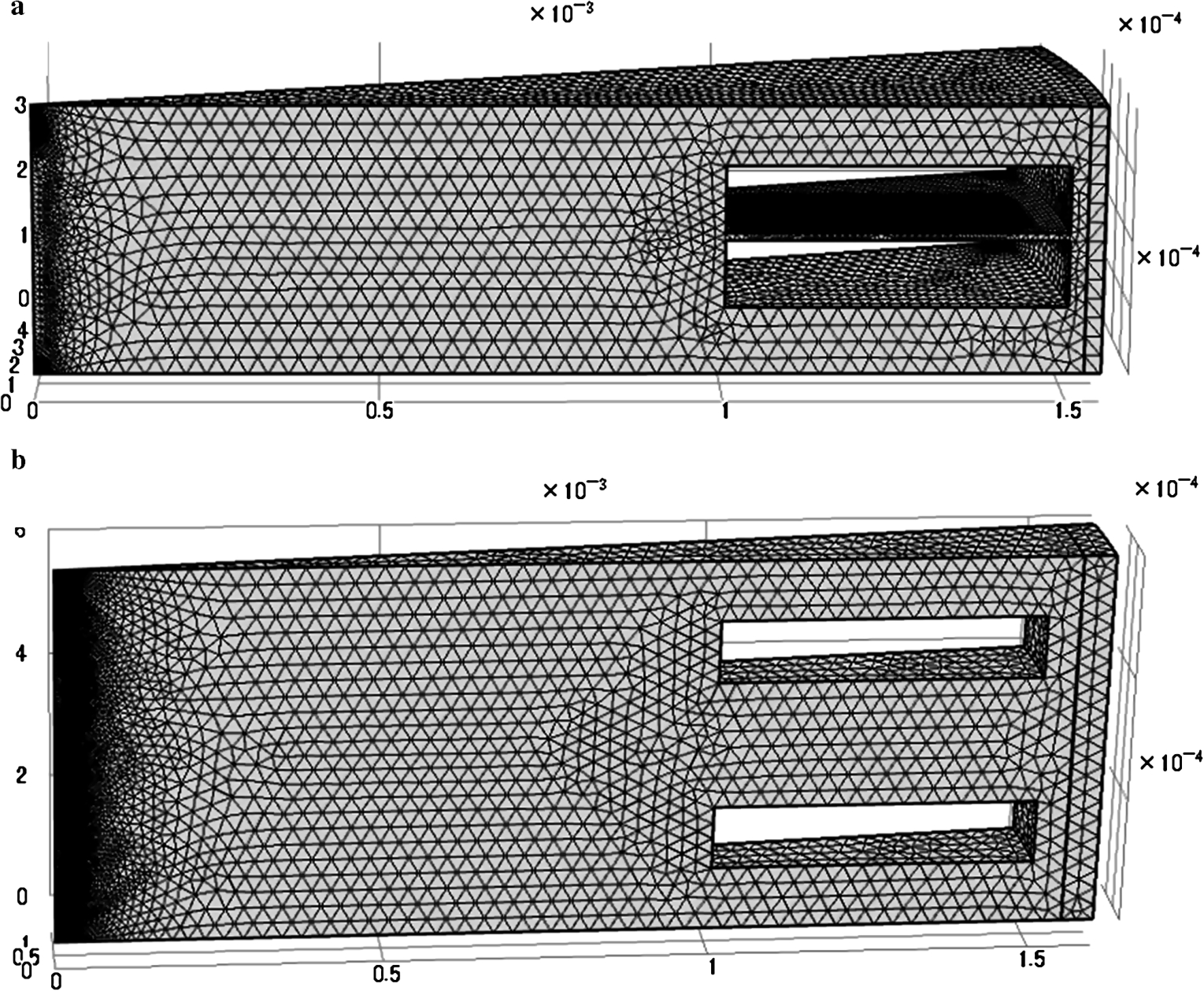
3D model used for FE simulation of flow between sheets of (a) 3DStack(S) with µ-pillar height responsible for the space between sheets and, (b) 3DStack(C) µ-pillars not shown because the space between sheets was determined by the height of the spacer (i.e., 100 µm). x-, y- and z-axis represents length in m.
The surface of each PET film was made hydrophilic by plasma treatment (18W, PDC-32G, Harrick Plasma Co., New York, USA) with oxygen for 5 min before nanoimprinting and 3DStack assembly.
FE simulation was carried out to confirm the flow pattern between sheets of the 3DStack(S) and 3DStack(C) using COMSOL Multiphysics® 5.0 software (COMSOL, Stockholm, Sweden) based on the rotating machinery, laminar flow application. The numerical parameters used in the FE simulation are summarized in Table 2. In the simulation, the height of the µ-pillars of 3DStack(C) was ignored since the space between sheets was equal to the height of the spacers (100 µm). The 3D models of the 3DStack used in the FE simulation is shown in Fig. 2.
Sample collection and ethical statement
In an ongoing collaboration for the serological monitoring of infectious diseases in Central Vietnam, a total of 272 serum samples from patients who visited the Outpatient Department of the Hue University Hospital between March and June 2014 were collected and screened for Rubella virus antibodies by Immunoglobulin G (IgG) ELISA (96-well ELISA). Two samples, serum containing Rubella virus antibody (Rubella virus antibody positive sample) and serum not containing Rubella virus antibody (Rubella virus antibody negative sample), one each respectively, were selected and subsequently used in the 3DStack ELISA. Serum samples collected from all patients were carried out under informed consent. All protocols and procedures were approved by the Research and Ethical Committee for the use of human subjects of the Hue University and the Tokyo Metropolitan University.
IgG ELISA
IgG ELISA was carried out using 3DStack(S) and 3DStack(C). For confirmation purposes, parallel testing by 96-well ELISA was carried out at each step. Briefly, the 3DStack(S), 3DStack(C) and 96-well ELISA plates were coated with 100 µl of predetermined optimal quantity (1:2) of Rubella virus Hemaglutination Antigen (HA) (Denka-Seiken, Tokyo, Japan) in sodium carbonate buffer at 4°C overnight. After overnight incubation, blocking was performed using 200 µl of 0.05% Tween-20 phosphate buffer solution (PBS-T) containing 5% skimmed milk (PBST-M) for 1 h at room temperature (RT). Primary antibodies that consisted of 100 µl of test samples and reference antiserum (positive and negative) (Denka-Seiken, Tokyo, Japan) at a 1-point dilution of 1:400 was added to each well for 1 h at RT. The positive and negative reference antiserum was used to confirm the specificity of the binding and to determine the cut-off values for the assay. For validation purposes primary antibodies were diluted in PBST-M four-folds from 1:100 to 1:6400.
100 µl of 1:1000 horseradish peroxidase conjugated goat anti-Human IgG (Invitrogen, Camarillo, CA, USA) diluted in PBST-M which served as the secondary antibody was added to each well for 1 h at RT. Finally, 100 µl of substrate solution containing ABTS [2, 2′ azinobis (3-ethylbenzthiazolinesulfonic acid)] solution (Roche Diagnostics, Mannheim, Germany) was added to each sample well. The wells were incubated for 30 min at room temperature and optical density at 405 nm (OD405) was measured against a reference of 490 nm using a Model 680 Microplate Reader (Bio-Rad, Hercules, CA, USA).
Three rounds of washing were performed in between all steps with 300 µl of PBS-T per round. In the 3DStack ELISA, each step was carried out by placing the 3DStack in a new well of a 96-well plate as support/base and to neglect the ELISA efficiency contribution from the 96-well plate. Additionally, where incubation consisted of at least 1 h using the 3DStack, the flow between sheets for the first 15 min was enhanced by placing the 96-well plate containing 3DStack on a magnetic stirrer. The 3DStack was removed following incubation with the substrate solution before OD405 measurement.
Results
Physical evaluation
Each 3DStack consisted of 10 layers of circular shaped transparent sheets stacked on one another with the bottom most sheet resting on the magnetic base of a central support core. The individual sheets of 3DStack(S) were assembled to rest on one another while in the 3DStack(C) the sheets were separated by spacers (Fig. 3). Average measurements taken from three 3DStack [3DStack(S); 3DStack(C)] were: outermost diameter (5.20; 5.28 mm), central opening diameter (2.05; 2.13 mm) and total height (1.93; 2.27 mm). These measurements were comparable to those established in the 3DStack designing stage with an error in dimension of ±0.29 [3DStack(S)] and ±0.22 [3DStack(C)] mm, respectively.

3DStack prototype showing (a) top-most layer composed of circular sheets with central opening assembled on the central support core and side view of 3DStack(S) (upper panel) and 3DStack(C) with spacers (lower panel), and (b) placement of 3DStack in the 96-well plate. (Colors are visible in the online version of the article;
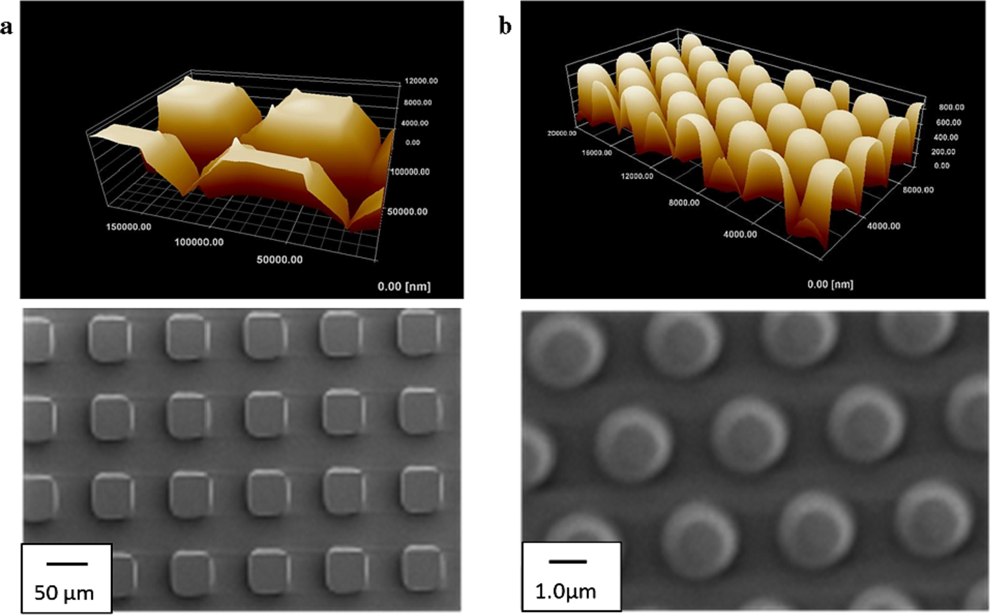
AFM image showing height of µ-pillars (upper panel) and SEM images (lower panel) showing diameter of- and distance between µ-pillar of (a) 3DStack(S) and (b) 3DStack(C). (Colors are visible in the online version of the article;
The 3DStack(S) and 3DStack(C) were patterned with square- and circular-shaped µ-pillars, respectively. The µ-pillar measurements based on the AFM and SEM images were: [height, diameter and distance between pillars (µm)]: 3DStack(S) 9.88, 45.5 and 48.8; 3DStack(C) 1.834, 2.017 and 2.985, respectively (Fig. 4).
FE simulation
The flow between sheets of the 3DStack is important for analytes to reach the reaction zones to allow antigen-antibody binding. In the 3DStack(S), the influence of both centrifugal and viscous forces on the flow pattern was observed. The flow pattern in the 3DStack(S) appears well distributed in between the sheets (convection) and along the surface (diffusion) while in the 3DStack(C) a radial flow pattern was observed in between sheets with limited flow along the surfaces (Fig. 5). The radial flow in the 3DStack(C) is probably due to the presence of the spacers (height of spacer is greater than the µ-pillars) which caused the sheets to diverge at the periphery. The flow between sheets of the 3DStack(S) due to the centrifugal and viscous forces were important in allowing the transport of analytes to the reaction zones and the removal of non-specific proteins, both of which are important considerations in the development of efficient ELISA-based and other microfluidic devices [11].

FE simulation showing the flow pattern and flow velocity in (a) 3DStack(S) with µ-pillars responsible for the interval height between sheets and (b) 3DStack(C) µ-pillars not shown because the space between sheets was due to the height of the spacer (i.e., 100 µm). Scale bar shows flow rate in m/s. (Colors are visible in the online version of the article;
An IgG ELISA for Rubella virus antibodies was carried out at a one-point dilution of 1:400 to compare the ELISA efficiency of the 3DStack prototypes (Fig. 6). In the 3DStack(S), the Rubella virus antibody positive and negative samples showed an OD value above and below the cutoff values, respectively. The OD values for the Rubella virus antibody negative sample and negative reference antiserum for the 3DStack(S) and 96-well ELISA were comparable, suggestive of the specificity of the 3DStack(S) ELISA. The results obtained from the 3DStack(C) ELISA were inconclusive (OD405: Rubella virus antibody negative sample > negative reference antiserum). Although high OD405 values were observed using the 3DStack(C), this is probably because of a high background. The limited flow between sheets of the 3DStack(C) as shown in the FE simulation may have hindered the thorough removal of non-specific proteins during the washing steps contributing to the higher background observed [1,2,11].
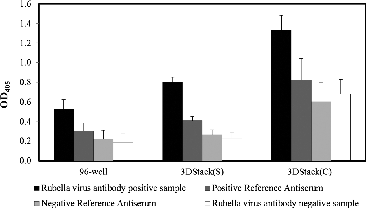
IgG ELISA for Rubella virus antibody. IgG ELISA for Rubella virus antibody was performed at a 1-point dilution of primary antibody (1:400). (OD405: Optical density measured at 405 nm; values represent mean of OD405 data obtained from 2 independent experiments.)
For validation purposes, serum samples were titrated fourfold from 1:100 to 1600. The OD405 value for the Rubella virus antibody positive sample was higher using the 3DStack(S) at all serum dilutions compared to the 96-well {OD405 [3DStack(S)] vs. 96 well}: 1:100 (2.0 vs. 1.5); 1:400 (1.2 vs. 0.8) and 1:1600 (0.55 vs. 0.2). The sensitivity of detection for the Rubella virus antibody positive sample using the 3DStack(S) ELISA was 1.3-fold (1:100), 1.5-fold (1:400) and 2.8-fold (1:1600) (average: 1.9-fold) higher compared to the 96-well ELISA. Student’s t-test analysis showed that the sensitivity based on the OD405 values obtained for the Rubella virus antibody positive sample at these serum dilution points using the 3DStack(S) were higher than that of the 96-well (

Validation by IgG ELISA for Rubella virus antibody at 1:100 to 1:1600 dilution of primary antibody (OD405: optical density measured at 405 nm). Points represent mean of OD405 values of 5 measurements (
Since the 3DStack(S) protypes were removed after incubation from the ABTS substrate solution placed in wells of the 96-well plate, loss of the ABTS substrate solution may not have been totally excluded. However, the signal variations observed at each dilution point using the Rubella virus antibody positive sample by the 3DStack(S) ELISA was smaller than that of the 96-well suggesting minimal loss of this solution (Fig. 7).
The ability of high surface-to-volume ratio diagnostics to produce important biochemical results due to the dominance of surface over volume effects were important consideration in the performance of the 3DStack ELISA [18]. The rapid development of the 3DStack design using readily available and low cost fabrication technologies to increase the surface area for reaction by stacking multiple nano-imprinted sheets were potential contributors to a more sensitive ELISA system compared to the 96-well ELISA. In addition, the prototype which was designed to fit snuggly into the well of a 96-well plate allowed the rapid performance of an initial assessment (IgG ELISA) without the need of numerous optimization steps (e.g., reducing time and reagent volumes) and modifications to standard institutional ELISA practices to be made. Although this study did not use reduced volumes or time, the improved detection sensitivity achieved using the 3DStack with an increased surface area compared to 96-wells will enable future optimizations to be explored. The consistency in the development of prototypes using nanoimprinting and press stamping as shown in this study were important in ensuring the consistency of the results obtained and in producing a more sensitive ELISA system [30,35].
While increasing the surface area was an important consideration in the development of the 3DStack, the flow of reagents and analytes to and from (i.e., during washing) the reaction zones are equally important to allow appropriate antigen-antibody binding and for the removal of non-specific proteins which may give false positive results [36]. In this study, the space between sheets determined by the µ-pillars in the case of the 3DStack(S) or the use of spacers in 3DStack(C) was responsible for the pattern of flow observed between sheets. The flow pattern observed due to the viscous and centrifugal force generated by rotating the 3DStack(S) allowed the removal of non-specific proteins and ensured the appropriate immunologic reaction to occur. Although the surface area of the 3DStack(C) was higher than that of the 3DStack(S), the flow pattern observed between the sheets of the 3DStack(C) was mainly radially-directed with minimal flow in the circumferential direction. This flow pattern, which was limited at the surface (i.e., reaction zone) may have reduced the diffusion of antibodies to the reaction zone. In addition, this flow pattern also hindered the removal of non-specific proteins which resulted in falsely elevated OD405 values. A combination of the different characteristics of 3DStack(C) (i.e., high surface area) and 3DStack(S) (i.e., flow pattern) should be explored in future work.
The flexibility offered by these readily available fabrication technologies in producing high surface-to-volume ratio devices and the improved detection sensitivity even using highly diluted samples as noted in this study provides future opportunities for readjustments to be made, including potentially miniaturization to reduce time and amount of reagents needed for such ELISA-based diagnostics [12,33–35]. The impact of these technologies in the use of ELISA systems for the rapid and accurate pathogen identification such as in the monitoring of infectious diseases during outbreaks or in the care of individual patients and its potential to reduce cost and time are crucial factors to consider especially in resource-constraint settings [33,34]. Additionally, the common principles shared by the different disciplines such as health, food industry, environmental, chemistry, biomedical and engineering and the flexibility in designing-product development-feedback cycle offered through these fabrication technologies provides future opportunities to be explored in the development of ELISA-based diagnostics [28].
Conclusion
The ease and rapidity of the 3DStack development and the achievable accuracy using readily available, low cost fabrication technologies namely nanoimprinting and press stamping in the rapid prototyping of a high surface-to-volume ratio provided an opportunity for a rapid assessment of the diagnostic performance of a 3DStack ELISA in the serological diagnosis of infectious diseases. The increased surface area and the flow pattern between sheets due to the viscous and centrifugal force generated by rotating the 3DStack contributed to an improved sensitivity of up to 1.9 fold higher compared to the 96-well ELISA. This study provides room for expansion of these technologies in the development of high sensitivity, high surface area miniaturized ELISA-based to reduce the time and volume of reagents required in immunological diagnosis for infectious diseases and its application in other disciplines.
Footnotes
Acknowledgement
We extend our appreciation to the doctors, nurses and laboratory staff at the Hue University Hospital who assisted us by collecting blood samples from the volunteers in this study. This study was financially supported by Grants-in-Aid from the Ministry of Health, Labor and Welfare Science Research Grant(s) (H24-Shinko-Ippan-013, H25-Shinko-Ippan-004) and the Tokyo Metropolitan Government (Asian Human Resource Fund).