
Abstract
Biocompatible metals have been suggested as revolutionary biomaterials for bone-grafting therapies. Although metals and their alloys are widely and successfully used in producing biomedical implants due to their good mechanical properties and corrosion resistance, they have a lack in bioactivity. Therefore coating of the metal surface with calcium phosphates (CaP) is a benign way to achieve well bioactivity and get controlled corrosion properties. The biocompatibility and bioactivity calcium phosphates (CaP) in bone growth were guided them to biomedical treatment of bone defects and fractures. Many techniques have been used for fabrication of CaP coatings on metal substrates such as magnesium and titanium. The present review will focus on the synthesis of CaP and their relative forms using different techniques especially electrochemical techniques. The latter has always been known of its unique way of optimizing the process parameters that led to a control in the structure and characteristics of the produced materials.
Introduction
During the past 50 years there has been a major progress in the development of biomedical materials that have been used in medical applications. Of these materials, metal implants represent an important category for their excellent mechanical and biomedical properties. Using modified metallic as orthopedic tools was an urgent need for Fixation and reformation the defects and fractures of bones and also to maintain the mechanical integrity. However, there are relevant problems associated with using metals when they are implanted as replacements for hard tissues. Biocompatibility is a foremost challenge confronting metals during their function as bioimplant. Coating of metal with a bioceramic layer such as calcium phosphate is a unique trial to face the above challenge. Generally, coating of the metal surface is to enhance and accelerate the process of osseointegration and also will increase its corrosion resistant [1].
Based on the literature titanium and stainless steel are ones of the best examples of metallic implants. Metallic alloys of cobalt, chromium and nickel base are also durable candidate biomaterials. Although the above metallic materials have one or more of the above mentioned advantages, there have been a number of disadvantages during real application including the discontinuity of these materials [2]. Those metals are suffering from toxic corrosion products [3] and allergens [4]. Difference in elastic modulus (EM) between metals and natural bone is another concern. For instance, whilst the values of EM of pure titanium is reported to be between 103–107 GPa [5], while those of trabecular/cancellous and cortical bone are 3–14.8 and 18.6–27, respectively [6]. These variations in EM between orthopedic metals (e.g., Ti and Fe-based alloys) and bones can cause stress shielding effect on bone and consequently led to ultimately severe medical conditions such as osteopenia [7]. Also, despite Ti is relatively inert metal and considered to be a satisfactory candidate for implantation, its long-term implantation may lead to an electrochemical corrosion. Therefore its corrosion products can cause inflammatory reactions [8]. In this case, a second surgery may be demanded for removal of the implant. Dual problems may happen due to the above infection conditions: financially and psychological effects. In that case, the solution for the above situation required using better biodegradable implanted metals such as magnesium or magnesium based alloys (see Section 4).
Several methods are used for coating metals to improve their integration to the host bone. These coatings are deposited by different techniques including (but not limited to) physical vapor Deposition (PVD) [9], ion plating [10] and sputtering [11]. Using a variety of the above mentioned techniques, a wide range of bioceramic materials has successfully been deposited to improve significantly the mechanical properties of the materials on which they are deposited [12]. These biocompatible coatings not only provide the implant the necessary tribological properties and the desired corrosion resistance, but also provide them with the desired superior biocompatibility.
Of these coatings bioceramics, calcium phosphates with diverse forms represent an important category of biocompatible and bioactive materials that offer biological affinity towards natural bone and yet have wide applications in bone repair, augmentation and substitution [13]. More specifically hydroxyapatite (HAP) and its biological counterpart calcium-deficient hydroxyapatite (CDHA) can provide the desired bioactivity on the implant surface. The distinctive variations between these forms rely on the ratio between Ca/P. Several methods can be used for producing calcium phosphates. These involve sol–gel [14], hydrothermal synthesis [15], electrodeposition [16], microwave assisted methods [17], precipitation of emulsions [18], crystallization of solutions [19], chemical deposition [20] and mechanic-chemical synthesis [21].
Recently, biodegradable coated materials have been extensively studied and represented a revolutionary research topic in the field of biomedical research. Many journals articles and textbooks have been released on metallic biocompatible biomaterials which are expected to be used for implants in the orthopedic surgery [22]. Basically, this review will first shed the light on the recent publications about calcium phosphate coated bioactive/biocompatible metals. Second it will focus on the different coating techniques on the metals that can be used in biomedical applications.
Calcium phosphates as bioactive material
Researchers in the fields of tissue engineering and biomaterials usually struggle to explore ways to cure bone diseases, fractures and failures. The new generations of biomaterials with controlled morphology and composition are fabricated to meet the increased demands for the modern health care systems. In numerous studies, it has been reported that a wide range of ceramic materials was successfully deposited to significantly improve the mechanical properties of the metallic materials on which they were deposited [23]. Coating of biologically inert metallic implants with biologically active materials (e.g., bioceramics) combines the mechanical benefits of metals with the biocompatibility of these bioactive materials. Lots of these bioactive bioceramics including CaCO3 (aragonite), CaSO4 · 2H2O (plaster of Paris), and calcium phosphates were used in the field of biomedical applications. Of these calcium phosphates, hydroxyapatite is less bioactive due to its low solubility. Hard tissues rather, contain calcium deficient hydroxyapatite (CDHA) with CaP ratio varies between 1.5 and 1.67. The chemical formula for CDHA is Ca10−x(HPO4)(PO4)6−x(OH)2−x (
Based on the literatures these bioceramics, coated materials were currently prepared using different approaches which include chemical process, hydrothermal treatments sol gel process and solvothermal technique. Different approaches led to diverse properties of the coated materials. Other approaches for synthesis of nano/microparticle of biocompatible materials such as CaP include template-directed growth methods, using surfactants, ligands or solid membrane templates. Solvothermal technique is considered to be a conventional technique offering an effective route for the production of controlled morphology of calcium phosphate materials in the presence of templates or surfactants [26]. However, there is a concern in using template materials during conventional solvothermal synthesis which may lead to formation of unwanted organic residues being incorporated into the final products [27]. Furthermore, these processes require lengthy reaction times of many hours [28].
Coatings techniques for implanted metals
During the last three decades, several methods have been introduced for production of bioceramic coatings. In the literature, there are many articles dealing with coating metallic substrates with CaP or its composites. These include plasma spraying [29] sol–gel [30], flame spraying [31], MOCVD [32], sputtering [33], laser ablation [34], soaking in calcifying solution [35], or SBF (biomimicking) solution [36], hydrothermal treatment [37], electrophoretic [38] and electrodeposition [39]. Though so many methods have been used in clinic, many limitations still exist. These include the high-cost, toxic reactants, high temperature and long coating duration during the procedure of a coating fabrication, as well as the interfacial separation under repeated loading conditions [40].
Biomimetic
Compared to the high temperature methods of coating, “biomimetic coating” or “biomimicking” is a unique process. It emerged as highly promising technique that enables in vitro HAP formation in simulated body fluid. The formation of HAP using biomimetic approach specifies the pronounced capability to form bone-like, carbonated hydroxyapatite layer in vitro. Generally, the coating process is achieved by immersing metallic substrates into a simulated body fluid (SBF) solution held at 37°C. SBF is a solution with ion concentration closely mimic the ionic species of human body plasma, kept under mild conditions of pH and physiological temperature of 37°C.
Kokubo [41] is considered to be one of the authors initiating the use of the SBF. The composition of the developed SBF has been modified by different workers. Tas [42] revised this original SBF composition using tris-(hydroxymethyl) amino methane (TRIS) as a buffer and increasing the HCO3 − concentration to 27 mM, the exact value of human plasma. Simultaneously, Bigi et al. [43] modified the original SBF formulation by using Hanks’ balanced salt solution (HEPES) as a buffering agent, instead of TRIS. Later, Dorozhkina et al. [44] showed that keeping the carbonate ion concentration at a low level (i.e., 4.2 mM) of HBSS led to the coating of less dense, irregular coating layers, as compared to a concentration of 27 mM. Quite recently, Kokubo [45] also revised his SBF recipe, using HEPES as the buffer to level the pH at 7.4 at 37°C, and brought the SBF ion concentrations almost equal to those found in human plasma. All of these trials being devoted to modify the SBF were effectively influenced the coating calcium phosphates. It was found that the coating thickness, density, as well as its porosity and the overall micromorphology of the calcium phosphates nucleating and growing on the implant substrate in the biomimetic coating process are controlled by the dynamically changing ionic concentration of SBF solution.
Pretreatment of the metal substrate is an essential step in this technique to activate the substrate surface. Different media have been used for the above purpose: alkaline solutions, generally NaOH to varying concentrations [46], or acidic solutions such as HF [47], and HCl [48]. Alternatively, studies have focused on the temperature of the pre-treatment in biomimetic protocols [49] or acidic or alkaline post-treatments [50]. In general, the biomimetic method has been demonstrated as an appropriate methodology for application of CaP on metal substrates. In this context, these protocols require further refinement if it is to be utilized as a successful CaP coating technique on different substrates.
The biomimetic technique is relatively cost-effective and is a way of creating homogeneous coatings on the target substrate. During the course of such process, the CaP phases are precipitated out of the solution and allowed to grow to a desired level on the coated substrate [51]. Images for CaP coatings using this technique are shown in Fig. 1. The morphology and crystal structure of the coated materials produced in physiological conditions have a high similarity to those of bone minerals [52]. The type of phase and crystallinity of the produced CaP were controlled by the variation of the pH, temperature [53] and the surface nature of the coated substrate [54].
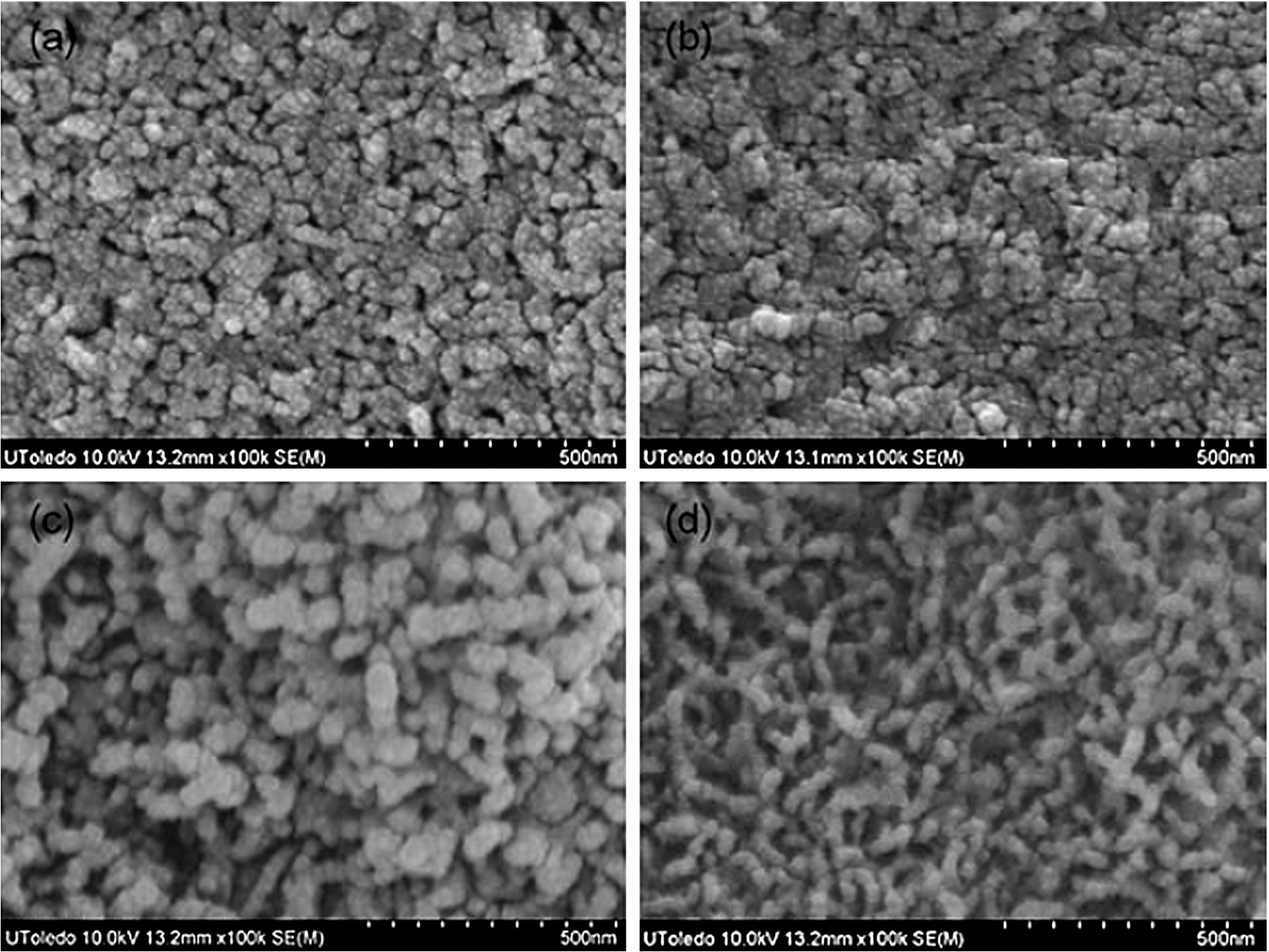
Scanning electron micrographs of coatings of carbonated calcium deficient hydroxyapatite (CDHA) developed by using a biomimetic coating process on Ti alloys. SEM images of the surfaces of CDHA coatings deposited from 1.5X t-SBF solutions with different concentrations of alendronate sodium (AS): (a) 0, (b) 10−6, (c) 10−5, and (d) 10−4 M (Ref. [52]).
Sol–gel technique has attracted much attention to the researchers due to its well-known inherent advantages in generating ceramic powders [55]. Sol–gel approach is the process of introducing coated surface into implant at a relatively low temperature that tolerates the deposition of calcium phosphate materials during the formation of coated layer. Sol–gel process has several advantages. The first, mixing of components on a molecular scale led to an increase in the homogeneity The second, the reduction of firing temperature to get small particle size with high surface area. The third: the ability is to produce uniform and fine grained structure [56]. During the sol–gel transformation, the viscosity of the solution gradually increases as the sol becomes interconnected to form rigid, porous network gel. With further drying and heat treatment, the gel converts into dense ceramics or glass particles.
Sol–gel methods have been used in many applications for coating metals in situ with the preparation of CaP sol which has been carried out by using precursors of calcium (most often calcium nitrate) and phosphorus (phosphorus pentoxide or triethylphosphite) in addition to couple of solvents, often water and ethanol. The phosphate precursors dissolved in ethanol [57]. Even as in some circumstances a little amount of water is added to the solution to achieve hydrolysis of the subsequent sol [58]. The calcium precursor was dissolved in ethanol and then added to the hydrolyzed phosphate sol in a drop wise manner. Mixture was refluxed at various temperatures which led to evaporation of solvents to get more viscous solution [57]. Normally, before accomplishing the sol–gel coating process, the surface of the metal implant should be polished and cleaned carefully, then washed with special chemical reagents to be ready for coating process. This pretreatment process is crucial stage to motivate the implant substrate surface.
Although the sol–gel is a popular research technique involving immersion of the implant (Mg, Ti or its alloys) into a liquid which is a concentrated to a gel-like texture. A little research has focused on the application of sol–gel coating on metals for clinical use and therefore the biocompatibility of such coatings has not been determined. Metal samples dipped into the sol–gel several times to acquire a CaP coating, then cured at high temperatures (in the range of 350–700°C) to increase the coating–substrate adhesion and accomplish apatite structures within the applied coatings [59]. The curing temperatures should not exceed the melting point of the metal or metal alloys [60] to avoid the changes in the surface integrity of the substrate. One advantage of the sol–gel technique over biomimetic coatings is the strength of the coating–substrate adhesion [61].
Plasma spray coatings
While there is numerous coating techniques developed with the purpose of combining the advantages of the high strength of Ti or Ti-based alloys with the bioactivity and osteoconductivity of CaPs [62], plasma spray coating used as a commercially available technique for hydroxyapatite coatings on titanium implants. These implants were successfully used in clinical practice for improving the initial healing and long term stability of orthopedic hip prostheses. However, the method of the plasma spray takes place at extremely high temperatures with high potential to affect the structure of Ti and the homogeneity of the CaP coating [63]. Furthermore, coating by plasma spray method does not allow the incorporation of bioactive molecules within the Ca–P layer [64]. These bioactive molecules like bone morphogenetic proteins or antibiotics could enhance bone healing or prevent local infection around prostheses [65]. Moreover, the plasma spraying method does not allow accurate control of the chemical composition, crystallographic structure and crystallinity of the coating. In addition, it cannot produce a uniform coating of devices with complicated shapes [66]. High temperature exposure may lead to undesirable phases such as CaO [67].
Summary of the different electrochemical deposition conditions on different substrates. These conditions include: the bath composition, temperature ranges, pH and types of cathodes and anodes
Summary of the different electrochemical deposition conditions on different substrates. These conditions include: the bath composition, temperature ranges, pH and types of cathodes and anodes
SBF: 0.185 g CaCl2, 0.4 g KCl, 0.06 g KH2PO4, 0.1 g MgCl2 · 6H2O, 0.1 g MgSO4 · 7H2O, 8.0 g NaCl, 0.35 g NaHCO3, 0.48 g Na2HPO4 and 1.00 g d-glucose in 1 L of Milli-Q water.
Normal treatment: Means mechanical polishing.
316LSS: C: 0.03%, Mn: 2%, Si: 0.75%, Cr: 7.98%, Ni: 9.34%, Mo: 2.15%, P: 0.045%, S: 0.03%, N: 0.1%, Fe: 67.575% (%wt).
Electrodeposition (ED) of calcium phosphate coatings on metal implant is an alternative process that uses aqueous solutions at low temperatures. It cannot affect the structure of the implant and can be applied to complex shapes. Electrodeposition technique produces superior properties, such as quick and uniform coating and it is possible to control the thickness and chemical composition of the coating [68].
Electrodeposition of calcium phosphate coatings can overcome the problems associated with the plasma spray technique. ED uses aqueous solutions at low temperatures, which does not affect the structure of the implants. The ED method is affected by controlling the operating parameters such as temperature, bath composition and current density. The effects of temperature and deposition time on the electrodeposited coatings where studied using titanium substrate as the working electrode in a simulated body fluid (SBF) and a voltage of −2 V (SCE). Bath composition considered as a primary parameter, as Mg2+, K+ and HCO3− ions were removed from the SBF. The resulting coating was investigated at 52–92°C using two current densities of 1.3 and 12.9 mA/cm2 [69]. Higher temperature ranges of 90–200°C have also been used with current densities of 5–25 mA/cm2 and deposition times of 10–120 min to study the morphological regulation and crystal growth of calcium phosphate coatings on titanium using a SBF solution without the MgCl2 · 6H2O, KCl and NaHCO3 [70]. In another work [71], CaP coatings were performed using an electrolyte consisting of CaCl2 and NH4H2PO4, at 36°C and current densities ranging from −0.5 to −10 mA/cm2 for 5 s to 60 min. The effect of pressure on the electrodeposition process has also been studied [72]. A limited number of metals has been coated using electrochemical deposition. Table 1 depicts and summarizes various studies that deal with coating different substrates using electrodeposition.
It is worth mentioning that despite the extensive research efforts in coating methods, only limited techniques have made it to clinical trials and commercialization (mainly plasma spray deposition), whereas most of the other techniques (such as biomimetic deposition, electrochemical and sol–gel, etc.) are still in the pre-clinical phase [87–90]. Properties such as adhesion, cost-effectiveness and mechanical and biochemical stability are properties that are related to industrial upscaling and real clinical applications [87–89]. However, and for instance, a study [90] showed that CaP coatings, deposited on Ti–6Al–4V implant either by plasma spray technique or electrochemically, increases the mechanical fixation and bone ongrowth compared with an uncoated titanium control in a canine cancellous bone implant model. The plasma spraying technique seemed to stimulate more bone ongrowth.
Magnesium as a biocompatible material
A variety of metals has been demonstrated as orthopedic implants [40]. This include (but not limited to); stainless steel, magnesium, titanium, cobalt–chromium and their alloys. They are traditional orthopedic device materials [91]. The long-term implantation stability in the inner environment like corrosion [92], toxic ions releasing [93] and degradation [94] limits their clinical applications. Biocompatible protective coatings are an optimum option for metallic implants [95]. Coatings can provide a barrier between the metal substrate and its environment and improve the bioactivity of the metal surface [96]. Of these metals, magnesium (Mg) and its alloys appear to be attractive candidates due to their non-toxicity and their similarity in mechanical properties with those of bones compared to other metals. Being light, biocompatible, Mg-based metallic implants have several advantages over other implantable metals that are currently in use. One of these are the eliminating of the stress shielding and there is no need for a second surgery after metal implant. Unfortunately, the fast degradation rates of Mg and its alloys in the aggressive physiological environment impose limitations on their clinical applications. This necessitates development of implants with controlled degradation rates to match the kinetics of bone healing [96].
Magnesium is considered to be low dense metal and exhibits high strength/weight ratio [93]. One main problem that can be solved by introducing magnesium metal as implanted biocompatible metal is the elastic modulus (EM). The elastic modulus of Mg has been reported as 45 GPa [97]. Therefore, in comparison with the other used metals, the EM value of Mg is far closer to that of natural bone. This Magnesium has advantages over these implants where it could decrease the possible stress shielding. Mg also has the advantage of biodegradation, and thus if corrosion rate is controlled, the metal would slowly degrade, removing the necessity for second removal surgeries, thereby decreasing health risks, costs and scarring. It has been documented that the corrosion products of Mg have potential benefits to the patient [98]. Conversely, other metals showed toxic corrosion products [99], a thing which gives another advantage of Mg as a substrate. The corrosion rate of Mg in physiological solutions is relatively high and this calls for controlling such rate in considerable matching with the time of healing time. The inhibition of Mg corrosion may be achieved via coatings with a biocompatible material such as calcium phosphates [100]. Electrochemical coating for Mg and Mg-based alloys using different bath compositions has been reported [101].
Future plans
Although one of the advantages of electrochemical deposition is producing CaP coatings on porous and non-regular surface where there is no significant shear force and loads, any migration of particulate CaP to articulating surfaces of an artificial hip could lead to reasonable concerns associated with excessive wear. Consequently, some authors have investigated a number of means by which electrodeposited calcium phosphate coatings can be stabilized (see Table 1). We may conclude that ED is a promising technique for coating of biodegradable metals with CaP. However, in the cathodic electrodeposition procedure we may obtain loose, porous and low adhering coatings on applying a constant potential. This was attributed to the formation of polarization concentrations where mass transfer from the bulk to the metal (main body) is considered to be slow. Also, hydrogen gas formed as result of cathodic reduction of H2O causes a lower adhesion of the CaP coating on the metal surface. We suggest using flow system for solving such problems. Flow-by planar electrochemical reactors offer both high mass transfer rate and sweeping of the H2 gas bubbles from the substrate surface [102]. See Fig. 2 which illustrates the proposed arrangement.
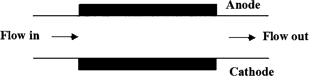
Flow-by planar electrochemical reactor.
The article reviewed recent progress in different coating techniques for biodegradable and biocompatible metals (or alloys) by calcium phosphate for possible application in orthopedic. These methods include: biomimetic, sol–gel, and plasma-spray coating and electrochemical techniques. A special concern has been paid to the electrochemical coating techniques with a summary of the different metal substrates, different coating baths and different experimental conditions. An important table has been introduced which summarize different experimental conditions for different substrates (either metal or alloy). A future work is introduced to solve possible problems in electrochemical coating. For instance, flow system is suggested for solving of gas bubble formation which can affect the quality of the CaP nanoparticles.
Conflict of interest
The authors have no conflict of interest to report.