



Abstract
Background:
The electrical potential, which is generated in bone by collagen displacement, has been well documented. However, the role of mineral crystals in bone piezoelectricity has not yet been elucidated.
Objective:
We examined the mechanism that the composite structure of organic and inorganic constituents and their collaborative functions play an important role in the electrical properties of human bone.
Methods:
The electrical potential and bone structure were evaluated using thermally stimulated depolarized current (TSDC) and micro computed tomography, respectively. After electrical polarization of bone specimens, the stored electrical charge was calculated using TSDC measurements. The CO3/PO4 peak ratio was calculated using attenuated total reflection to compare the content of carbonate ion in the bone specimens.
Results:
The TSDC curve contained 3 peaks at 100, 300 and 500°C, which were classified into 4 patterns. The CO3/PO4 peak ratio positively correlated with the stored charges at approximately 300°C in the polarized bone. There was a positive correlation between the stored bone charge and the bone mineral density only.
Conclusions:
It is suggested that the peak at 300°C is attributed to carbonate apatite and the total bone mass of human bone, not the three-dimensional structure, affects the stored charge.
Introduction
Wolff found the phenomenon, when the internal architecture of the trabeculae undergoes adaptive changes, secondary changes of the external cortical portion of the bone occur [1]. After that time, bone piezoelectricity was propounded [2]; the mechanical stress of the mineralized tissue induces an electrical potential, which is generated by collagen piezoelectricity [3,4]. The areas of bone under compression develop negative potentials, whereas those under tension develop positive potentials. Indeed, the negative and positive potentials affect bone remodeling because living bone becomes thicker on its compressed concave side and thinner on its tensed convex side [5,6]. Additionally, we reported that bone healing is enhanced near the negatively charged surface of implanted hydroxyapatite (HA) ceramics [7,8]. These reports suggest that a polarized region exists in human bone, which is an electret. The electrical potential, which is generated in bone by collagen displacement, has been well documented [9]. However, the role of mineral crystals in bone piezoelectricity has not yet been elucidated. In this study, we examined the mechanism that the composite structure of organic and inorganic constituents and their collaborative functions play an important role in the electrical properties of bone. For examination, we used human bone specimens that were obtained during surgeries of femoral neck fracture (FNF), which is one of the most frequent fragile fractures in the elderly population. First, we characterized the electrical properties of human bone specimens and then analyzed the cancellous bone structures and the mineral crystals, especially in carbonate apatite, to evaluate the faculty as an electret. Furthermore, we attempted electrical polarization of living bone to progress its ability as an electret that enhances new bone formation.
Materials and methods
Preparation and characterization of human bones
In this study, we used human femoral neck bone that was harvested during FNF hemiarthroplasty surgeries from 2012 to 2014 at Kawakita general hospital (Fig. 1(A), (B)). The research protocol was approved by the ethics committees of the institutions that the authors are affiliated with and was conducted according to the Declaration of Helsinki. Informed consent was obtained from each candidate. Thirty-eight cases of FNF, including 5 men (mean age of 80.0 ± 12.9 years) and 33 women (mean age of 82.5 ± 9.3 years), participated in this study. After temporary storage at −20°C (Fig. 2(A)), the specimens were sliced into 1 mm thick pieces using a diamond saw (IsoMet® Low Speed Saw, Buehler, Illinois, USA), washed with distilled water, dried at 40°C and used for the experiment (Fig. 2(B)).
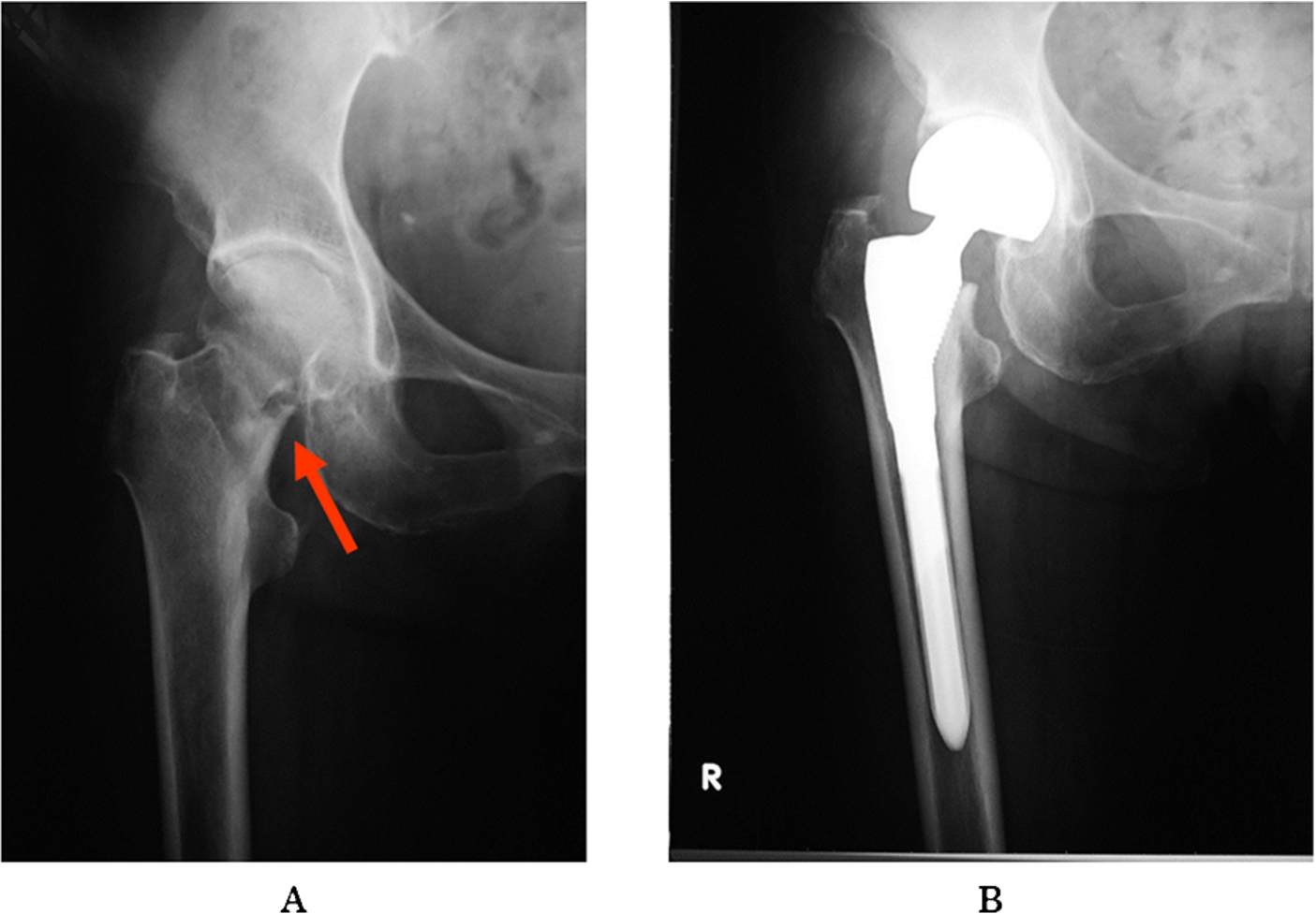
The femoral neck human bones are harvested during femoral neck fracture hemiarthroplasty surgeries. (A) Radiograph of the femoral neck fracture, which is indicated with an arrow. (B) Radiograph after the hemiarthroplasty.

Preparation of the femoral neck human bones. (A) The harvested femoral neck bone. (B) The bone specimen that is used for the experiment is sliced into 1 mm thick pieces, washed with distilled water and dried at 40°C. (C) The structure of human femoral neck bone constructed by micro CT scan.
The bone specimens were tightly clamped with a pair of platinum electrodes and were covered with alumina plates to form a bilayer. The thermally stimulated depolarized current (TSDC) measurements were performed using an electric current measuring unit (6514 system electrometer, Keithley, Ohio, USA). The sample temperature was increased at a heating rate of 5.0°C/min from room temperature (25°C) to 650°C, and the depolarization current was measured. The total stored charges in the bone samples were calculated using the following formula:
Bone specimens, in which the extremely small or large TSDC electric current density of less than 10−12 A · cm−2 or more than 10−6 A · cm−2 was recorded, were eliminated from count.
Micro computed tomography scanning
The specimens, which bordered the sliced bones that were used for the TSDC measurements, were scanned to evaluate the cancellous bone structures using micro computed tomography (CT) (smx100, Shimazu, Tokyo, Japan) (Fig. 2(C)), which was analyzed using computer analysis software (3D-BON, LATOC, Tokyo, Japan). Trabecular bone was examined using the parallel plate model. The primary parameters that were evaluated included the bone mineral content (BMC), the bone volume (BV), the tissue volume (TV), trabecular thickness (Tb. Th), trabecular number (Tb. N), trabecular separation (Tb. Sp) and trabecular spacing (Tb. Spac). The secondary parameters that we calculated included the BV/trabecular bone, the bone mineral density (BMD) and the BMC/TV.
The population variance among the secondary parameters calculated using the trabecular bone analysis and the stored charge were compared using the Bartlett test. The existence of a difference in the population mean was determined using one-way analysis of variance (one-way ANOVA). Thereafter, the statistical significance between the total stored charge and each secondary parameter was detected using the Pearson product-moment correlation coefficient (
Polarization treatment of the human bone to evaluate the faculty as an electret
Other bone specimens, which were clamped with a pair of platinum electrodes, were electrically polarized in direct current (DC) electric fields of 5 kV/cm in air at 25°C (
Quantification of the carbonic acid to estimate carbonate apatite in the bone
Attenuated total reflection (ATR) spectra of the bone specimens were recorded in the range of 400 to 4000/cm using an ATR spectrophotometer (Thermo Scientific, Nicolet is50, MA, USA). There were several absorbing regions for PO4 and CO3 ions in the apatite spectrum. The peak intensities of the carbonate and phosphate ions were recorded at 871/cm and 559/cm, respectively, and were analyzed using the instrument software (Thermo Scientific, OMNIC, MA, USA). The ratio of the two peak intensities was calculated to estimate the ratio of carbonate ions to phosphate ions in the bone specimens.
Results
Electrical characterization of the human bones
The representative patterns of the TSDC curve of the bone specimens are shown in Fig. 3. Each TSDC curve had peaks at approximately 100, 300 and 500°C, and each curve was classified into 4 patterns according to the distribution of these peaks. Type A had one peak at 500–600°C (
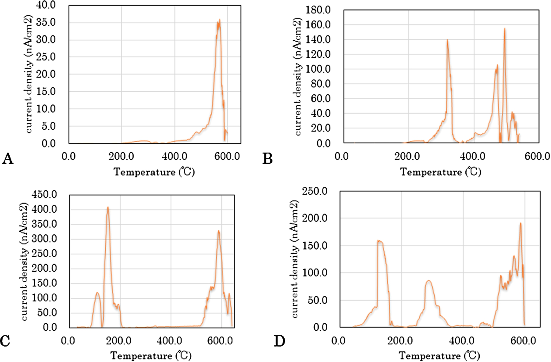
Representative TSDC curve pattern. (A) Type A; (B) Type B; (C) Type C; (D) Type D.
The average of total charge estimated by TSDC and BMD in the non-treated bone specimens, and age of donor in each type
Average ± SD.
The average of calculated parameters of bone structure of all specimens
BMD: bone mineral density, BV: bone volume, TV: tissue volume, BMC: bone mineral content, Tb. Th: trabecular thickness, Tb. N: trabecular number, Tb. Sp: trabecular separation, Tb. Spac: trabecular spacing. Average ± SD.
The average total charge, which was estimated using the TSDC and BMD in the non-treated bone specimens, and the donor age of each type are shown in Table 1. There were no significant differences among these parameters in the groups. The averages of the calculated parameters of the bone structure of all of the specimens are shown in Table 2. There was a positive correlation between the total bone charge and the BMD (

Correlation between the stored bone charge and the BMD. There is a positive correlation between the total bone charge and the BMD (
The TSDC patterns of the electrically polarized 21 bone samples were classified into 4 types, as shown for the non-polarized samples. There was one sample each of type A and C, 3 of type B, and 16 of type D. The mean stored charge was 56.9 ± 50.5 µC/cm2 for polarized bone, which was significantly larger compared with 16.1 ± 11.2 µC/cm2 for non-polarized bone (
Quantification of the carbonic acid to estimate carbonate apatite in the bone
The human bone TSDC curve had 3 peaks at approximately 100, 300 and 500°C, and the peaks at 300°C is expected to be characteristic for human and derived from carbonate apatite. Therefore, we attempted to quantify the carbonic acid by ATR spectra to estimate carbonate apatite in the bone specimens.
A representative ATR spectrum of a bone specimen is shown in Fig. 5. There were several peak intensities for the carbonate and phosphate ions. Because the protein C–C peak masked the peaks at 1410 and 1540 cm−1 for the v3 CO3 bands, we used 871 cm−1 for the v2 CO3 band. All of the specimens showed the CO3 peak, which is characteristic of type B carbonate apatite [10]. Phosphate ions exhibited peaks at 559 and 599 cm−1 for the v4 band and at 961 cm−1 for the v1 band. The peaks were comparable to those obtained by LeGeros for a type B carbonate HA (562 and 602 cm−1 for v4 and 957 cm−1 for v1) [11]. Because the 559 cm−1 peak correlates with the amount of phosphate, we adopted this peak. We compared the carbonate content in the bone specimens with the CO3/PO4 peak ratio.
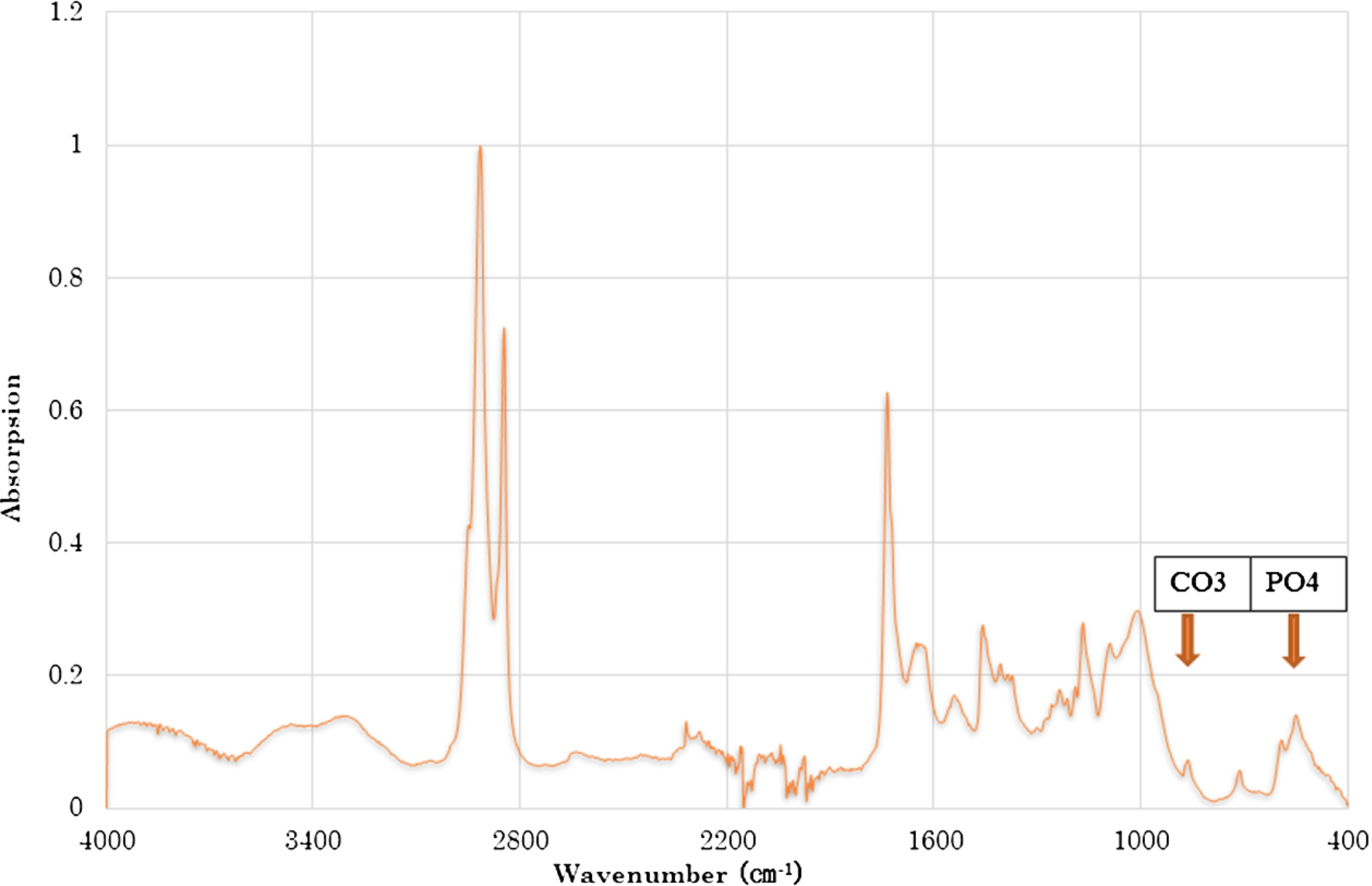
Representative ATR spectrum of the bone specimen.

Amount of stored charge at approximately 300°C and the CO3/PO4 peak ratio. The CO3/PO4 peak ratio positively correlates with the stored charge at approximately 300°C in the polarized bone (
The CO3/PO4 peak ratio did not correlate with the stored charges at approximately 300°C in the non-polarized bone. However, it had a positive correlation in the polarized bone (
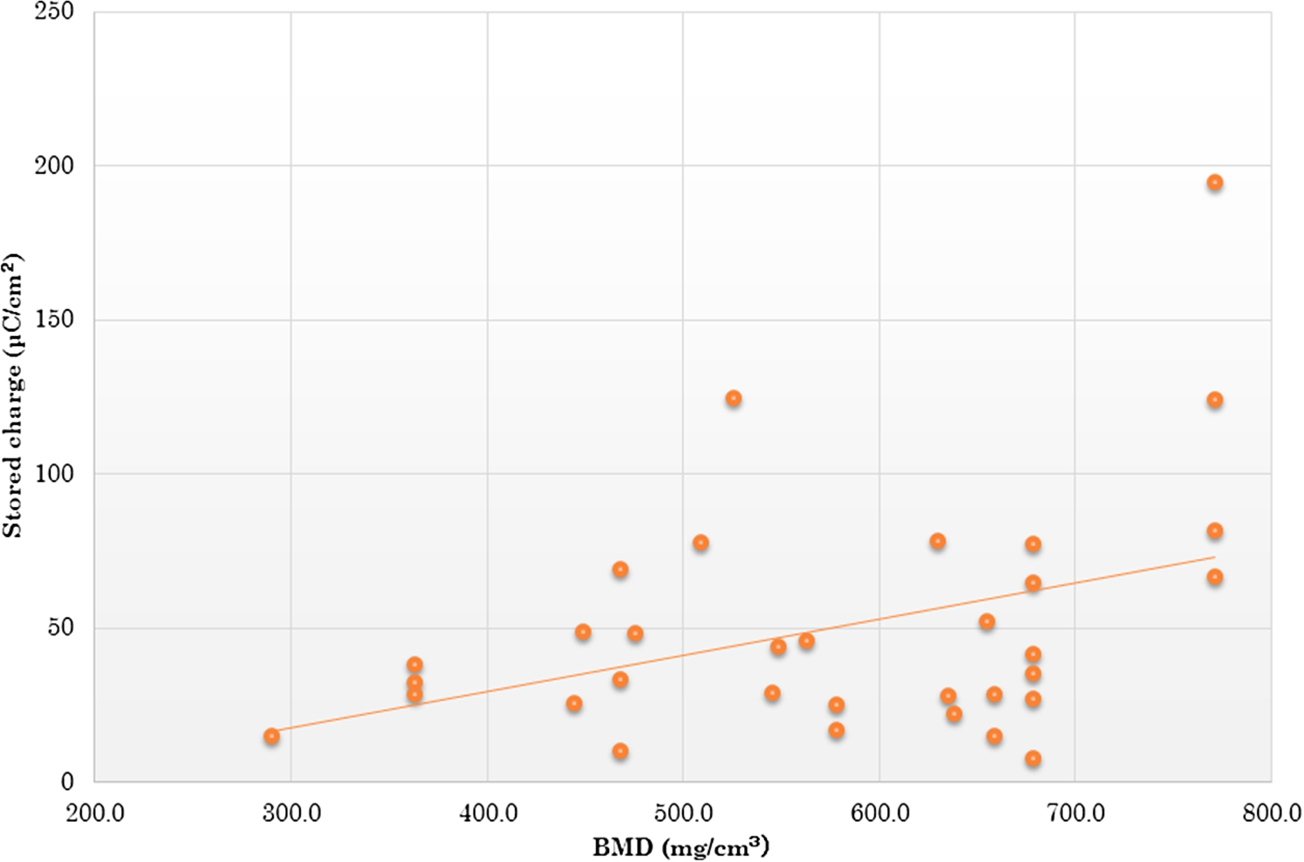
There is a positive correlation between the stored charge and the BMD in the polarized bone.
Moreover, the BMD positively correlated with total charge in each polarized bone specimen (
These results suggest that the carbonate ions in the bone influence the stored charge.
Looking into Wolff’s law and the discovery of bone’s piezoelectricity, we have considered the storage of electric charges in bone through mechanical stress for bone remodeling, and our very recent study successfully proved the bone’s being electrically charged state, which is called an electret. We reported that the rabbit and bovine bone TSDC curves have two significant peaks: at approximately 100°C and 500°C [12]. The peak at approximately 100°C is attributed to collagen fibrils because the ethylenediaminetetraacetic acid (EDTA)-treated animal cortical bone, which therefore lacked minerals, showed depolarization of this peak. It is reasonable that the shear forces of the collagen fibrils create stress-generated potentials in the bone piezoelectricity [2]. In this study, no or very small peak at 100°C was observed. This is because the peak of organic substance including collagen is recorded under 100°C and very small compared with that of inorganic substance. Therefore, it becomes difficult to see when the peaks of inorganic substances are large. The peak at approximately 500°C is attributed to apatite minerals because the animal calcined bone, which had the collagen removed, exhibited depolarization of this peak and had a similar activation energy to the synthesized apatite. Hence, in this study, the human bone TSDC curve had 3 peaks at approximately 100, 300 and 500°C. Considering that bone primarily consists of an organic matrix, such as collagen, and an inorganic matrix, including apatite minerals, it is expected that the peaks at 300 and 500°C are attributed to 2 types of apatite minerals. We previously reported that X-ray diffraction (XRD) and FTIR suggest that the minerals in calcined bone specimens convert to hydroxyapatite with a loss of organic content and carbonate ions during the calcification [12]. Therefore, the peak at 500°C is attributed to hydroxyapatite. However, calcium phosphate and calcium carbonate are the primary mineral components of human bone. Accordingly, the peak at 300°C is derived from carbonate apatite. The substitution of carbonate ions in bone minerals has been reported to increase with age [13]. This suggests that differences in the amount of carbonate ions, specifically the carbonate apatite content, contributes to the formation of the 300°C peak in the TSDC curve. In this study, there was no correlation between the CO3/PO4 peak ratio and the stored charges at approximately 300°C in the non-polarized bone. However, there was a positive correlation between these values after polarization. This indicates that the peak at approximately 300°C is attributed to carbonate apatite, and the small amount of stored charge in the natural bone is amplified by the polarization treatment such that it is detectable.
This study is the first report that has succeeded in electrical polarization of human bone in which stored charge was increased as a result. Autogenous bone graft is gold standard clinically to fill up bone defect, however, it is expected when electrically polarized living bone can be used as a bone filler its ability to enhance new bone formation may facilitate bone healing. Although there was a positive correlation between the stored bone charge and the BMD, no significant correlation was found between the stored charge and the other investigated parameters. These results suggest that the total bone mass of human bone, not the three-dimensional structure, affects the stored charge. Therefore, we propose the new method to prepare a bone filler, that is polarization treatment of human bone after crumbling for packing into bone defect. We can expect enhanced new bone formation for treatment of comminuted fractures or reconstruction after resection of bone tumor with this method.
Conclusion
It is suggested that human bone is an electret and its TSDC curve has three peaks at 100, 300 and 500°C which are attributed to collagen, carbonate apatite and hydroxyapatite, respectively and that the stored charge in the bone is affected according to total bone mass.
Conflict of interest
The authors have no conflict of interest to report.