
Abstract
The method of formation of bioactive calcium-phosphate coating on medical titanium alloy Ti–6Al–4V (3.5–5.3% V; 5.3–6.8% Al; balance –Ti) by plasma electrolytic oxidation (PEO) has been developed. Evaluation of osteogenerating properties of the coating at fractures of the shaft of the femur on Wistar line laboratory rats has been performed. It has been established that the calcium-phosphate PEO coating accelerates osteogenesis and promotes the formation of a pronounced periosteal callus in the fracture area. The presence of calcium phosphates in the PEO coating surface layer significantly accelerates the growth of bone tissue on the titanium surface.
Keywords
Introduction
Titanium and its alloys are extensively applied for manufacturing dental and orthopedic implants and artificial joints, plates, and screws used in fracture fixation due to a unique combination of their physical, electrochemical, and mechanical properties (high ultimate strength limit, excellent corrosion resistance, wear resistance, high hardness, and low density) [1]. Ti–10Cu alloy shows even antibacterial properties [2]. About 30% of all implants (and the most part of dental implants) [3] are manufactured from Ti–6Al–4V. It is also considered as a promising biomaterial in its porous form improving mechanical compatibility of the implant to the bone tissue [4]. A passive oxide layer on the alloy surface ensures good biocompatibility and protects the metal substrate from an aggressive biological medium [5,6]. The authors of [7,8] evaluated in vitro different medical alloys of titanium and revealed the release of V2+ and Al3+ ions into different simulated solutions. The presence of such elements in soft tissues promotes the increase of the amount of released inflammation mediators, which could participate in the osteolysis process [9]. To improve biocompatibility, bioactive layers containing bone tissue components, including hydroxyapatite, which is extensively studied [10–12] can be formed. The following methods are applied for the coatings formation: sparc anodization [13,14], plasma spraying [15], thermal processing [16], pulse laser sintering [17], electrophoretic deposition [18], biomimetic deposition [19] etc.
Despite various advantages of the developed and applied methods, one should mention that hydroxyapatite, whose cost is rather high, is used as an initial reagent in the coating formation. Therefore, to provide a sufficient progress in this direction, it is required to develop new economically sound methods of synthesis and deposition of hydroxyapatite on the surface of titanium implants. Such a surface modification would enable one to ensure better implant biocompatibility with the bone tissue and reduce negative side effects as compared to metal implants without coating.
Among the available electrochemical methods of coatings deposition on the metal implant surface, the most significant advantages are concerned with the method of plasma electrolytic oxidation [20–24]. The PEO process yields the electrochemical synthesis of coatings containing both the oxidized metal and the electrolyte components on the metal surface.
As it is well known, that in the surgery practice there are cases when titanium implant must not to extract from bone after fracture healing. The objective of the present work was to perform in vivo study of osteogenerating properties of calcium-phosphate coating on permanent implant by comparative study of internal fixation by coated and uncoated implants at fracture of the shaft of the femur on Wistar line rats. The fracture fixation was performed by intramedullary nailing (which modeled the nonremovable implants) using screws made of Ti–6Al–4V titanium alloy without coating and with calcium-phosphate coating obtained using the PEO method on the surface of the treated alloy material.
Objects and methods
To perform the experiments, 2 types of implants were used: Ti–6Al–4V titanium alloy screws (grade VT-6 (Russian analog of Ti–6Al–4V) manufacturer – SPA (Scientific Production Association) “Deost”, “Osteomed”, Russia) and Ti–6Al–4V screws with calcium-phosphate PEO-coatings. The alloy composition was: 0.015% H; 0.05% N; 0.1% C; 0.1% Si; 0.2% O; 0.3% Zr; 0.6% Fe; 3.5–5.3% V; 5.3–6.8% Al; balance Ti. The screw sizes (in series) depended on the size of the medullary canal of the femur of experimental animals. In most cases, screws of a length from 26 to 30 mm and a diameter of 2.0 and 2.3 mm were used. The roughness factor of the tested specimens with and without coatings was in the range of 1.2 ± 0.2 µm.
Plasma electrolytic oxidation was carried out in a bipolar mode [20,21] in the electrolyte containing 30 g/l of calcium glycerophosphate ((C3H7O6P)Ca·2H2O) and 40 g/l of calcium acetate (Ca(CH3COOO)2·H2O). A reversible thyristor rectifier was used as the power supply. The polarizing pulses frequency was equal to 300 Hz. The polarizing signal duty cycle equaled to 0.5. The electrical parameters were controlled using an automated control system coupled with the power supply through a computer with respective software.
The phase compositions of coatings were studied on an automatic D8 Advance X-ray diffractometer (
The experiment was carried out on adult male rats of the Wistar line of a weight of 200–250 g held under standard vivarium conditions in accordance with “Sanitary regulations for management, equipment, and maintenance of experimental clinical biological facilities” (No. 1045-73 of 06.04.1973). All the experiments were carried out in accordance with the rules of fair treatment of laboratory animals and of performance of works and use of experimental animals (Appendix 3 to the order No. 755 of 12.08.1977 of the USSR Ministry of Public Health). The experimental routine order was approved by the Ethics Committee of PSMU of the Russia Ministry of Public Health. Rats’ feeding was carried out in accordance with norms approved by the order No. 163 of 10.03.1986 of the USSR Ministry of Public Health.
Animals were divided into 2 groups (18 animals each): control and test ones. In the control group, implants with standard commercial parameters made of titanium of the grade VT-6 without coating were used for internal fixation. In the test group, similar implants with deposited calcium-phosphate coating were used.
For the euthanasia, the general anesthesia by intraperitoneal injection of 3% sodium thiopental was used. After that, the animals were decapitated.
Within the scopes of the present study, a closed diaphyseal femur fracture was simulated. Fixation of the fracture fragments was carried out by the method of closed reamed retrograde intramedullary nailing using a screw (Fig. 1).

Simulation of fracture (1) and fixation (2). 1 – fracture of the middle one-third of the shaft of the femur, 2 – head of the screw placed in retrograde way through intercondylar space of the femur.
Animals were put out of the experiment on 28th and 42nd days after operation. For each period, 9 rats from each group were examined.
Operation was carried out at aseptic conditions under single-component inhalation narcosis (sevoflurane). Upon treatment of the operation area, the frontal longitudinal access to the intercondylar space was realized: the wound size was about 0.5 cm. Here, the lower extremity was bent in the knee joint at an angle slightly larger than 90°. The patella appeared to be above the entry point of the screw, while the patella ligament was split in the longitudinal direction to further preserve the shin extensor mechanism. Through the intercondylar space, the entry point into the femur medullary canal was formed using a thin trocar. Reaming of the medullary cavity was carried out using Kirschner wires of diameters of 1.5 and 1.8 mm (alternately). Thereafter, a screw of a diameter of 2.0 mm was used for fracture fixation (Figs 2, 3).
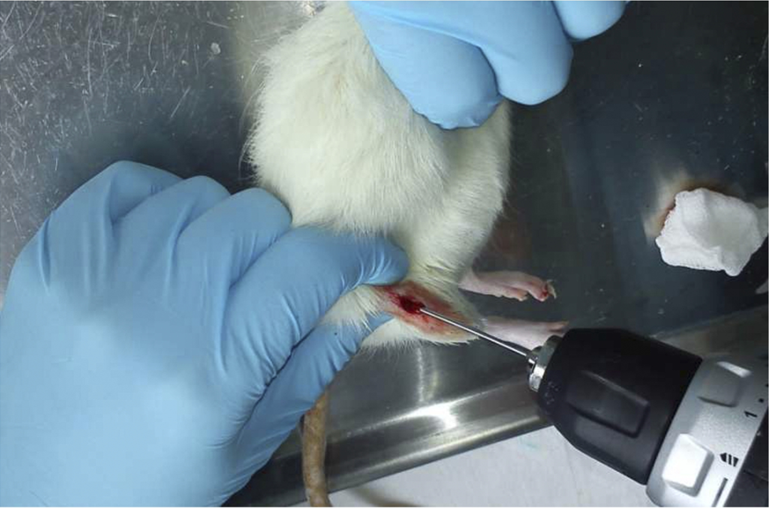
Reaming of the femur medullary canal.
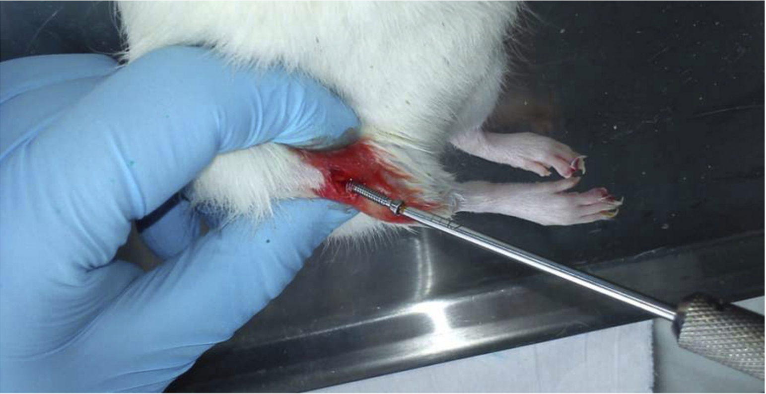
Screw introduction into the medullary canal.
If reaming using 1.5-mm wire was carried out without noticeable efforts, the cavity was further extended by 2.0-mm wire. Thereafter, a screw of a diameter of 2.3 mm was used. The entry point in the interconylar space was extended by a core drill for subsequent dipping of the screw head into the femur condylar subchondral layer. The screw was introduced half-length to the canal, so that in the middle one-third of the femur its end serves as end stop. Then, a manual bending load was used to simulate the closed femur fracture emerging at the boundary of the screw introduced until the middle of the femur. Fragments reduction was also carried out by the closed manual method. Upon reduction, the screw was introduced into the central fracture fragment until complete head ‘dipping’ into the bone to recover the sliding moment in the knee joint. One suture was used for wound closure. The wound was treated using an antiseptic. Evaluation of the results of fracture recovery was performed by the X-ray method the day when the animal was put out of the experiment. The experiment was carried out on 10 rats: 5 rats each in control and test groups. On the 28th day 2 animals each of both types were put out of the experiment; on the 42nd day 3 animals each for two types of used implants were put out as well.
The authors of [19,20] suggested the way of formation of coatings containing calcium phosphates, including hydroxyapatite, on commercially pure titanium using the PEO method. The performed series of physical–chemical studies demonstrated good prospects for application of this material in the implant surgery. The presence of calcium phosphates in the PEO coating surface layer significantly accelerates the growth of bone tissue on the titanium surface. It was established that the bone tissue of a thickness up to 50 µm formed on the surface of calcium-phosphate coatings within 45 days after subcutaneous implantation into laboratory microorganisms. No growth of the bone tissue was observed within the same period of time on the surface not treated using the PEO method, which indicates to high bioactivity of the obtained coatings.
In the present work, the technique of formation of the calcium-phosphate coating was applied for the implant introduced directly to the damaged bone part. Unlike the subcutaneous implantation [20,21,24], the former method is the most similar to real surgery practice.
According to X-ray diffraction data, the coating’s chemical composition comprises hydroxyapatite Ca10(PO4)6(OH)2 along with titanium oxide and calcium phosphates. The quantitative element composition is, according to the X-ray fluorescence analysis data, as follows (at.%): O – 52; Ca – 20.3; P – 14.2; Ti – 7.5; Al – 3.3; V – 1.2; Na – 1.5.
As seen from the X-ray study results, in case of one animal with titanium implant without coating, on the 28th day one observed a weak periosteal reaction in the fracture area, but fracture healing wasn’t expressed (Fig. 4).

Fracture in the middle one-third of the diaphysis (see arrow); weak periosteal reaction, no fracture healing (28 days after operation).
In case of the test group of animals with titanium implants having calcium-phosphate PEO-coating on the surface, the complete union fracture took place for both rats within 4 weeks (Fig. 5). One should mention that as early as within 2 weeks rats of this group started to actively use the leg, unlike test animals having the implant without coating.

X-ray image of complex fracture of the shaft of the femur at using the implant with calcium-phosphate surface coating. The main fracture line spreads in the supracondylar area (1), the large fragment reaches the diaphysis middle one-third (2). One can see good periosteal callus (28 days after operation).
In the series of test group animals put out of the experiment on the 42nd day, for all the rats one detected expressed X-ray indications of complete union fracture – expressed bone callus and absence of visible fracture line (Fig. 6).

Fracture X-ray image on the 42nd day after operation for test group rats (implant with calcium-phosphate coating). Bone callus is clearly seen; fracture line is not visible.
For the control group of animals having the implant without coating, in two cases the bone callus was not expressed, but the union fracture was evident (Fig. 7).

X-ray image of the shaft of the femur of the test group rat on the 42nd day after operation (implant without coating). Bone callus is not expressed; fracture line is shown by the arrow.

X-ray image on the 42nd day after operation for one of three rats of the control group (implant without coating). Non-union fracture is shown by the arrow.

Macro-preparation of one of three animals of the control group on the 42nd day after operation. Non-union fracture. Fracture line is shown by the arrow.
For one control group rat, one can clearly see the appearance of non-union fracture along the frontal shaft outline (Fig. 8).
On the macro-preparation of this animal (Fig. 9), one can clearly see that the fracture area is filled all-around with the healing tissue: no sign of bone callus was found at this tissue removal. In the latter case, the classic case of non-union femur fracture was observed.
Thus, the operation reproduced the closed less invasive type of internal fixation providing the indirect fracture healing. Such fixation and bone healing types are optimal for fractures of the shaft of the femur.
The obtained results are in full agreement with those of the earlier studies [20,21,24] related to favorable effects of bioactive coatings containing hydroxyapatite on growth and recovery of the bone tissue.
As was established in experiments in Wistar line rats, the calcium-phosphate coating obtained by the PEO method has a positive effect on osteogenesis during recovery of the fracture of the shaft of the femur. Hydroxyapatite contained in the bioactive coating serves as a growth initiator (precursor) in the process of bone tissue formation, thus significantly reducing the healing period.
Footnotes
Acknowledgement
The work was financially supported by the Russian Science Foundation (project No. 14-33-00009) and the Russian Federation Government (Federal Agency for Scientific Organizations).
Conflict of interest
The authors have no conflict of interest to report.