
Abstract
Background:
Chitosan and alginate are two natural and accessible polymers that are known to be biocompatible, biodegradable and possesses good antimicrobial activity. When combined, they exhibit desirable characteristics and can be created into a scaffold for cell culture.
Objective:
In this study interaction of chitosan-alginate scaffolds with mesenchymal stem cells are studied.
Methods:
Mesenchymal stem cells were derived from human umbilical cord tissues, characterized by flow cytometry and other growth parameters studied as well. Proliferation and viability of cultured cells were studied by MTT Assay and Trypan Blue dye exclusion assay.
Results:
Besides chitosan-alginate scaffold was prepared by freeze-drying method and characterized by FTIR, SEM and Rheological properties. The obtained 3D porous structure allowed very efficient seeding of hUMSCs that are able to inhabit the whole volume of the scaffold, showing good adhesion and proliferation. These materials showed desirable rheological properties for facile injection as tissue scaffolds.
Conclusion:
The results of this study demonstrated that chitosan-alginate scaffold may be promising biomaterial in the field of tissue engineering, which is currently under a great deal of examination for the development and/or restoration of tissue and organs. It combines the stem cell therapy and biomaterials.
Introduction
Mesenchymal stem cells (MSCs) are capable of self-renewal and have the ability to differentiate into several distinct mesenchymal lineages, such as bone, cartilage, adipose tissue, muscle, and marrow stroma, etc. [1]. MSCs are found in a variety of tissues, including the umbilical cord [2–4], which is the focus of this study and provides a noninvasive source of abundant stem cells. Three main elements, scaffolds, cells and signals, make up tissue engineering. The interactions among those three elements have been linked together [5]. Tissue engineering evolved from the field of biomaterials expansion and refers to the practice of combining scaffolds, cells, and biologically active molecules into functional tissues. The goal of tissue engineering is to accumulate functional constructs that restore, maintain, or improve damaged tissues or whole organs. One of the important component is scaffold material.
The 3D scaffolds have to be degraded and absorbed, with non toxicity during degradation and good biocompatibility [6]. In recent years a wide range of polymeric biomaterials with numerous properties have been developed for bioengineering and tissue engineering applications. These biomaterials should be biodegradable, bioactive and biocompatible [7]. The biomaterials architecture is also important and affects the cell matrix interaction [8], which is of significance in cellular behaviors such as cell adhesion, proliferation and differentiation. In this context, chitosan and alginate both has been found a fascinating candidate for cell culture, tissue engineering applications and in directed tissue regeneration.
Chitosan (CS), as a polycationic polymer, is achieved from crustacean shells by partial and full alkaline deacetylation. As a result of its biodegradability, nontoxicity and antimicrobial activity, CS has been frequently used for biomedical applications, such as tissue engineering scaffolds, drug delivery, wound dressings and antibacterial coatings [9–11]. Alginate has carboxyl groups which may introduce negative charge to the polymer at appropriate pH [12]. Due to its negative charge, it is able to chemically bond with positively charged chitosan, forming a stronger scaffold material [13,14].
The combination of natural polymers like chitosan and alginate might overcome some of the disadvantages, such as, low mechanical strength, of single polymer like pure chitosan based scaffolds, and lead to a material with fascinating characteristics to be used in the biomedical field [15]. Chitosan alginate scaffolds have been used for variety of applications. These scaffolds have been shown to be effective in regenerating cartilage [15] and bone [13]. These scaffolds may be implanted in the body to fill a defect and encourage tissue regeneration [16,17].
In our previous studies, two types of scaffolds, including chitosan and chitosan-alginate (CA), have been demonstrated to be suitable for invivo applications in tissue engineering as they showing excellent antibacterial activity. In this study, we tried to seed mesenchymal stem cells isolated from Human umbilical cord into scaffolds of chitosan-alginate which were highly compatible with seeded cells, further supported possible translation of these materials.
Materials and methods
Materials
All materials were purchased from Himedia unless otherwise stated. chitosan (from shrimp shells) DD (degree of deacetylation) was >75%, calcium alginate (alginic acid calcium salt) was also obtained from Himedia Laboratories Pvt. Ltd, Mumbai, Acetic acid (Merck Ltd, Mumbai), NaOH pellets (Merck Ltd, Mumbai) were used as such. Reagents such as cell culture medium and fetal bovine serum were obtained from Himedia Laboratories Pvt. Ltd, Mumbai.
Methods
Chitosan alginate scaffolds synthesis
The chitosan-alginate (CA) scaffolds were prepared as previously reported [12]. Briefly, a 4 wt% chitosan and 2 wt% acetic acid solution was mixed under constant stirring in a blender for 7 minutes to obtain a homogeneous chitosan solution. A 4 wt% alginate solution was added to the chitosan solution, and mixed in a blender for 5 minutes to obtain a homogeneous CA solution. The CA solution was cast in the beaker and frozen at −200°C for 8 h. The samples were then lyophilized. Different combinations of scaffold were made such as CA1, CA2, CA3, CA4, CA5, CA6, CA7, CA8 respectively.
Characterization of CA scaffolds
Fourier Transform Infrared Spectroscopy (FTIR) and Surface scanning studies
The infrared spectrum of scaffolds was recorded on FTIR (Fourier Transform Infrared Spectroscopy) spectrophotometer. Morphological analysis was carried out by a scanning electron microscope (SEM) JEOL Model JSM-6360LV at an accelerating voltge 10 kV and at 500, 1000 and 5000 resolution.
Biodegradation of chitosan-alginate scaffolds
The initial dry weight of scaffolds was determined and recorded as W1. The scaffolds were sterilized in PBS buffer at 121°C for 15 minutes. After cooling, the free solution was removed from the sterilized scaffolds and incubated in 7 ml of sterilized PBS buffer containing 10 μg/ml lysozyme. All incubations were done in 6 well plates at 37°C in a CO2. Media were replaced weekly with freshly prepared lysozyme solution. After fourteen days of incubation the samples were removed from the degradation media, washed with distilled water and freeze-dried. The weight of the freeze-dried scaffolds was recorded as W2. The percentage degradation of the scaffolds was calculated using equation given below;
Rheological experiments
Rheological experiments were performed using a controlled stress rheometer (AR2000, TA Instrument Ltd). 2 cone steel plates (20 mm diameter) were used and the 500-lm gap was filled with tested colloidal gel. A solvent trap was used to prevent evaporation of water. The viscoelastic properties of the sample were determined at 20°C by forward-and-backward stress sweep experiments. The viscosity (g) was monitored while the stress was increased and then decreased (frequency 1/4 1 Hz) in triplicate with 10 min between cycles. The gel recoverability was assessed using defined time breaks between cycles. All samples were analyzed in triplicate.
Isolation of umbilical cord-derived MSCs (UC-MSCs)
hUMSCs were harvested from the umbilical cord obtained after normal full-term delivery of patient and isolated as reported elsewhere [18,19]. Six umbilical cords, app. length 20 cm, were stored aseptically in cold PBS with 100 μg/ml Pen/Strep, and 2.5 μg/ml Amphotericin B (Himedia Laboratories), within 6–24 hours from partum. The umbilical vein and arteries were dissected from the tissue, which was then cut into fragments (2–3 mm3). Several pieces of tissue were placed in a Petri dish with a low amount of GM to allow attachment to the plastic and later on were covered completely with culture medium. GM was changed every second day. After 7 days, when the cells grew out of the explanted tissue, the cord fragments were removed and attached cells were cultured until reaching confluence. MSCs were successfully isolated from two out of six specimens. After reaching confluence, cells were subcultured routinely in GM, using Trypsin/EDTA solution (Hi-media). The populations of MSCs were expanded for up to 15 passages for UC-MSCs.
Seeding of stem cells on scaffolds
The scaffolds were sterilized by autoclaving and presoaked in a 12-well cell culture plate containing DMEM culture medium for a couple of days before cell seeding. Approximately 1 million huMSC in 1 ml Dulbecco’s Modified Eagle Medium (DMEM) were seeded on each scaffold disc. The DMEM medium contained 10% fetal bovine serum (FBS), 50IU ml−1 penicillin, and 50 μg ml−1 streptomycin. The scaffolds were transferred to another cell culture plate after 3 hours of cell seeding. The culture medium in the plate was changed in 24 hours for the first time and every three days thereafter. A hemocytometer was used to determine the concentration of cells in cell suspension. The morphology of cells cultured on scaffolds was examined scanning electron microscopy.
Cell proliferation
Cell proliferation was assessed using the Almar Blue Assay. Scaffolds were seeded with cells in DMEM cell culture medium. After 24 hours of cell seeding, the scaffolds were washed with PBS and placed into 12-well culture plates. 2 ml of 10% v/v Almar Blue in DMEM medium was added to each well containing scaffolds and the plates were incubated for 4 hours at 37°C. Absorbance of the solution in each plate, which is proportional to the number of cells attached to the sample, was measured with a micro plate reader (Biorad, imark, USA) at wavelengths of 570 and 600 nm. A calibration curve generated from a known number of huMSC reacting with the Almar Blue was used to calculate the number of cells. Samples were assayed at 3, 7, 14 and 21 days, respectively after cell culture.
Cell viability
The MTT assay is used to quantify spectrophotometrically the amount of living cells, due to the reduction of MTT to purple formazan by the mitochondria of the living cell. For the MTT test, the mesenchymal stem cells were seeded at a concentration of 2 × 10⁵ cells/mL in respective medium for 24 h in a 96 well microtiter plate. After 24 h of incubation, the old media were replaced with media containing different concentrations of MNPs, and the cells were exposed for an incubation time of 48 h. Blue formazan crystals, from the metabolism of MTT in the mitochondria of viable cells, were washed with PBS and were dissolved in 50 mL of dimethylsulfoxide and measured at 550 nm by the plate reader. The experiments were repeated three times and the data was graphically presented as the mean. The percentage of cell viability (%) compared with the control well containing cells without scaffold is calculated by the equation,
Histology
Scaffolds seeded with huMSc were washed twice with PBS and fixed with 4% neutral buffered formalin for histological assay. The scaffolds were embedded in paraffin and cut into 5 mm thick sections using a microtome. The sections were stained with hematoxylin-eosin. The satined sections were then mounted on glass slides, and examined with a Nikon e200 microscope.
Results and discussion
CA scaffolds exhibited microporous structures and shape retention
In tissue engineering, scaffold should have high porosity and inter connected pore structure to enhance a compatible biological condition for cell attachment, proliferation and differentiation. The porous structure of scaffolds are achieved by freeze-drying method. During the freezing process, ice crystals are formed. After removing these crystals by lyophilisation, a porous material is formed. The kind of porosity depends, to a greater extent, on the freezing conditions. Both the temperature, the thermal gradient and the cooling rate have an effect on the pore structure [20,21]. In the present study, surface morphology was examined from micrographs taken with a SEM. The scan micrographs of CS, SA and CS–SA scaffolds are shown in Fig. 1. scaffold CA1 and CA2 exhibits flat lamellar phases on which a large number of protruding microfibrils are evident, while scaffold CA3 clearly shows porous smooth surface having highly interconnected pores.

SEM pictures of chitosan (CA1), chitosan-alginate (CA2, CA3a, CA3b), alginate (CA8) scaffolds with 500 and 1000 resolution.

FTIR spectra of (a) chitosan (CA1), (b), (c), (d) chitosan-alginate scaffold (CA3), (CA4a and CA4b), and (e) alginate CA2.
The significant increase in Young’s modulus and yield strength for the chitosan-alginate scaffold can be attributed to the strong ionic interactions between chitosan and alginate to form a chitosan-alginate complex, as confirmed with FTIR analysis (Fig. 2). The characteristic peak of alginate (Fig. 2(e)) is seen at 1620 cm−1 corresponding to carbonyl (C=O) bond(6). In the spectrum of chitosan (Fig. 2(a)), peaks 3422 cm−1, 1653 cm−1, 1592 cm−1, 1173 cm−1 and 1071 cm−1 are due to O–H stretch, amide II band, C–O stretch of acetyl group, amide II band, N–H stretch, skeletal vibration involving the bridge C–O stretch. The peaks at 1424 and 1070 cm−1 in both alginate and chitosan scaffold correspond to carboxyl –COOH and C–O stretching bands, respectively. In the chitosan-alginate spectrum (Fig. 2(b), 2(c) and 2(d)), amide II peaks were significantly intensified, the amide I peak shifted from 1643 to 1654 cm−1, and the peak of the amino group (1173 cm−1) was absent. These changes suggest the formation of the chitosan-alginate complex as a result of the ionic interaction between the negatively charged carbonyl group (–COOH) of alginate and the positively charged amino (–NH2) of chitosan.
Biodegradation of chitosan-alginate scaffolds
The ultimate goal for the application of chitosan-alginate 3D porous scaffolds in tissue engineering were the hope that it could disintegrated naturally along the cells growth. As a result, the time to degradation would affect the condition of the cells growth.
In vitro biodegradation is a crucial parameter to be considered in tissue engineering. The biodegradation of scaffolds provides space for tissue growth and matrix deposition. The lysozyme was used as degradation enzyme to investigate through time course of degradation conditions of CA 3D porous scaffolds. Table 1 shows the degradation weight (%) of CA scaffolds with various CA concentrations.
Degradation weight of CA scaffolds after 4 days incubation
Degradation weight of CA scaffolds after 4 days incubation
In the degradation process of CA scaffold by lysozyme, the enzyme must enter the scaffolds and react with the chitosan and alginate polymers. The scaffolds all have pore sizes of about 30–70 μm, which provides a large enough area for lysozyme to enter, but the morphology of the scaffold substrate layers is different in each one. Therefore, CA6 scaffold degrades faster as compare to other combinations of chitosan and alginate which containing lower chitosan conc. and moderate alginate conc. But as conc. of alginate increases, its affects degradation rate (CA7 & CA8). It is also observed that scaffolds containing higher chitosan conc. taking time to degrade as compared with CA6–CA8 combinations.

In Vitro degradation of CA6 scaffold.

Graph represents degradation rate of chitosan scaffold after 4 days (%), showing higher degradation rate of CA6.

Viscosity and shear-thinning behavior of colloidal gels mixed at different ratios for accelerating (solid symbols) and decelerating (open symbols) shear force.
Rheological studies were employed to probe the differences in viscoelasticity of colloidal gels. Mass ratios of 4% chitosan and 3% alginate yielded the highest viscosity gel and improved reversibility compared to other ratios (Fig. 5). As expected, colloidal gels containing more positively-charged particles (CA6) exhibited higher viscosity, while colloidal gels composed of excess negatively charged particles (CA5) exhibited more fluidity. Pure suspensions of chitosan and alginate exhibited minimal shear-thinning behavior (C100 and A100). Delaying shear cycles for more time may enhance the recovery of gel viscosity. All the results suggested that the colloidal gels were desirable for injectable applications.
For colloidal gels, the strength of the cohesion depends upon the interparticle interactions such as electrostatic forces and van der Waals attractions [12]. These interparticle interactions were controlled by the composition of the colloidal gels, such as concentration and ratio of the two oppositely charged particles. chitosan and alginate scaffolds self-assembled through interparticle interactions resulting in a stable 3D porous network. Under static conditions, the viscosity and structure of colloidal assemblies leading to a stable structure exhibiting high viscosity at equilibrium. If the particle-particle equilibrium is disrupted, for example by external force applied to disrupt the interparticle interactions, the colloidal system will demonstrate shear-thinning behavior. Once the external force is removed, the strong cohesive property of the colloidal gel is recovered and the 3D porous structure reconstructed. This reversibility makes the gel an excellent material for applications in molding, extrusion, or injection of tissue scaffolds and drug delivery systems.
Isolation and culture of different MSCs
Depending on the starting amount and type of tissue used to establish the primary cultures, cells that morphologically resembled MSCs could be seen as early as 7 days post-plating for the UC-MSCs up to 15 days post-plating for the PB-MSCs, and the confluence could be reached within 10–15 days. Although during the initial culture period some morphological heterogeneity in the adherent fraction could be seen, as the cultures were passaged, morphological homogeneity was gradually achieved, cells were fibroblast-like and no spontaneous differentiation was noticed (Fig. 6).

Morphology of UC-MSCs cultures. Inverted microscopic images of UC-MSCs (B) monolayer cultures at passage 4. In monolayer culture, the cells assumed a polymorphic, fibroblast-like morphology, which was maintained throughout the passaging process.

Cell proliferation as measured by the Almar Blue assay. PC stands for pure chitosan scaffold, CA for chitosan-alginate scaffolds. Data show the average and its level of statistical significance (
Mesenchymal stem cells isolated from human umbilical cord was seeded onto chitosan-alginate scaffolds. Cell proliferation on both chitosan and chitosan-alginate scaffolds as a function of time was assessed using the Almar Blue assay, and the results are shown in Fig. 7. The cells on both scaffolds were doubled within the first week of cell culture and continued to increase over time. Three samples in triplet were tested and the results were analyzed. No significant difference in cell number was observed between the chitosan and chitosan-alginate scaffolds at day 3 and 7 after cell culture. However, the cell number on the chitosan-alginate scaffolds became notably higher than that on the chitosan scaffold at day 14 and day 21.
Cell viability
Despite having antibacterial activity, the suitability of material for tissue engineering applications depends that scaffolds must not have intolerable toxic effects in human body. The effect of scaffolds on the viability of huMSC was studied as the reflection of their suitability and the values obtained were shown in Fig. 8. After 48 hours of incubation, sample CA 3 shows slightly higher absorbance as compare to other samples at every concentration (approx >80%) indicated the presence of more number of cells on sample. Therefore, these scaffolds can be considered as biocompatible product having nontoxic effects. The cyto-compatibility of chitosan and chitosan-alginate were already been proved [22,23]. Therefore, considering good biocompatibility, chitosan-alginate scaffolds could be used for tissue engineering materials.

Cell viability versus concentration of CA Scaffold for CA1, CA2, CA3 and CA8 samples on huMSC after 48 h.

SEM microphotographs of mesenchymal stem cells grown on (a, b) chitosan-alginate and (c, d) chitosan-alginate scaffolds after 14 days cell culture, and on (e, f) chitosan-alginate scaffolds after 21 days cell culture with 1000×, 3000× & 6000× magnification respectively.

(Continued.)
To observe the cell morphology inside the micro and the constructs were cross-sectioned in thick slices and observed by SEM Fig. 3 (CA1-b, CA4-d, CA8-f). Figure 9 shows SEM images of huMSC grown on chitosan-alginate scaffolds after 14 and 21 days of cell culture. Figures (e & f) show the images taken at day 21 at a higher magnification in order to detail the cell morphology. At day 14, huMSC on scaffolds proliferate well and exhibited a spherical morphology. At day 21, however, the cells on the chitosan-alginate scaffolds became more flattened and started to assume a fibroblast-like morphology. As can be seen, cells spread on the surface of the scaffold and after 21 days culture the whole surface was covered. The observed results suggest that the material system promoted intercellular contacts and the spatial arrangement of the cells.
Microscopic images of huMSC’s on (a) pure chitosan and (b & c) chitosan-alginate scaffolds after 21 days of cell culture.
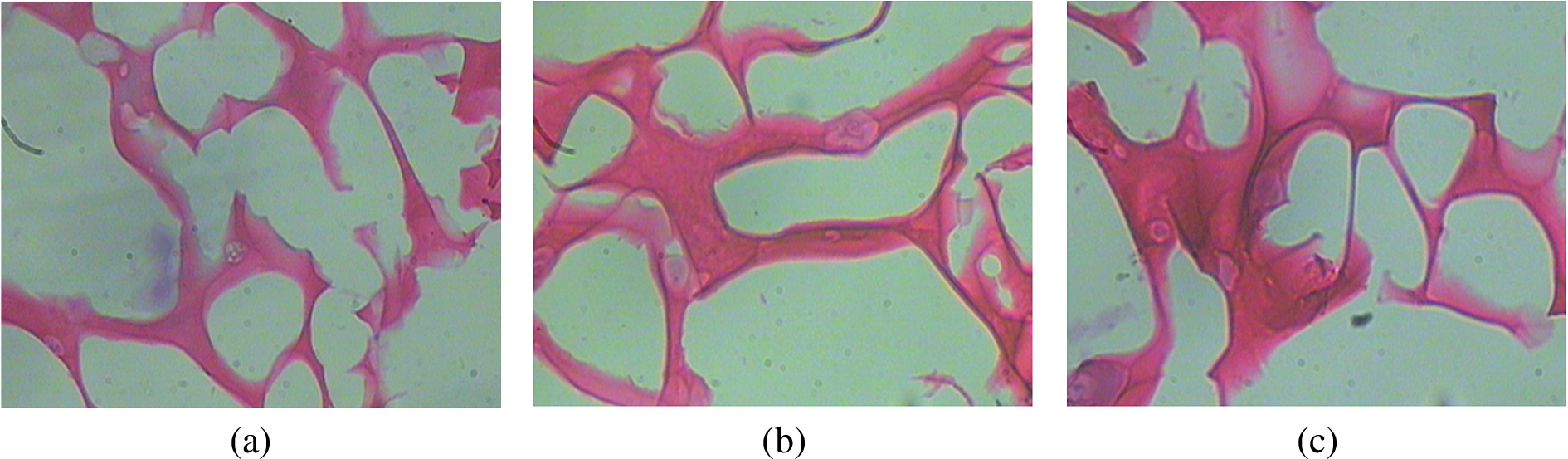
Hematoxylin & Eosin was used to stain the harvested scaffolds in order to study the interaction of cells with the materials. Figure 10 shows the microscopic images of huMSC’s stained with H & E and grown on chitosan-alginate scaffolds after 21 days of cell culture with the cell nuclei appearing in violet and cytoplasm and materials in pink. The cells on the chitosan-alginate scaffolds had smaller nuclei and larger cytoplasm. Furthermore, the cells on the chitosan-alginate scaffolds tended to form flattened fibroblast-like morphology.
Discussion
In the present study, chitosan-alginate and chitosan-alginate scaffolds were fabricated and investigated. SEM results demonstrated that the ternary scaffold has thin walled uniform pore dimensions randomly dispersed in the polymer matrix. The cohesive strength of these material resulted from the interparticle interactions between the oppositely charged chitosan-alginate biomaterials. The shear sensitivity to external force and recoverable pseudoplastic property made it an excellent injectable biomaterial. hUMSCs seeded in CA scaffolds with micro and macropores are able to attach and gradually proliferate along 3 weeks of culture. SEM images showed that the substrate surface becomes completely covered by a dense layer of cells and viable cells are uniformly distributed in the pore structure. These preliminary studies prove the feasibility of chitosan-alginate scaffold and it can serve as a potential material for tissue engineering applications.
Above mentioned work is applied for the patent entitled “Incorporation of mesenchymal stem cells on the CA Scaffolds for Biomedical Applications.” (Application number 3247/MUM/2015.)
Footnotes
Acknowledgements
The authors would like to thank Nowrosjee Wadia College, Biotechnology Department, for providing lab facilities for Research Work. Authors are thankful to Department of physics and Department of chemistry, University of Pune, Pune for providing the SEM and FTIR results respectively. The authors are grateful to Dr. J. K. Pal and Dr. Rajendra Patil Department of Biotechnology, University of Pune, Pune for providing the facility of freeze drying.
Conflict of interest
The authors have no conflict of interest to report.