
Abstract
In this work, we present an evolutive trabecular model for bone remodeling based on a boundary detection algorithm accounting for both biology and applied mechanical forces, known to be an important factor in bone evolution. A finite element (FE) numerical model using the Abaqus/Standard® software was used with a UMAT subroutine to solve the governing coupled mechanical-biological non-linear differential equations of the bone evolution model. The simulations present cell activation on a simplified trabeculae configuration organization with trabecular thickness of
Introduction
It is well know that bone density distribution evolve as a function of its mechanical environment [1] but the rules governing this evolution are not well understood and are depending mainly on the bone biology. This has been well accepted and developed since the famous Wolff’s Law [2]. Nowadays, detailed understanding of these phenomena has a real interest for the prevision of bone remodeling and bone reconstruction in many orthopedic and surgical applications.
Bone remodeling is a mechanobiological phenomenon occurring at different length scales from the macroscopic to the nanoscopic. Modeling the existing links between these scales is sometimes complex [3] but building precise bone remodeling numerical models requires multiphysics [4] and multiscale [5–7] approaches in coordination with the cells activity since they are the main biological driving force of bone remodeling. In addition, bone is considered as a poroviscoelastic material sometimes grafted with bioresorbable materials when fractured [8–14] with a kinetic evolution depending both on the mechanical properties and on the feeding of biological nutrients [15,16].
At the mesoscopic scale, the trabeculae are important in the remodeling of bone where osteoblasts and osteoclasts are acting on their surfaces. We use here the coordination of the three types of bone cells being the osteoblasts, osteoclasts and osteocytes [17] to propose a finite element model of bone remodeling at the trabecular level solving a set of ordinary differential equations coupling the mechanics and biology [18–25]. According to the mechanical information, the surface of the trabeculae will either be resorbed by osteoclasts or (re)built by osteoblasts [26]. The simulation is used to predict the bone evolving density and microstructure during remodeling.
Numerical model development
Bone remodeling model
We develop a continuous theoretical numerical model where the osteocytes perceive a mechanical information, and translate it into biological signals [18–24] so that for an increasing stimulus, osteoblasts produce bone and on the contrary, for a low stimulus, osteoclasts degrade bone. The developed model is based on a strain energy density function where the elastic deformation energy influences directly the biology of the bone. The biological stimulus S received at any point x of the geometry and time t is given by
Trabecular surface search algorithm
Spongy bone is constituted by trabeculae, small beams interconnected and filled with bone marrow. The bone remodeling process begins at the trabeculae surface by the action of osteoclasts receiving the signal transmitted by the osteocytes forming a sealing zone and dissolving bone [25,26]. Then osteoblasts are recruited by the increase of the osteocytes signals sent through the osteocytic canalicular network to the surface where reconstruction occurs. This phenomenon is directly integrated in the bone model remodeling equations acting on the trabecular surface of the bone.
We present here the algorithm, to detect the trabeculae boundary elements (see Fig. 1(a)). This algorithm (developed in the Abaqus UMAT subroutine) enables the localization of the bone cell activation on the trabeculae surface (and therefore will better control the macroscopic bone density evolution). The first step of the algorithm is to create an array that lists in our model the adjacent element of each element (who share two nodes at least). Then we set the existing physical bone material with a given numerical value (1) and empty spaces (void) within the trabeculae with another numerical value (0) as presented on Fig. 1(b) according to:
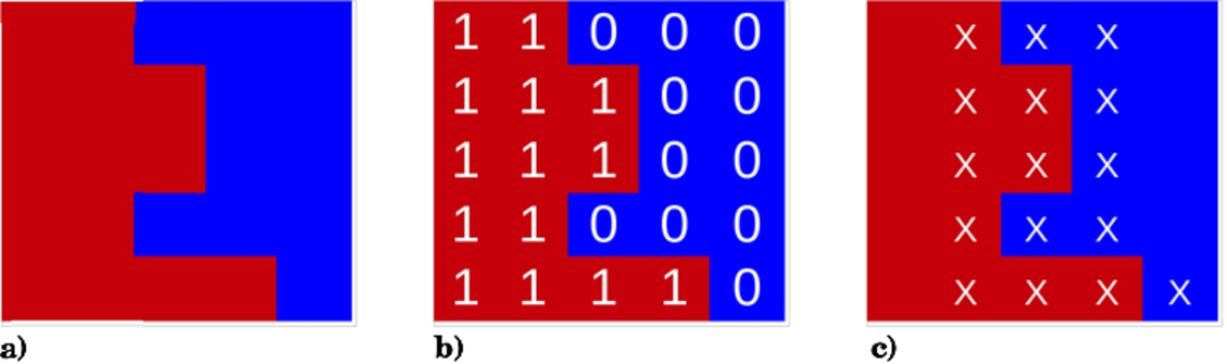
Theoretical definition of the boundary element search algorithm: (a) General configuration. Red illustrates bone element and blue void element. (b) Configuration after flagging. (c) Active element (marked X).
The constitutive bone remodeling equations are then calculated only for the active area elements so remodeling will occur only on these elements. The numerical model calculates the new bone density for each element at each time increment and the algorithm of boundary element search is used again in order to determine the new active area for the next iteration.
A simplified bone microstructure is presented here as an arbitrary non oriented grid. This configuration is loaded on each of the four sides at 45° degrees (Fig. 2, top left corner). Only bone remodeling is allowed within the geometry without any external displacement.
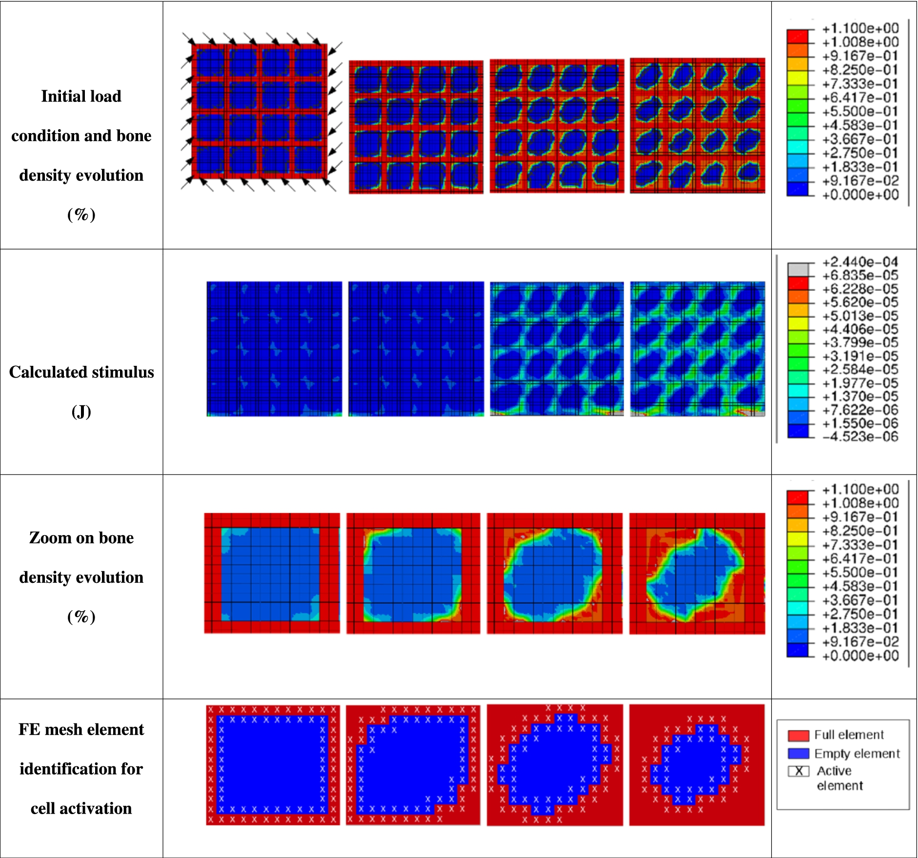
Trabeculae kinetics. First and second lines: evolution of the bone density (%) and of the stimulus (J) according to time with a 45° load applied on the 4 sides. Third and fourth lines: zoom on bone density (in %), and drawing of the algorithm evolution.
First and second line of Fig. 2 show the bone density and the stimulus evolution as a function of time (from left to right). Using the defined detection algorithm enables to observe good correlation of the bone density evolution regarding the trabeculae orientations in the applied mechanical load directions. Localization of the cell activation is clearly identified through detection of the active zone when the stimulus is specifically localized in this area.
Cell activity is highlighted on lines three and four of Fig. 2 through the algorithm activation where evolutions are shown as a function of time (from left to right). Bone reconstruction (3rd line) occurs specifically on the trabeculae surface. On the fourth line, the algorithm detects the activation zone and FE model elements are identified through an interpolation of the bone density evolution. This defines the new trabeculae boundaries for the new increment of density calculation.
Using this approach shows the principal difference between a macroscopic evolution model (accounting for an average bone density evolution through the entire geometry) and a more localized evolution (accounting for localized biological effects). In turn, more patient specific problems can be determined accounting for their own biology and materials kinetics (prosthesis, illness, …).
Phenomenological models available in the literature provide mathematically optimized bone microstructures as a function of phenomenological parameters not being able to account for patient specific microstructure evolutions. In this paper, an algorithm implemented in an Abaqus/Standard® UMAT subroutine is presented for known trabecular bone orientation. Results show a reorientation of the trabeculae network in the main principal applied mechanical stress. Both the local mechanical information and cells activation are identified and can provide accurate bone reconstruction specific to each patient issue.
Conflict of interest
The authors have no conflict of interest to report.