
Abstract
Background:
Inorganic ions released from bioceramics and bioactive glasses have been reported to influence osteogenic cell functions. Cell responses depend on types of the ions provided, for example, silicate ion has been found to up-regulate their proliferation, differentiation and mineralization.
Objective:
Mouse osteoblast-like cells (MC3T3-E1) were cultured in media containing silicate and calcium ions with/without magnesium ion to evaluate their combined effects on the cell’s functions.
Methods:
The cells were cultured in the media containing the extract of silicate-containing vaterite (SiV) and magnesium- and siloxane-containing one (MgSiV) and normal medium and then their adhesion, proliferation, differentiation and mineralization were evaluated.
Results:
The adhesion of the cells was enhanced when they were cultured in the medium containing MgSiV-extract. Their proliferation and differentiation were up-regulated in both media containing MgSiV-extract and SiV-extract. In particular, the MgSiV-extract significantly enhanced their differentiation than the SiV-extract. This was supported by the mineralization test’s results, which showed a large amount of mineral deposit was observed in the cells cultured in the MgSiV-extract medium.
Conclusions:
Providing the three kinds of ions was effective for up-regulating the cell’s mineralization compared to providing silicate and calcium ions without magnesium ion.
Introduction
Ions released from bioactive glasses and ceramics have been reported to influence functions of cells, such as osteoblasts and mesenchymal stem cells, and regarded to play one of the important role in bioactivities of such materials in vitro and in vivo [1]. Up-regulation of cell activities by the ions have been explained in resent reports. In particular, the ionic dissolution products released from bioactive glasses and glass-ceramics have enjoyed an increasing interest in bioceramics research field [2–6]. A trace amount of silicate ions released from bioactive glasses have been found to enhance proliferation, differentiation and mineralization of osteogenic cells. Calcium ions (Ca2+) are one of the component materials of bone and teeth and also enhance such functions of osteoblasts [7]. In our previous research, siloxane-containing vaterite (SiV) particles were developed as materials providing silicate and calcium ions [8–11]. Although SiV releases silicate ions with
Cell responses to the ions depend on ion types. For example, copper was reported to advance synergetic stimulating effects on angiogenesis and zinc was found to possess anti-inflammatory effect and stimulate bone formation [1]. Magnesium enhances cell adhesion onto materials and angiogenic function of endothelial cells [12,13]. Zreiqat et al. reported that human bone-derived cells possessed the up-regulated expression of integrin family, which is adhesion proteins, when they were cultured on magnesium-modified alumina compared with unmodified alumina (Mg-free) [12]. Cell adhesion to biomaterial surfaces is one of the important processes for achievement of satisfactory metabolisms of cells on the surfaces, especially for adherent cells like osteoblasts. To promote cell adhesion would be effective for achieving enhanced proliferation and following biological reactions of such cells. We, therefore, hypothesized that bioceramics having the ability of providing silicate, calcium and magnesium could be good candidate for materials used in bone regeneration. In our previous work, magnesium and siloxane-containing vaterite (MgSiV) was developed [14]. MgSiV particles were developed as a new type of vaterite-based inorganic particle with the ability of enhancing osteoblast’s activities and bone formation. MgSiV is synthesized by a conventional carbonation process in the same way for SiV. The results of the immersion tests for MgSiV and SiV using Tris-HCl buffer solution (pH 7.4) described that MgSiV released Mg2+ ion and almost the same amounts of silicate and Ca2+ ions as those of SiV for more than 7 days [14]. The silicate ions released from MgSiV are believed to stimulate the functions of osteoblast-like cells like SiV, since they are supposed to have a similar ionic state to that released from SiV.
The aim of the present work is to estimate the responses of MC3T3-E1 cells to the ionic products released from MgSiV and SiV and examine the combined effects of several kinds of ions on the functions of the cells. Although individual effects of each ion on the cell functions have been reported by many researchers, there were few reports on the combined effects of silicate, Ca2+ and Mg2+ ions on the functions. As described above, MgSiV and SiV release three and two kinds of ion in combination through their degradation, respectively. We focused on the combined effects of Mg2+ ion with Ca2+ and silicate ions on the adhesion, proliferation, differentiation and mineralization of MC3T3-E1 cells.
Materials and methods
Preparation of MgSiV particles
SiV particles were prepared by a carbonation process in methanol solvent, following the published report [9]. MgSiV particles were prepared by the same method apart from adding Mg2+ ion source [14]. As Mg2+ ion source, 0.2 mol of magnesium hydroxide (Wako Pure Chemicals Inc., Osaka, Japan) was added to a solvent consisting of 2.0 L methanol (Wako Pure Chemicals Inc.) and 200 mL distilled water while stirring at 21°C. Carbon dioxide (CO2) gas was bubbled to the slurry at a feed rate of 2.0 L/min for 20 min. Subsequently, 1.8 mol calcium hydroxide (Kishida Chemical Co., Ltd., Japan) and 60 mL 3-aminopropyltriethoxysilane (APTES, Dow Corning Co., USA) were mixed into the slurry under CO2 gas flow. The gas was added over 40 min, resulting in the formation of a precursor gel. The resulting gel was aged at room temperature for 12 h and then dried at 110°C for 24 h to remove the residual solvent. The obtained agglomerated powder was ground to form MgSiV particles. SiV particles containing no magnesium were also prepared as a control in the same manner. SEM images of MgSiV and SiV particles obtained were shown in Fig. 1.

SEM images of (a) MgSiV and (b) SiV particles.
The SiV or MgSiV particles were sterilized with ethylene oxide gas at 44°C for 20 h. The ion extracts were prepared by immersing 300 mg of the particles in 30 mL of α-modified minimum essential medium (α-MEM; MEMα with l-glutamine and phenol red, Wako, Japan) supplemented with 10% fetal bovine serum (FBS) and incubated in 5% (v/v) CO2 at 37°C for 24 hours. An α-MEM which contains no particles was also incubated under the same conditions. The ion extract derived from SiV or MgSiV is denoted by “SiV-extract” or “MgSiV-extract”, respectively. The concentrations of Ca, Si, Mg and P elements in α-MEM and the ion extracts were measured by inductively coupled plasma atomic emission spectroscopy (ICP-AES; ICPS-500, Shimadzu, Kyoto, Japan). This measurement was repeated for a total of three repetitions.
Cell culture test
The effects of the ion extracts on the initial adhesion ability of osteoblasts were determined with adhered cellular numbers on a culture plate and their morphology. Murine osteoblast-like cells (MC3T3-E1 cells) suspended in α-MEM or each ion extract at a density of
For evaluation of proliferation ability, MC3T3-E1 cells suspension was prepared using α-MEM at a density of
To evaluate the differentiation ability, the cells were pre-cultured in the same condition with the evaluation of proliferation. After 7 days, the culture media in the wells were replaced with a differentiation medium, which is α-MEM supplemented with 0.05 mM L-ascorbic acid-2-phosphate, 100 nM dexamethasone and 10 mM β-glycerophosphate as differentiation agents and 1% penicillin/streptomycin, or the medium containing the ion extracts. Subsequently, the media were replaced with fresh ones once in two days up to 28 days. Alkaline phosphatase (ALP) activity of the cells was analyzed using a p-nitrophenyl phosphate (pNPP) tablet sets (Lab Assay ALP Kit; Wako, Japan) 7, 14, 21 and 28 days after the replacing with the differentiation medium. ALP activity was evaluated after measuring the absorbance of p-nitrophenol product formed at 405 nm with the spectrophotometer. The value of ALP activity was expressed as µmol of pNPP/min/cells.
The mineralization of MC3T3-E1 cells were evaluated by an Alizarin red (Alizarin Red S; Wako, Japan) staining method. The Alizarin red forms insoluble orange-red lakes, identifying points of insoluble cations that are incorporated within mineralized tissues. The cell culture was conducted in the same condition with ALP assay for 28 and 35 days. After the above time points, the cultured samples were washed with phosphate buffer solution (PBS) and subsequently fixed with 70% ethanol for 60 min at 4°C. The samples were stained with the Alizarin red solution dissolved in 50 mM acetic acid for 10 min and washed 5 times with deionized water. The resulting samples were observed with an optical microscope.
Statistical analysis
The data were presented as the mean ± standard error of the mean (S.E.M.). Statistical analysis was performed using Student’s t-test and single-factor ANOVA (SPSS 21 software; IBM, USA) followed by Tukey’s multiple comparison test. Values of
Results and discussion
Ion concentrations in the extracts
After soaking for 24 h, Ca, Si, Mg and P concentrations in MgSiV-extract were originally 146, 65, 76 and 35 ppm, respectively. In our previous report, most of Mg and Si in MgSiV and SiV were released in 1 day after soaking in Tris buffer solution (
Concentrations (ppm) of Ca, Si, Mg and P elements in α-MEM and the ion extracts used for the cell culture analysis
Concentrations (ppm) of Ca, Si, Mg and P elements in α-MEM and the ion extracts used for the cell culture analysis
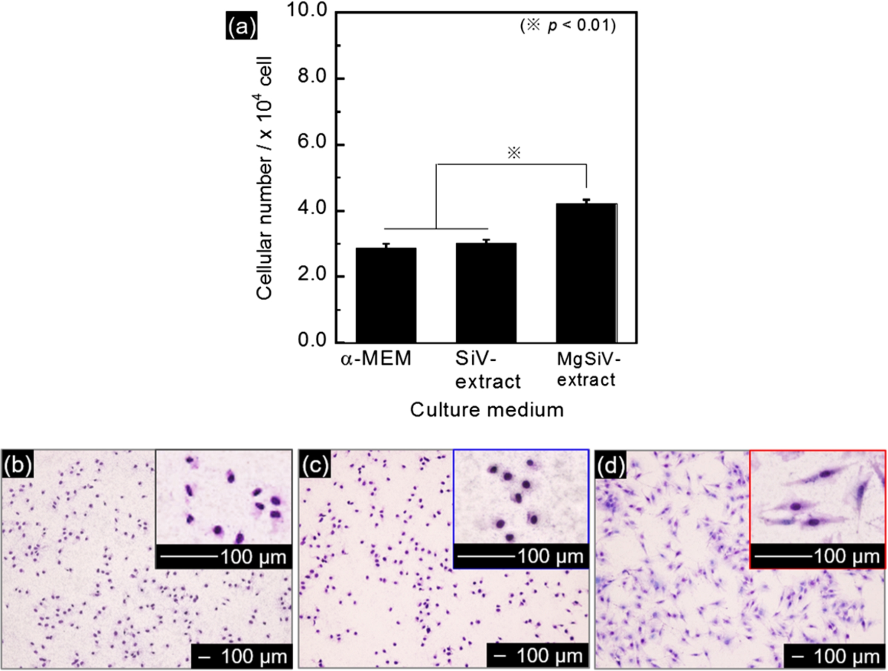
(a) Numbers of the cells adhered on culture-wells in 3 hours. (b)–(d) Morphology of the cells adhered on culture-wells in (b) α-MEM, (c) SiV-extract and (d) MgSiV-extract in 3 hours.
Figure 2(a) shows the cellular numbers adhered on culture-wells in 3 hours. The adhered cellular number cultured in SiV-extract was approximately
The morphology of the cells adhered in α-MEM and the ion extracts were indicated in Fig. 2(b)–(d). The cells cultured in α-MEM and SiV-extract exhibited less-spread and circular shape (Fig. 2(b) and (c)), while those cultured in MgSiV-extract exhibited spindle-like elongated shape (Fig. 2(c)). The cells cultured in MgSiV-extract might have good extensibility of their pseudopodium. These results indicate that Mg2+ ions released from MgSiV promoted the initial cellular adhesion.

(a) Numbers of the cells cultured in α-MEM and the extracts for 1–7 days. (b) ALP activities of the cells cultured in α-MEM and the extracts for 7–28 days.
Figure 3(a) shows the results of the proliferation assay for MC3T3-E1 cells cultured for 7 days. The cellular numbers increased with a culture time for all samples. The numbers of live cells cultured in SiV- or MgSiV-extract were larger than that in α-MEM at every time point during 7 days. At the initial stage of proliferation (from 1 day to 3 days), the cells in MgSiV-extract exhibited the highest proliferation ability, although there were no significant differences in the values between MgSiV and SiV-extracts. Mg2+ ions released from MgSiV influenced the cellular proliferation, especially at initial stage. The cellular numbers cultured in SiV-extract for 7 days were almost the same as those in MgSiV-extract. Appropriate extracellular Mg2+ ion levels, such as 19 ppm (the physiological concentration of the ions is 19 to 24 ppm), were essential to the stimulations of osteoblast proliferation by platelet-derived growth factor (PDGF), whereas Ca2+ ion levels do not influence the stimulation [15]. PDGF has been found to promote proliferation and migration in a variety of cell types including osteoblastic cells. Although it is still unclear how the stimulation of osteoblast proliferation by PDGF changes when larger amounts of Mg2+ ions are provided, the up-regulated proliferation in the early stage in the present experiment might be relate to the PDGF stimulations. Further investigation is needed to clarify more detail of the proliferation behaviors influenced by the ratio of Ca2+ and Mg2+ ion concentrations in the extracts.
Alkaline phosphatase (ALP) activity
The ALP activities in MC3T3-E1 cells cultured in α-MEM and the ion extracts for 28 days were indicated in Fig. 3(b). ALP activity is one of the established markers for the osteoblast phenotype. It is generally accepted that increase in the ALP activity reflects a shift to a more differentiated bone cells and the activity reaches its peak directly before start of mineralization of the cells [16]. The cells cultured in SiV- and MgSiV-extract exhibited significantly higher ALP activity than that in α-MEM at every time point during 28 days. It is considered that the differentiation of MC3T3-E1 cells was strongly enhanced by the extracellular silicate ions. Moreover, the activity level of MgSiV-extract reached its peak at 14 days of culture, while that of SiV-extract reached at 21 days. This suggests that the differentiation of MC3T3-E1 cells was substantially promoted by the additionally released Mg2+ ions in addition to silicate ions from MgSiV.
Mineralization
Figure 4 shows the results of Alizarin red staining of mineralized nodules that were formed by MC3T3-E1 cells after the cultivation for 28 and 35 days. The degree of redness in the images represents the quantity of Ca2+ ion incorporated into the cells. After the cultivation for 28 days, all the samples possessed almost no mineralized tissues (Fig. 4(a), (c) and (e)) On the other hand, the cells cultured for 35 days in MgSiV-extract showed well-mineralized nodules (Fig. 4(f)), while those in α-MEM and SiV-extracts indicated no red-stained tissues (Fig. 4(b) and (d)). An enhancement of mineralized nodule formation in the cells was observed only when they were cultured in MgSiV-extract.
Magnesium has been regarded to control bone calcification and crystallization processes [17,18]. Wuthier et al. reported that the highest contents of magnesium were contained in calcified cartilage, trabecular, and cortical bone. This must indicate that magnesium plays an active role in calcification and mineralization processes. Leidi et al., however, revealed that the culture medium containing higher amount of magnesium ions (120 ppm) compared to a conventional one (24 ppm) inhibited the differentiation and mineralization of osteoblast-like cells (SaOS-2) [19]. Serre et al. reported that magnesium-substituted hydroxyapatite and collagen composites leaded a decrease of proliferation and calcification activities of osteoblast-like cells. In particular, the composites containing higher amount of magnesium ions possessed a toxic effect on the cells [20]. These reports seem to show completely opposite results to those obtained in the present work. This might be because there are differences in the cell type used and the amount of magnesium ion in culture media among these reports. For example, Yoshizawa et al. reported that the mineralization of human bone marrow stromal cells was enhanced by providing ∼240 ppm of magnesium ions, whereas 2,400 ppm of the ions did not up-regulate them [21].
Another possible reason why there are such differences in the cell responses is a combination of ions provided to the cells. Silicate, Ca2+ and Mg2+ ions were provided to the cells at the same time in the present work, the other reports did only Mg2+ ion. Conventional culture media contain Ca2+ and Mg2+ ions. The present work aimed to clarify what would happen to the cells when additional amounts of these ions were provided to them with silicate ion, since some bioactive glasses and bioceramics, such as SiV and MgSiV, release not only single ion but also several ones at the same time through their dissolution or degradation in vitro and in vivo. The differentiation and mineralization of the cells were up-regulated by providing both Mg2+ and silicate ions, as shown in Figs 3 and 4. This must indicate combined effects of the two ions on the cell functions. We will examine the combined effects of several kinds of ions on cell functions with systematic procedure in future work.
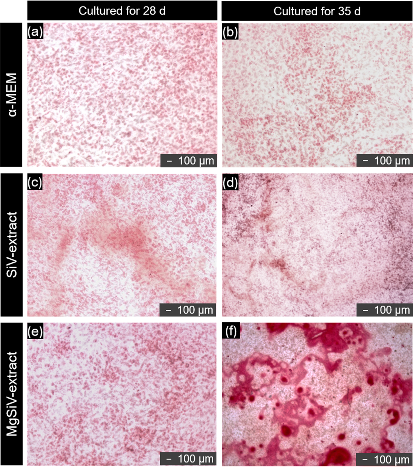
Optical micrographs of Alizarin red stained mineralized nodules formed by MC3T3-E1 cells after the cultivation for 28 and 35 days. (a) cultured in α-MEM for 28 days, (b) cultured in α-MEM for 35 days, (c) cultured in SiV-extract for 28 days, (d) cultured in SiV-extract for 35 days, (e) cultured in MgSiV-extract for 28 days and (f) cultured in MgSiV-extract for 35 days.
The differences in the initial cellular morphology between MgSiV- and SiV-extracts were shown in Fig. 2(b)–(d). Cellular morphologies are known to be related to gene expression [22]. Several factors, such as roughness, wettability and functional groups on substrate surfaces on which cells are cultured, have been reported to influence cellular morphologies. These factors, however, would be the same for all conditions in the present work, because we used the same well-plates. The cells cultured in MgSiV-extract which showed well-elongated shapes resulted in good mineralization ability. This means the additional amount of Mg2+ ions promoted the elongation of the cells followed by the up-regulated mineralization. Thus, to enhance cell elongation might be effective for up-regulating mineralization. We, however, also expect that combined effects of the ions would be involved in the up-regulation of mineralization observed in the present work, since the proliferation and the ALP level were enhanced for not only the MgSiV-extract but also the SiV-one, as shown in Fig. 3.
Cytocompatibility of magnesium- and siloxane-containing vaterite (MgSiV) was examined by culture tests using MC3T3-E1 cells for its ion extract. At the initial stage of cell culture test, the cells in MgSiV-extract showed better elongation of their pseudopodium and had excellent adhesion ability compared with that in conventional α-MEM and siloxane-containing vaterite (SiV)-extract. The proliferation ability of the cells cultured in MgSiV-extract was higher than that in SiV-extract after 3 days of culture, while the ability in the two extracts resulted in the same level after 7 days. The level of ALP activity of the cells cultured in MgSiV-extract increased at the earlier time point of the culture than those in α-MEM and SiV-extract. This result implies that the differentiation of the cells was promoted in MgSiV-extract. Moreover, the cells cultured in MgSiV-extract exhibited well-mineralized nodules by Alizarin red staining after culture for 35 days. The combination of Si, Ca and Mg provided was effective for up-regulating the cell’s mineralization.
Footnotes
Acknowledgement
This work was supported in part by JSPS KAKENHI Grant Number 26820304.
Conflict of interest
The authors have no conflict of interest to report.