
Abstract
Background:
Material properties of the scaffolds as well as their microstructure are vital in determining in vivo cellular response. Three-dimensional (3D), highly porous scaffolds are used in tissue engineering to provide a suitable microenvironment and to support regeneration of bone. Both pore sizes and their architecture, in particular interconnection density, impact functionality of scaffold during its biomedical applications.
Objective:
In this paper a comparative study of the microstructure of highly porous hydroxyapatite scaffolds produced via gelcasting of foamed slurries and replication of polyurethane sponge were carried out.
Methods:
Quantitative stereological analysis of the microstructure was conducted using transmission X-ray computed microtomography (μCT) and scanning electron microscopy (SEM). Application of the X-ray microtomography allowed obtaining the 2D cross-sectional images of examined samples, and then the 3D reflection of individual samples.
Results:
In our studies we proved that the distribution of pores in HAp bioceramics can be controlled by selection of the manufacturing method. In the case of material produced by the gelcasting method, the porosity of the samples was about
Conclusions:
The results of quantitative description of microstructure allowed determining the differences between porous hydroxyapatite bioceramics obtained via replication of porous organic matrix and gelcasting of foamed slurry. The stereological analysis demonstrated, that bioceramics prepared via gelling of foamed slurry has a lower pore size and grains (1.1–1.9 μm) than the material obtained by the method of replication of polyurethane sponge (2.1–2.3 μm). Based on morphological analysis the porosity of tested materials was determined. In the case of material produce by the gelcasting, porosity of the samples was about
Introduction
In the field of tissue engineering, one of the most popular approaches is the use of implant materials whose function is to guide and support the growth and migration of cells. Main efforts of scientists have been focused on the development of three-dimensional porous scaffolds for bone replacement [1–4]. Among the many different types of bone graft materials bioceramics based on hydroxyapatite (HAp) still attracts great attention. It is mainly due to its biocompatibility and bioactivity resulting from the similarity of synthetic HAp to the inorganic component of bone and tooth tissues [5–8].
Hydroxyapatite bioceramics is applied in bone substitution as dense and, porous blocks, in the form of granules, or coatings on metallic implants [9–12]. Materials with different degrees of porosity, pore size and architecture can be used in orthopedics, dentistry and craniofacial surgery. The mechanical properties of hydroxyapatite depend on many factors including: the grain size, crystallinity, porosity and chemical composition. Different process of fabrication HAp materials significantly influence the above factors [13–18]. The scaffolds serve as a template for cell interactions and the formation of bone-extracellular matrix to provide structural support for the newly formed tissue.
A direct relation between pore architecture and bone formation is assumed, since it provides both surface and space for cell adhesion and bone ingrowth. The basic criterion for the suitability of hydroxyapatite bioceramics is appropriate open porosity and pore size distribution. It is known that the development of osteons within the bone implant is possible when inner pore diameters range from 100 to 500 μm and interconnections between the pores have at least 50 μm [19–21]. Furthermore, the process of combining HAp implants with bone tissue is strongly conditioned by the development of the surface of porous ceramics. During infiltration of pores by living tissue the biological fixation is formed what allows for implant stabilization in the cavity and prevents its loosing. Thanks to bioactivity of HAp on the bone/implant interface the chemical bond is created.
There are many methods of preparing porous bioceramic scaffolds with desired internal architecture, such as incorporation of pore-creating additives, replication of polymer foams by impregnation, sintering of the ceramic powder at high temperatures or lyophilization [5,22–26]. The method of replication of porous organic matrix, mainly polyurethane foams, is one of the most popular and simplest fabrication technique for engineering of highly porous bone implants [27]. The gelcasting method of foams has also became popular, mainly due to the possibility of obtaining spherical macropores (with diameter up to several hundred microns), connected by micropores (called “windows” of diameter up to one hundred micrometers) to enable passage of body fluids and gases [28–30]. According to Jones and Hench [31], application of gel-casting technique allows obtaining final microstructure of scaffold closely mimicking the structure of trabecular bone.
To optimize biological efficiency of implant materials a good control of scaffold microstructure is required. Computer microtomography (μCT) is a radiological imaging method which yields transverse tomographic images reflecting with high accuracy the spatial distribution of X-ray attenuation. The creation of μCT images includes making and saving the images of tested objects, made from several angles and then forming a three-dimensional image of the photos. μCT is an excellent, non-destructive imaging technique widely used in the field of materials science, micro-mechanics, electronics, geology and biology. Quantitative assessment of the material microstructure allows finding relationships between scaffold’s architecture and physicochemical as well as biological properties. Furthermore, connections between microstructure and technological parameters of process used for the production of porous implants can also be determined [32–34]. An essential element investigations is image analysis, which may supply information about the quantities characterizing geometry of two-dimensional specimen’s microstructure. Basic stereological equations connect parameters of the spatial structure with the values obtained from image analysis.
The aim of this study was to carry out stereological i.e. quantitative analysis of the microstructure of highly porous hydroxyapatite scaffolds obtained by the method of replication of porous organic matrix (by impregnation of polyurethane foams) and gelcasting of foamed slurry. In order to reproduce the spatial structure and determine the amount, size, shape and distribution of pores in the studied materials X-ray microtomography was applied.
Materials and methods
Preparation and characterization of initial HAp powder
The hydroxyapatite initial powder was obtained by co-precipitation method [35]. The H3PO4 (AR Lachema, Czech Republic) and the CaO (cz.ad Loba, Germany) were used as substrates. During the synthesis pH value of the mixtures was stabilized at
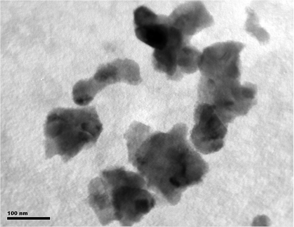
TEM micrograph of calcined hydroxyapatite powder.
The highly porous hydroxyapatite bioceramics was prepared by two methods: gelcasting of foamed slurry and replication of pours polyurethane matrix. Schematic representations of both methods are presented in Fig. 2.
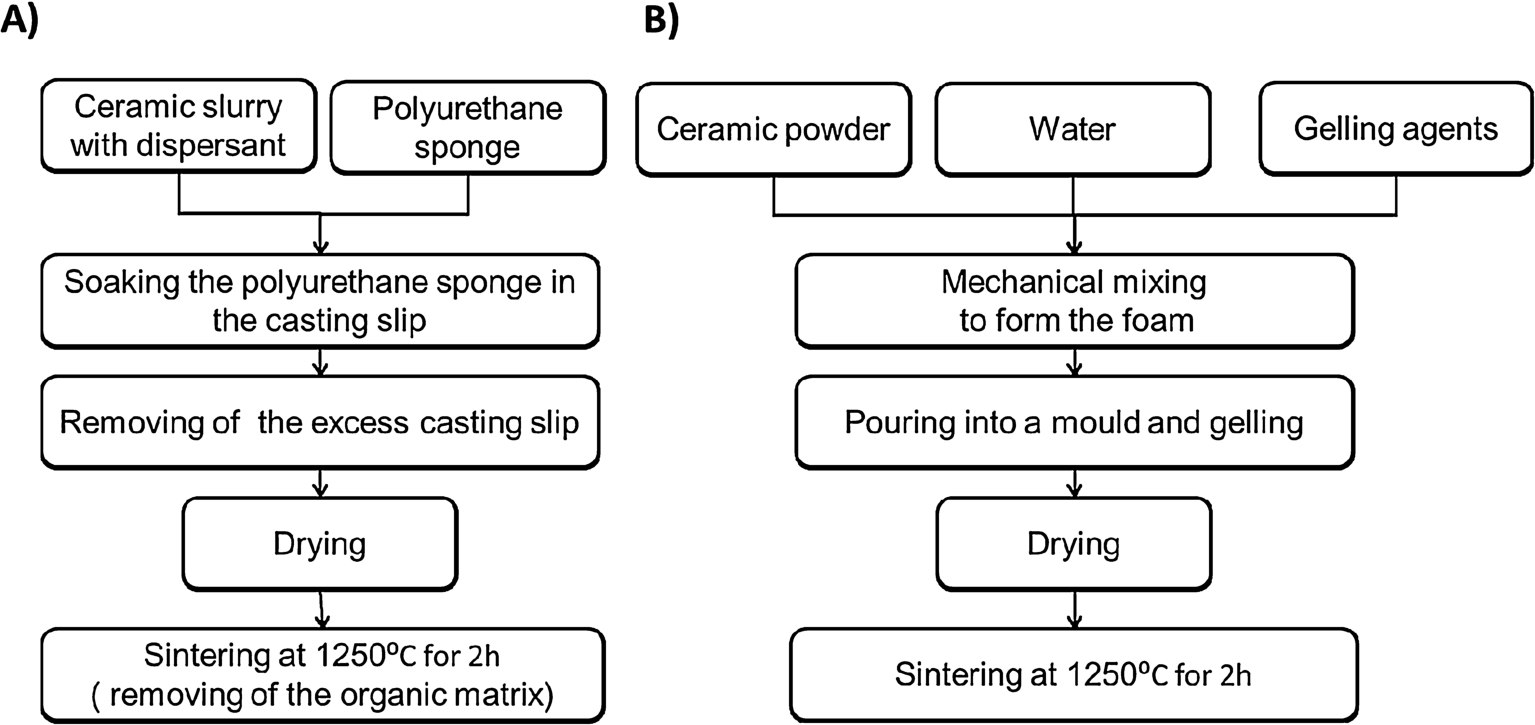
Schemes for the preparation of porous hydroxyapatite bioceramics: (A) the method of replication of porous organic matrix (B) the gelcasting of foamed slurry.
In preparation of highly porous hydroxyapatite scaffolds via the method of replication of porous organic matrix: the polyurethane foam (Eurofoam Co.) was used to mapping the porous microstructure. The cylindrical samples with a diameter of 14 mm and a height of 5 mm were cut from the sponges and impregnated with the HAp slurry. The suspension composed of: hydroxyapatite powder calcined at 800°C, 27.5 wt.% of water, 2.5 wt.% Dolapix PC-67 (Zschimmer-Schwarz) – used as dispersant and 0.05 wt.% of binder (0.5 wt.% methylcellulose, Fluka). The amount of slurry for soaking of 1 cm3 of the polyurethane sponge was
In the case of gelcasting technique [36]: ceramic water suspension with 49 vol.% of HAp powder as a solid phase was prepared by adding 0.64 wt.% of dispersant (Darvan 811, R.T. Vanderbilt). The 2.5 wt.% agarose solution was prepared by mixing agarose powder (POCH) with distilled water (in autoclave at 121°C) and added to the HAp slurry maintaining the temperature at 60°C. The suspension was foamed using the high-speed mixer. Tergitol TMN-10 (Sigma-Aldrich) and Simulsol SL-26 (Seppic) were introduced into the reaction system as stabilizers of foams. Detailed descriptions of the developed foaming process were presented in our earlier work. The green samples were dried at room temperature and then sintered at 1250°C for 3 hours at the heating rate 120–240°C/h with 2 hours soaking time.
Microstructure observations of both highly porous HAp bioceramic samples were made using a scanning electron microscope Hitachi S-3500 together with BSE imaging mode allowing for observation of specimens without coating them with conductive layer.
Quantitative description of the scaffolds microstructure was made on the basis of SEM images of randomly selected areas on the samples using computer image analysis applying the program Micrometer [33,34]. This method allows obtaining information about the actual size and distribution of pores in the material. SEM analysis of images included: processing, measurements and interpretation of results. Microstructure observations were performed using magnification:

Graphical interpretation of parameters describing image of the pore:
In addition, the microstructure of the sinters was examined using Computed Tomography Xradia Micro XCT-400. Scanning of porous HAp samples was made at a voltage of 50 kV and 10 W lamp. The table on which the samples were placed was rotated at 1° in the range of 0–360°. Exposure time for a single sample was four hours. Each of the investigated samples was scanned at the same distance from the X-ray source and with the same parameters of the lamp what guaranteed the same resolution for individual materials. The cross sections and the 3D reconstruction of tested samples were shown on photographs and on their basis a change of total porosity (open and closed) as a function of the sample height was determined.
Figure 4 shows the SEM micrographs of the fractured hydroxyapatite material obtained via replication of pours organic matrix. The spherical pores, forming interconnected system covering the entire volume of HAp sample – from the surface to the interior were observed. Presence of such a system with the open pores (Fig. 4(a)) facilitates bone ingrowth and promotes the strong bone bonding ability. It was found that pore walls revealed a good degree of sintering (Fig. 4(d)).
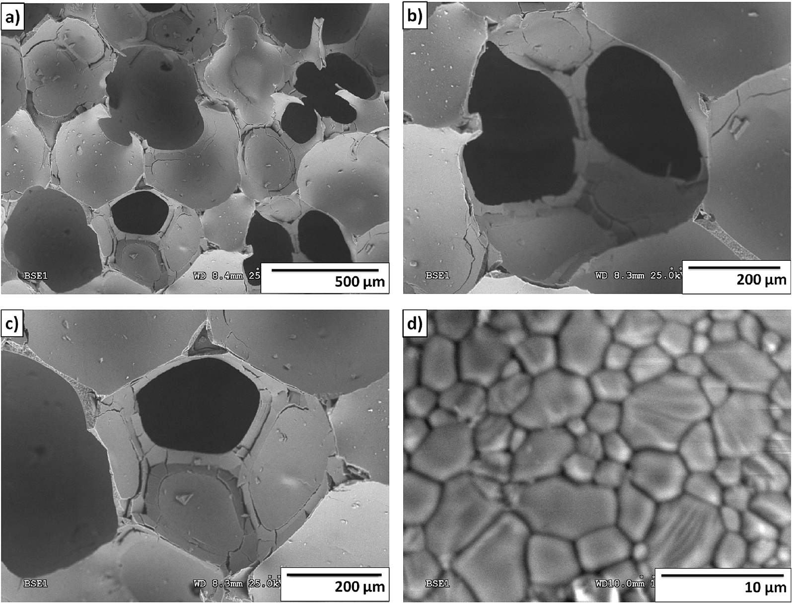
Microstructure of the HAp material produced by replication of pours organic matrix.
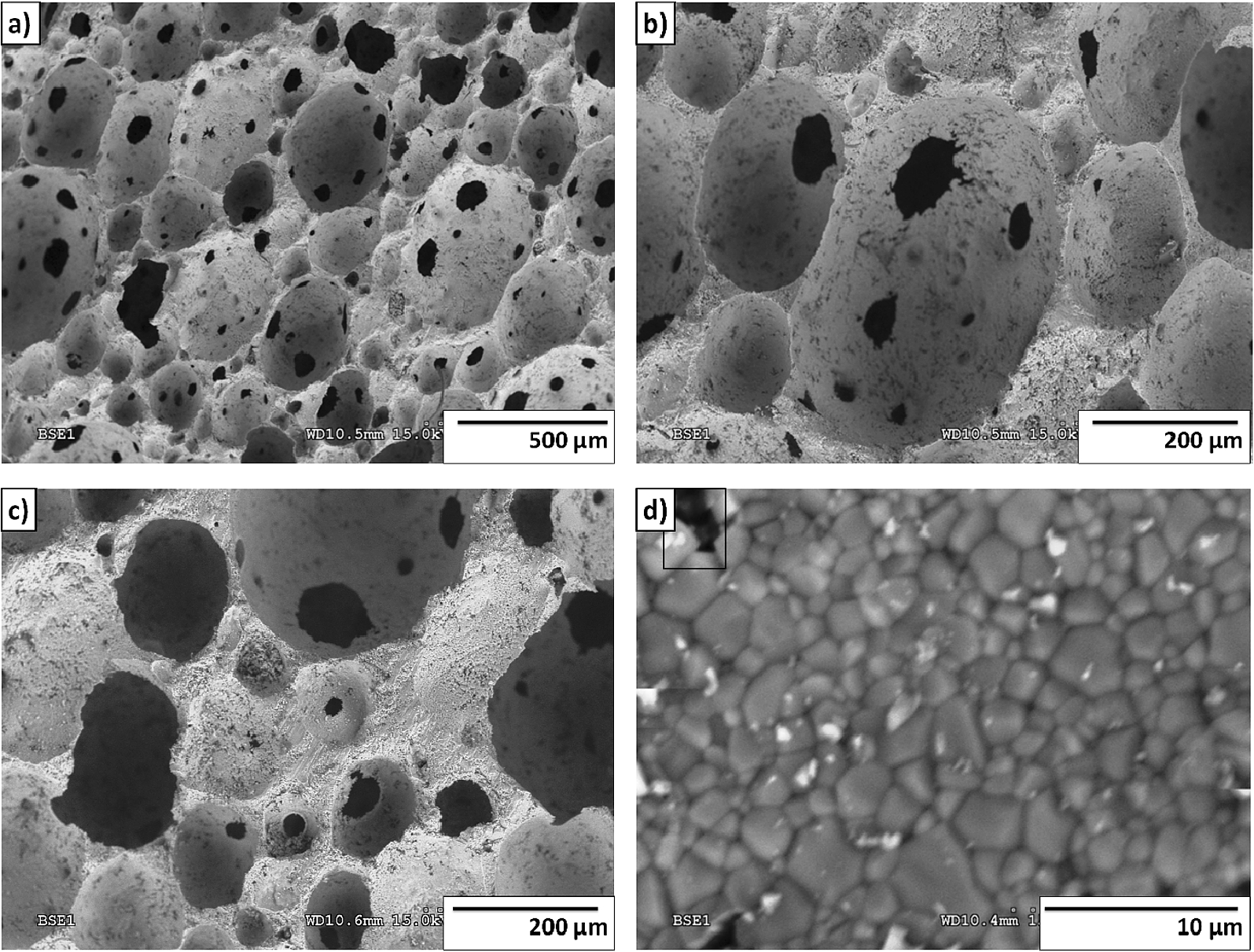
Microstructure of the hydroxyapatite bioceramics produced by gelcasting technique.
Figure 5 shows the SEM micrographs of the fracture of the hydroxyapatite foams obtained via gelcasting applying agarose. It has been shown that materials prepared by this method are characterized by two types of pores: the spherical macropores and pores of smaller size also with spherical shape. The spherical macropores occurring in these samples are referred to as “foam cells”, while the smaller pores connecting “foam cells” and forming an interconnection system between them are called “windows” [36]. Observation of the microstructure showed that the macropores are distributed uniformly throughout the volume of the sample. It was observed, that in the sintered ceramic walls, there are micro-voids of approximately 1 μm (Fig. 5(d)). On the basis of scanning electron images, it was confirmed that the amount of occurring voids is relatively lower than the amount of macropores and “windows”.
Results of stereological analysis of the macropores are presented in Table 1.
The stereological analysis demonstrated, that bioceramics prepared by gelling of foamed slurry has lower pore size, than the material obtained by the method of replication of polyurethane spongy. It was stated that the method of gelcasting of foams allowed obtaining hydroxyapatite bioceramics with the macropores ranging from 95 μm to 158 μm (the modal value of 120 μm). However, when the method of replication of polyurethane sponge was applied macropores from 295 μm to 337 μm (the modal value of 300 μm) were obtained. Additionally, in the case of bioceramics obtained by gelcasting the small pores of size 34 μm–60 μm so called “windows” were observed on spherical walls of macropores. Micropores of size 0.6 μm–1.3 μm were also visible in sintered areas.
Based on the results of stereological analysis, which were used to determine the shape factors, it was found that the macropores present in both series of samples were characterized by a similar shape. This is evidenced by comparable values of determined parameters (elongation, surface development, convexity) (Table 1).
Parameters describing shape factors of pores present in the HAp material
X-ray microtomography analysis of the interior of porous sinters was done. Two-dimentional cross-sectional images were taken (Fig. 6) to determine pore size distribution and to create a virtual 3D models of tested materials.
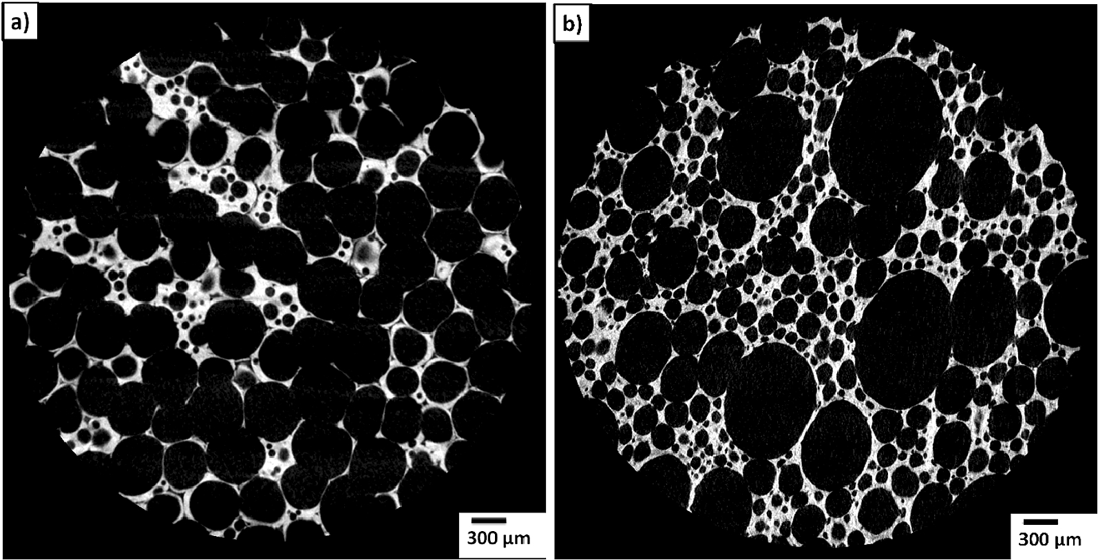
Reconstructed 2D cross-sectional images: (a) the hydroxyapatite ceramics produced by gelcasting technique, (b) the hydroxyapatite ceramics produced by replication of porous organic matrix.
Figure 6 presents 2D cross-sectional images of the studied highly porous hydroxyapatite bioceramics. The X-ray computed microtomography analysis of two hydroxyapatite materials, produced by two different methods, revealed that they varied in pore size distribution. Material obtained by gelling of the foamed slurry (sample A) could be described by a bimodal distribution of pore size. However, in the case of method of replication of porous organic matrix (sample B), the distribution of the pore size is multimodal. Based on the μCT images, 3D models of studied materials were created as shown in Fig. 7. It was observed that the sample A has a greater amount of mutually contacted spherical pores than the material B. On the basis of these observations, it was found that the distribution of macropores and micropores in the hydroxyapatite bioceramics can be controlled by selection of the manufacturing method.
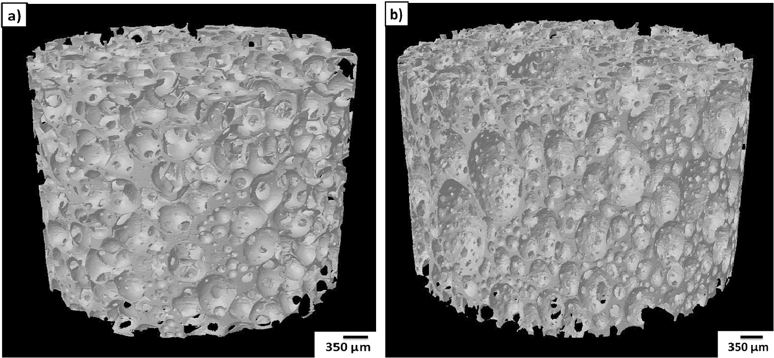
3D model of the sintered hydroxyapatite material: (a) produced by the method of gelcasting of foamed slurry, (b) produced by replication of porous organic matrix.
It was observed that the grain sizes in sintered areas ranged from 2.1 μm to 2.3 μm – in the case of scaffold obtained via replication of polyurethane sponge, and from 1.1 μm to 1.9 μm for the bioceramics prepared by gelling method.
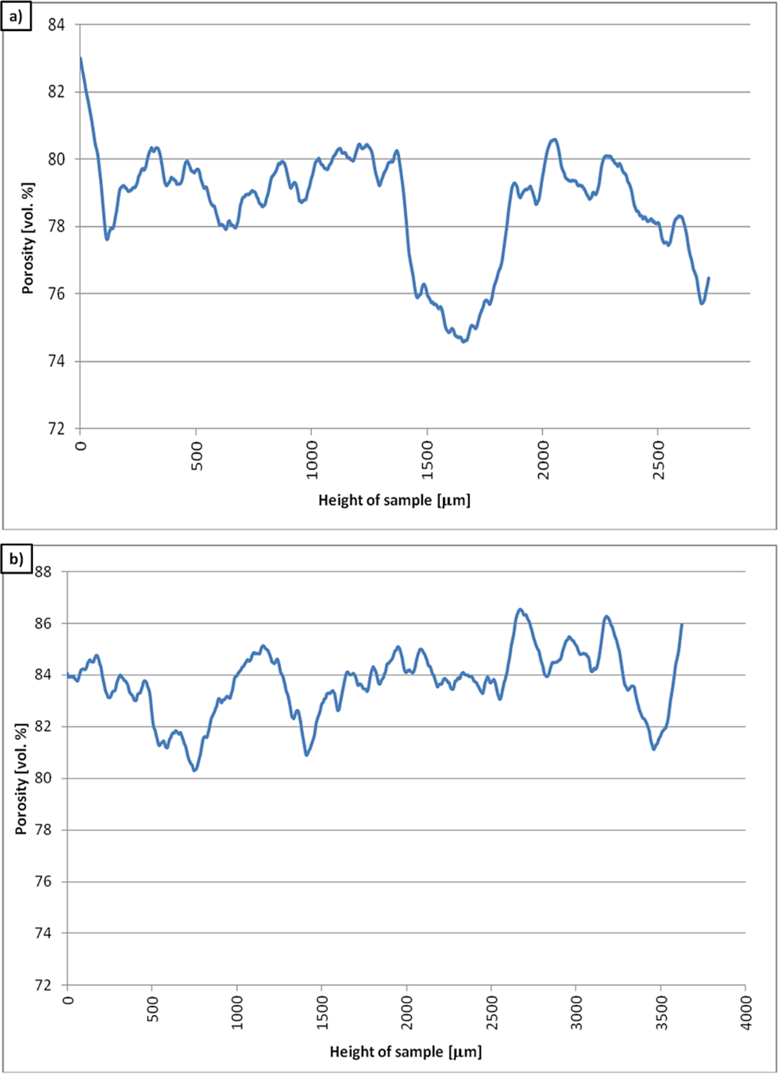
Distribution of porosity [vol.%] as a function of height of the sample [μm]: (a) material produced by the method of gelcasting of foamed slurry, (b) material produce by method of replication of porous organic matrix.
The morphometric analysis allowed determining porosity changes depending on the height of tested sinters (Fig. 8). Porosity is calculated as the ratio of the volume occupied by hydroxyapatite phase to the sum of volumes of the solid material and voids (pores).
Based on morphological analysis the total porosity of tested materials was determined. In the case of material, produced by gelcasting method, the porosity was
Porosity, pore size distribution, and pore connectivity are the key parameters in the process of designing scaffolds for tissue engineering and regenerative medicine. In this respect, to characterize the porosity it is helpful to use X-ray microtomography, which gives more information about the microstructure of scaffolds than classical mercury intrusion porosimetry (MIP) or gas adsorption/condensation method. In this study application of μCT method allowed obtaining the 2D cross-sectional images of examined materials, and then their 3D models. The results of quantitative description of microstructure allowed determining the differences between porous hydroxyapatite bioceramics obtained via replication of porous organic matrix and gelcasting of foamed slurry. The stereological analysis demonstrated, that bioceramics prepared via gelling of foamed slurry has a lower pore size and grains (1.1–1.9 μm) than the material obtained by the method of replication of polyurethane sponge (2.1–2.3 μm). Based on morphological analysis the porosity of tested materials was determined. In the case of material produce by the gelcasting, porosity of the samples was about
It should be emphasized that, based on the study of size, shape and distribution of pores in the analyzed highly porous scaffolds, it is difficult to assess their in vivo behavior as bone grafting materials. The final evaluation of both bioceramics will be possible after conducting in vivo tests. Nevertheless, the results can be considered as very promising. Developed materials meet the criteria for porous bone substitutes and, due to their phase composition, with a great possibility will promote formation of the chemical bond on the bone-implant interface.
Footnotes
Acknowledgements
The authors would like to thank to dr Marek Potoczek for preparation of the hydroxyapatite bioceramics by the method of gelcasting of foamed slurry. The authors also, would like to thank Professor W. Święszkowski and his Team from the Faculty of Materials Science and Engineering of Warsaw University of Technology for their support during the course of work with X-ray microtomography.
Conflict of interest
The authors have no conflict of interest to report.