
Abstract
Background:
Polytetrafluoroethylene (PTFE) is poorly biocompatible due to its low surface energy and hydrophobicity, which cause weak cell attachment and proliferation and complicate its use in implants.
Objective:
NH3 plasma was used for surface modification and binding of amine groups on the PTFE surface. Collagen was immobilized on the plasma-treated PTFE in order to enable it to support enhanced cell adhesion and growth.
Methods:
PTFE was exposed to NH3 plasma and collagen was immobilized on the NH3 plasma-treated surface. ATR-IR, SEM, EDXA and contact angle were conducted to determine the composition, microstructure and wettability of samples. The cytocompatibility of the samples was assessed via the growth HUVEC cells using MTT assay.
Results:
Plasma treatment resulted in an incorporation of functional groups, containing N2 and O2 that caused the PTFE surface to become hydrophilic with contact angle 68°. Also, a reduction in F/C ratio was observed after collagen immobilization that indicates the presence of collagen. Cells proliferated in greater numbers on the collagen immobilized-PTFE as compared to the plasma-treated one.
Conclusions:
Plasma treatment incorporates functional polar moieties on the PTFE surface, causing enhanced wettability, collagen immobilization and cell viability. Collagen-immobilized PTFE may offer a valuable solution in biomedical applications such as vessel grafts.
Keywords
Introduction
Cardiovascular diseases are an important cause of mortality in developed countries and patients usually require open surgical procedures such as; bypass operations in which unhealthy vascular veins are transplanted or replaced via grafts. These grafts are not transplanted from biological native organs or tissues [1–4], because regular auto-grafts are not achievable [5] or are inappropriate for the operating procedure [6], while rejection reactions of allografts are likely to occur in such cases. Thus implants are made of artificial materials, for instance polyethylene terephthalate (PET), polytetrafluoroethylene (PTFE), silk, polyurethanes (PU), polyglycolic acid (PGA) and Polylactic acid (PLA) [1,2].
PTFE, commonly referred to as ‘Teflon’, is a stable, chemically and biologically inert polymer, having properties like hydrophobicity, high oxygen permeability and the capability to withstand enzymatic and microbiological attack [7–9]. PTFE is poorly soluble and resistant to oxidants, acids and alkalis [10] and can be fabricated in various forms [11]. Consequently, PTFE has clinical applications in cardiovascular, reconstructive vascular system (include vascular grafts and heart valves), abdominal and orthopedic surgery [1,12–14]. Unfortunately, the hydrophobicity and low surface energy of PTFE cause weak cell attachment and inefficient cell spreading on the surface. To solve these problems, alteration of the surface characteristics of PTFE is needed either by chemical or physical modification or by immobilization of natural or synthetic biomolecules onto its surface [15]. Some of these methods are Extreme ultraviolet (EUV) [10], plasma discharge [11], dielectric barrier discharge [16], surface coating [11,17,18] and biomolecule grafting [19].
Plasma surface modification is a commonly used procedure to increase surface biocompatibility and protein adhesion incorporation of various functional groups without influencing any bulk properties [7,11,20–23]. Surface properties of plasma-modified polymers depend on the plasma expose time, temperature and the composition of ambient atmosphere under which the modified specimens are stored [24]. Interfacial adhesion of PTFE can be enhanced by exposing it to air, Ar, O2, CO2, H2O and NH3 plasmas [21]. Until today, plasma deposition has been used to apply different coatings with diverse chemically reactive moieties such as: amine (–NH2), carboxyl (–COOH), hydroxyl (–OH), etc. [25]. The wettability of a PTFE surface can be altered and enhanced by using plasma [9,26]. Researchers used Ar + O2 plasma to produce superhydrophobic PTFE surfaces with high durability [4], but the use of Ar plasma has been shown to decrease the hydrophobicity and contact angle of this polymer surface [27,28]. Dielectric barrier discharge (DBD) plasma and radio frequency (RF) plasma treatment also cause a decrease in water contact angle of a PTFE surface [29]. In addition, low-pressure H2 and NH3 microwave Plasma modification of PTFE powder, also resulted in enhanced wettability [30]. In another report using Ar + CO2 plasma, it was shown that the resulting wettability of the PTFE surface depends on the plasma exposure time [31].
Collagen, a rope-like protein, comprises almost one thirds of all proteins in biological tissues and is an important element for the strength and toughness of tissues [32]. It is the main structural component of connective tissue and is a guide for cell growth and ell-cell interactions [33]. Collagen has numerous applications in biomedical, pharmaceutical, cosmetic and food industries, owing to its biodegradability, bioactivity, low toxicity and immunogenicity, weak antigenicity and high biocompatibility [34,35].
In the current research, NH3 plasma was used for surface modification and binding of amine groups on the surface of PTFE. Collagen was immobilized on the surface of the modified PTFE in order to assess enhanced biocompatibility. Finally, human umbilical vein endothelial cells (HUVEC) were cultured on the PTFE surface and the surface properties were characterized by various techniques.
Materials and methods
Material preparation
PTFE was purchased from ®Good Fellows Ltd., UK with a density of
Plasma treatment
Plasma enhanced chemical vapor deposition (PECVD) was used to modify the PTFE surface with NH3 gas. First, the chamber air was evacuated for 30 minutes to obtain a vacuum pressure of about
Plasma treatment on samples by a different operating power and exposure time
Plasma treatment on samples by a different operating power and exposure time
Before collagen grafted the plasma-treated samples with the minimum contact angle, they were soaked in acetone for 20 min and rinsed with deionized water to exchange the acetone. This treatment provides a wetted surface in order to enhance collagen binding to the sample surfaces. Subsequently, the samples were placed in 2.5% glutaraldehyde to interact with the grafts and to facilitate the interaction between the PTFE surface and collagen. After washing for 1 h in deionized water to remove unbound glutaraldehyde, the samples were soaked in 1 ml of collagen/deionized water solution at 4°C to allow the formation of chains of cross-linked collagen on the PTFE surface. After immobilization, free collagen was rinsed off with nitric acid [37]. The schematic diagram of the reaction sequence of collagen attachment to plasma-treated PTFE is shown in Fig. 1.

The schematic diagram of the reaction sequence of collagen attachment to NH3 plasma-treated PTFE surface.
Measurement of the water contact angle
To determine the wettability of untreated PTFE and the plasma-treated samples, the contact angles between water and PTFE sample surfaces were measured by a manual contact angle measuring device and a goniometer (®Krüss LBI02, Germany). To decrease the effect of gravity, small drops of 0.2 ml each were applied on the surface using an automated pipette. The measurements were carried out at a 22°C and 45% relative humidity (RH). The averaged value of the angles of each drop was determined from an average of 4 measurements on both sides with a standard deviation of ±2°.
Surface morphology and microstructure analysis
The surface morphology and elemental analysis of the untreated PTFE, plasma-treated and collagen-immobilized samples were investigated using a scanning electron microscope (SEM) and energy-dispersive X-ray analysis (EDXA) (®VEGA/TE SCAN, USA, SEM/EDXA). In order to improve the imaging quality and prevent charging of the samples during SEM operation, the samples were first coated with a thin gold film using a sputter coating device (®BIO-RAD-E5200, UK).
ATR-IR spectroscopy
Attenuated total reflection infrared spectra (ATR-IR) (®Equinox 55, Bruker, ATR-IR) were obtained to confirm the plasma modification and to characterize the chemical composition of the surfaces of the untreated PTFE, plasma-treated and collagen-immobilized samples at wavenumbers of
Cell proliferation and viability analysis
In vitro experiments were performed with human umbilical vein endothelial cell line (HUVEC), purchased from ®NCBI, Pasteur Institute, Tehran, Iran. The cells were grown in RPMI-1640 (®Gibco, USA) with 10% fetal bovine serum (®Gibco, USA), 100 IU penicillin/ml and 100 μg streptomycin/ml. The samples were cut into pieces, rinsed with alcohol and sterilized on both sides under UVC radiations for 30 minutes before the cell culturing. The samples were placed in 24-well plates and the cells were seeded on the samples at a density of 50,000 cells/well and grown on the samples in above-mentioned culture conditions for 24 h at 37°C. The viability and proliferation of the HUVEC cells, attached to the plasma-treated PTFE were examined by the methylthiazol tetrazolium (MTT) (®Invitrogen, Germany) assay before and after collagen immobilization. MTT solution (100 μL; at a concentration of
Cell morphology assessment
SEM was utilized to study the HUVEC cell attachment and morphology on the collagen-immobilized samples. A density of 4000–5000 cells was placed on sterilized disks in 1 mL culture media, followed by culturing for 3 days. In order to perform SEM, the samples were immersed into a 2.5% glutaraldehyde solution (®Merck, Germany) for 24 h in a refrigerator and subsequently dehydrated in graded ethanol (50%, 60%, 70%, 80%, 90%, 95% and 100) for 5 min. The dried and fixed samples were then gold-coated using a Sputter coating device (®BIO-RAD-E5200, UK), and viewed with an SEM.
Statistical analysis
The statistical analysis was carried out using SPSS software for Windows (®v 17.0; IBM, NY, USA). When the statistical differences were detected, a student’s t-test was performed. Data are reported as mean ± standard deviation at a significance level of
Results and discussion
Sample characterization
The PTFE substrate was modified by NH3 plasma discharge and the operational parameters (discharge power and time) were varied in order to deposit a varying number of amine groups on the surface of this polymer. Collagen was successfully immobilized on the surface of these NH3 plasma-treated samples. SEM microscopy was used to evaluate the surface morphology of untreated PTFE and plasma-treated samples (Fig. 2). It was found that the surface morphology of untreated PTFE sample was flat and smooth with no significant superficial structures. This morphology was significantly altered after plasma treatment, which is attributed to the etching reactions between the PTFE surface and plasma discharge which contains active species [31].

SEM micrographs of (a) untreated PTFE, and (b) plasma-treated sample.
EDXA results were obtained in order to assess the elemental composition of the PTFE surface before and after collagen immobilization. Table 2 shows the analysis results for plasma-treated samples. Carbon is the most abundant element on the PTFE surface with a concentration of 50.39%. The next abundant element is fluorine at 39.85%, which means that the F/C ratio is 0.79 in this sample. These two elements are expected to make up the majority of the polymer surface, as they are the structural elements of PTFE. Nitrogen and oxygen were incorporated onto the PTFE surface after NH3 plasma treatment, and the amount of nitrogen was almost twice that of oxygen. The presence of nitrogen is due to NH3 plasma operation, which means that the plasma radiation could successfully modify the sample surface and incorporate the nitrogen onto it as well. It should be noted that the presence of oxygen may be caused by air leakage in the plasma chamber or could have arisen when the samples were exposed to air immediately after plasma treatment [39].
Chemical composition of PTFE samples after plasma treating
The EDXA results of the collagen-immobilized samples are given in Table 3. After collagen immobilization, the concentrations of carbon, nitrogen and oxygen atoms increased while the percentage of fluorine atoms on the PTFE surface had a dramatic. With an abundance of 55.39%, carbon is still the most abundant element followed by nitrogen (18.36%) and fluorine (14.91%). The F/C atomic ratio thus decreased to 0.27, a 52% decline as compared to the plasma-treated samples before collagen immobilization. Thus, the biological modification and an increase in the amount of nitrogen and oxygen elements on the PTFE surface may be because of the C/O elements, present in the collagen structure. Similarly, an increase in the amount of PTFE-surface carbon confirms the surface modification of this polymer with collagen.
The bulk composition of the untreated, plasma-treated and collagen-immobilized PTFE samples was examined by ATR-IR (Fig. 3). In the asymmetric spectra of the untreated samples, only the characteristic bands of C–F and C–C bonds were observed, at about 1150 and
Chemical composition of plasma-treated samples after collagen immobilization

ATR-IR results of (a) untreated PTFE, (b) Plasma-treated samples (at 100 W for 180 sec), and (c) collagen-immobilized samples.
The ATR-IR test outcome of untreated PTFE demonstrates that the surface did not contain significant impurities before plasma treatment. After treatment, absorption features were present in the spectra, which is indicative of the chemical changes on the PTFE surface. As explained above, N–H, C–H and ammonium fluoride were observed in the treated samples, which proved the PTFE surface modification. Similarly, after collagen immobilization,
The measurement of the contact angle between water and a surface is one of the most straightforward ways to characterize the hydrophilicity and wettability of the surface. The evaluation of the water contact angle on the untreated PTFE and the plasma-treated samples, as a function of plasma exposure time, is shown in Table 4. The water contact angle of the untreated PTFE sample was measured to be 114°, revealing the hydrophobic nature of this surface. The contact angle decreased to approximately 68° after plasma treatment at 100 W for 180 sec. However, when the PTFE samples were plasma-treated for more than 180 sec, the contact angle started to rise, reaching a value of 92°. Adding hydrophilic nitrogen/oxygen moieties on the surface was expected to cause a decrease in hydrophobicity, because these elements allow the hydrogen-bond formation between electronegative atoms, present on the treated surface and the surrounding water molecules [39,40]. The water contact-angle measurements reveal a decrease in water contact angle in all plasma-treated samples, regardless of the duration and intensity of the plasma treatment compare to the untreated sample. Oxygen moieties were incorporated into the sample from the oxygen impurities, present in the plasma chamber or exposure of sample to air or water vapor after plasma treatment [28,41]. At constant operating power and increasing exposure time, a decrease in contact angle was observed. But, as the exposure time increased, the water contact angle also showed an increase. Although such a reversal of the water contact angle is not reported in related studies, it should be noted that plasma deposition can be used to either increase or decrease the contact angle [27,42,43]. This can be the effect of the embedment of nitrogen moieties in the substrate and the destruction of the amine groups on the PTFE surface after prolonged plasma treatment. In other words, the incorporation of hydrophobic moieties can be valid up to a certain limit.
Average contact angle in different exposure time and constant operating power
Average contact angle in different exposure time and constant operating power
A similar behavior was observed for the experiment in which the exposure time was constant, at increasing operating power. Our results show a rise in wettability, followed by a decreasing trend. Therefore, the hydrophilicity is shown to depend on the plasma treatment time as well as the plasma intensity (Table 5).
Average contact angle in different operating power and constant exposure time
To investigate the improved biocompatibility and cells interaction on samples, the MTT assay and SEM analysis were carried out. Figure 4 shows the HUVEC cell viability after 24 h of incubation with samples. The results demonstrate a promoted cell viability and proliferation functions without the onset of cell necrosis on the collagen-immobilized PTFE surfaces. Collagen immobilization increased cell viability to 99%, as compared to the plasma-treated samples, in which only 67% of the cells could. Therefore, the collagen-immobilized samples can be considered to be non-cytotoxic and highly cell-compatible.

The HVEC cell viability after 24 h of incubation with the samples.
SEM observations in Fig. 5 demonstrate the morphology of HVEC cells on the collagen-immobilized sample after 3 days of culture. Cells were flattened and well-spread across the surface and were adhered to the substrate with cellular micro-extensions (e.g., filopodia) that is indicative of live cells. Collagen immobilization can therefore be used as a convenient modification for artificial vessels and medical applications.
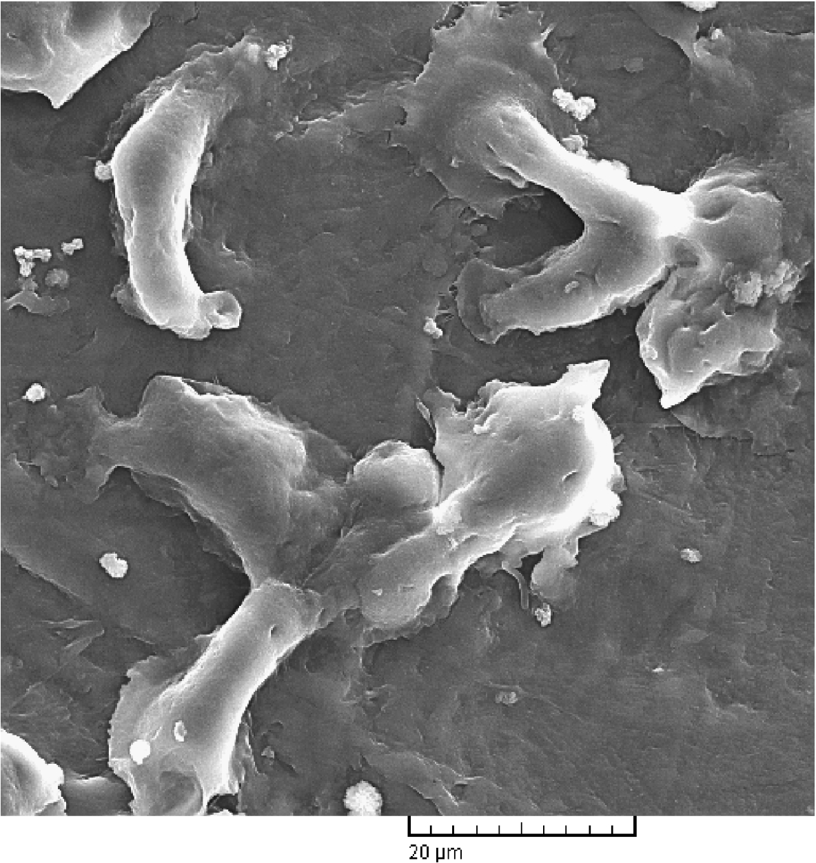
SEM micrograph of HVEC cells on collagen-immobilized sample after 3 days of culture.
This work presents a mechanism for increasing biocompatibility of PTFE polymer by using NH3 plasma treatment and subsequent collagen immobilization. The resulting contact angle was directly correlated to the exposure time and plasma power. While, the optimal parameters of plasma were the operating power of 100 W and the exposure time of 180 sec for polymer-surface treatment, which resulted in the achievement of minimum contact angle of 68°. Plasma treatment resulted in an incorporation of functional groups, containing N2 and O2 that caused the PTFE surface to become hydrophilic. Also, an increase in the amount of N2 and O2 and a reduction in F/C ratio were observed after collagen immobilization that indicates the presence of collagen and occurrence of defluorination phenomenon. In vitro experiments demonstrated that collagen immobilization can increase cell viability to 99% and that cells can homogeneously spread over the modified surface, having flattened shape and filopodia. Therefore, hydrophilic PTFE samples can prove to be suitable in a variety of medical applications.
Footnotes
Acknowledgements
The authors gratefully acknowledge the scientific help and assistance of Pasteur Institute of Iran (IPI) and Department of Biomedical Engineering, Islamic Azad University, Yazd Branch.
Conflict of interest
The authors have no conflict of interest to report.