



Abstract
Background:
Effective encapsulation of drugs into the delivery systems could increase the efficiency of nanoparticles in prevention and treatment of diseases.
Objective:
The purpose of this study was to compare the different methods for determination of encapsulation efficiency of a model protein in the PLGA nanoparticles.
Methods:
The various direct methods include dichloromethane, acetonitrile, modified acetonitrile and NaOH based extraction and radioactive methods were used to directly calculate the encapsulation efficiency of the loaded protein in the PLGA nanoparticles. Furthermore, indirect methods include BCA, Fluorescent and radioactive methods were compared.
Results:
The encapsulation efficiencies determined by indirect methods include dichloromethane, acetonitrile, modified acetonitrile, NaOH based extraction and radioactive methods were 12.62% ± 1.97, 17.43% ± 2.51, 64.69% ± 4.31, 86.36% ± 2.25 and 90.15% ± 1.78, respectively. Moreover, the encapsulation efficiencies determined by indirect methods include BCA, fluorescent and radioactive methods were 81.46% ± 1.92, 88.23% ± 1.15 and 89.6% ± 1.9, respectively.
Conclusions:
Among the results obtained by indirect methods, radioactive and fluorescent methods showed more reliable. Moreover, NaOH and radioactive methods were the most reliable methods among the direct methods.
Introduction
Among different colloidal drug delivery systems, polymer based nanoparticles are promising vehicles for targeted delivery to specified organs in the body at therapeutically optimal rate and dose [1,2]. Among various polymers, biodegradable polymers such as poly (lactic acid), PLA and poly (lactide-co-glycolide) and PLGA are suitable and versatile ones which has been increasingly applied in the nanomedicine [3,4]. PLGA polymers with unique properties such as biocompatibility, different degradation time, FDA approval and potential of surface modification are the most frequently used polymers for fabrication of delivery systems for drugs, proteins and various other macromolecules such as peptides, RNA and DNA [5,6]. Several factors such as lactide to glycolide ratio and Molecular weight of the polymer could affect its characteristics including solubility, degradation rate and release profile of the encapsulated drug [7]. The biodegradation rate of PLGA nanoparticles could be tailored from a few weeks to several months, depends on several factors including molecular weight, ratio of glycolide to lactide, stereochemistry and end-group functionalization [8]. Inside the body, the PLGA degrades to glycolic acid (GA) and lactic acid (LA) monomers and easily eliminated through the citric acid cycle [9]. Among the various methods to prepare PLGA nanoparticles, the single/double emulsion (oil–water/water–oil–water) are the most widely used methods to encapsulate hydrophobic and hydrophilic macromolecules like proteins [10].
From therapeutic and economic points of view, the rate of drug encapsulation is an important parameter that could impact on efficiency of nanoparticles in prevention and treatment of diseases [11]. Blanco et al. and Lamprecht et al. studied on different parameters affecting the entrapment efficiency in PLGA particles [12,13]. Moreover, Feczko et al. studied the combinations of variables and different parameters to describe extensive definition for optimize size and encapsulation efficiency [11].
However, determination of accurate encapsulation efficiency (the percentage of proteins that is entrapped into the nanoparticles) is usually challenging. For determination of loaded protein in PLGA nanoparticles both direct and indirect methods are encountered with some complexity. For direct methods the nanoparticles must be dissolved in an organic solvent, in which the protein precipitates and must be re-dissolved in a proper medium such as buffer, water or NaOH solution. Complete precipitation of protein, complete re-dissolving of precipitated protein, possible interaction of poly vinylalcohol (PVA), frequently used in preparation of PLGA nanoparticles, are among the challenges of this method. Complex preparation medium of PLGA nanoparticles, especially in emulsification-solvent evaporation method, possible interaction of surfactant, mainly PVA and detection of trace amounts of non-encapsulated protein diluted in the supernatant of nanoparticle suspension are amongst the challenges of indirect method. For achieving accurate and precise encapsulation efficiency, different methods have been studied [1,14,15].
Thus, in this study we evaluated and compared the validity and reliability of different methods for evaluation of encapsulation efficiency. Bovine serum albumin (BSA) was used as a model protein.
Experimental
Materials
Bovine serum albumin (BSA), PLGA (lactic to glycolic acid molar ratio of 50 : 50) and Poly (vinyl alcohol) (PVA, average MW 30,000–70,000) were purchased from Sigma Aldrich (USA).
Methods
Preparation of nanoparticles
Nanoparticles were prepared by double emulsion solvent evaporation technique described by Lamprecth et al. with some modifications [16]. Briefly, 200 μg of BSA was dissolved in 200 μl of PBS and added to 20 mg of PLGA dissolved in 700 μl of dichloromethane (DCM). The mixture was sonicated for 10 seconds to make W1/O emulsion. Then, 4 ml of 1.5% w/v solution of PVA was added to W1/O emulsion and sonicated in ice bath for 60 seconds. The resulting emulsion was added to 10 ml of PVA 0.3% w/v solution and stirred for 3 hours. NPs were isolated by centrifugation at 20,000g for 15 min and washed 3 times with deionized water. The resulted nanoparticles suspension was freeze-dried.
Physiochemical characterization of PLGA nanoparticles
The zeta potential and size of nanoparticles were measured by Dynamic Light Scattering (DLS) method (Malvern 3000, UK). Moreover, morphology of nanoparticles was studied by scanning electron microscope (Tescan, Czech Republic).
Determination of encapsulation efficiency
Direct methods
Dichloromethane based extraction
Ten mg of PLGA nanoparticles loaded with BSA were dissolved in 1 ml of DCM and stirred on shaker incubator for 30 min at 37°C. One ml of phosphate buffer saline (PBS) was added and vigorously voretexed to extract the BSA. This step was repeated for three times. The mixture centrifuged at 10,000g for 5 min and the polymer containing supernatant was discarded. Finally, the total protein in supernatant was determined by BCA (bicinchoninic acid) method (Thermo Fisher Scientific) according manufacturer’s instructions [1]. Briefly, 200 μl of working reagent added to 25 μl of standard and supernatant and incubated at 37°C for 30 min. Then, the absorbance at 562 nm was read on a 680Microplate reader (Bio-Rad laboratories, Hercules, CA). To validate the extraction procedures, Blank PLGA nanoparticles were spiked with a known amount of BSA.
Acetonitrile based extraction
Similar to DCM method, PLGA NPs were dissolved in acetonitrile and final extracted protein in solution was measured by BCA method [1]. Blank nanoparticles were spiked with a known amount of BSA.
Modified acetonitrile based extraction
Ten mg of PLGA NPs were dissolved in 1 ml of acetonitrile and incubated for 30 min at 37°C under continuous shaking. Then, 1 ml of phosphate buffer saline (PBS) was added and slowly shaken to extract the BSA. The suspension was centrifuged at 10,000g for 5 min and the polymer containing supernatant was discarded. The pellet containing BSA was dissolved in NaOH 0.1 N. The solution was neutralized by 1 ml of HCl and protein was measured by BCA method. For validation, blank nanoparticles were spiked with a known amount of BSA.
NaOH based extraction
One ml of NaOH 1 N was added to 10 mg PLGA nanoparticles, sonicated for 15 min and incubated at 37°C for 18 hours under continuous shaking. After neutralization with HCl 1 N, mixture was centrifuged at 10,000g for 5 min and the supernatant was analyzed by BCA method [17]. For validation, blank nanoparticles were spiked with a known amount of BSA.
Radioactive method
BSA was labeled with iodine 125 (
PLGA nanoparticles were prepared with Iodine-labeled BSA and radioactivity of nanoparticles loaded with
Indirect methods
BCA method
After preparation of PLGA nanoparticles, amount of unloaded BSA was measured in supernatant of preparation medium by BCA method. The encapsulation efficiency (EE) of BSA was calculated by the equation given as:
Radioactive method
The nanoparticles were fabricated with
Fluorescent method
BSA labeled by FITC. The method described by Crandall et al. was used for the preparation of FITC-BSA conjugate [20]. Briefly, 50 mg of BSA and 20 mg of FITC were dissolved in 2 ml of 0.9% NaCl and 2 ml carbonate buffer pH 9.6. The solutions were mixed and stirred overnight at 4°C. Unreacted FITC were removed by Amicon Ultra-4 Centrifugal Filter Devices (Milipore) and dialysis against carbonate buffer for five days.
The nanoparticles were loaded with FITC-BSA. After centrifugation, the supernatant was collected to measure the unloaded FITC-BSA using spectrofluorimetry method (excitation/emission wavelengths of 495/518 nm) by spectrofluorimeter (Synergy H4 hybrid Reader, USA) [15].
Results and discussion
PLGA based nanoparticles are frequently used for delivery of peptide and proteins, both as drug or as antigen [21]. Encapsulation efficiency of the loaded protein is an important characteristic, both economically and therapeutically [10]. Various methods have been used to determine the encapsulation efficiency. These could be divided in direct and indirect methods. While in direct methods, the efficiency of encapsulation is calculated directly from the loaded protein in the nanoparticles, in the indirect method, the non-entrapped protein or drug is considered [1]. The indirect methods could only be used during preparation process and could not be applied after lyophilization. Due to the interaction of surfactants such as PVA, detection of non-encapsulated protein is the main challenge of this method. Therefore, at the present study, PLGA nanoparticles were encapsulated with BSA as a model protein and different direct and indirect methods were used and compared for determination of its encapsulation efficiency.
Physicochemical characterization of nanoparticles
The SEM images revealed spherical and regular shape for PLGA nanoparticles (Fig. 1). Particle size of the BSA loaded PLGA nanoparticles was 207.6 ± 4.8 nm, while the polydispersity index (PDI) was 0.112. Moreover, the zeta potential of PLGA nanoparticles was −35 ± 1.3 mV (
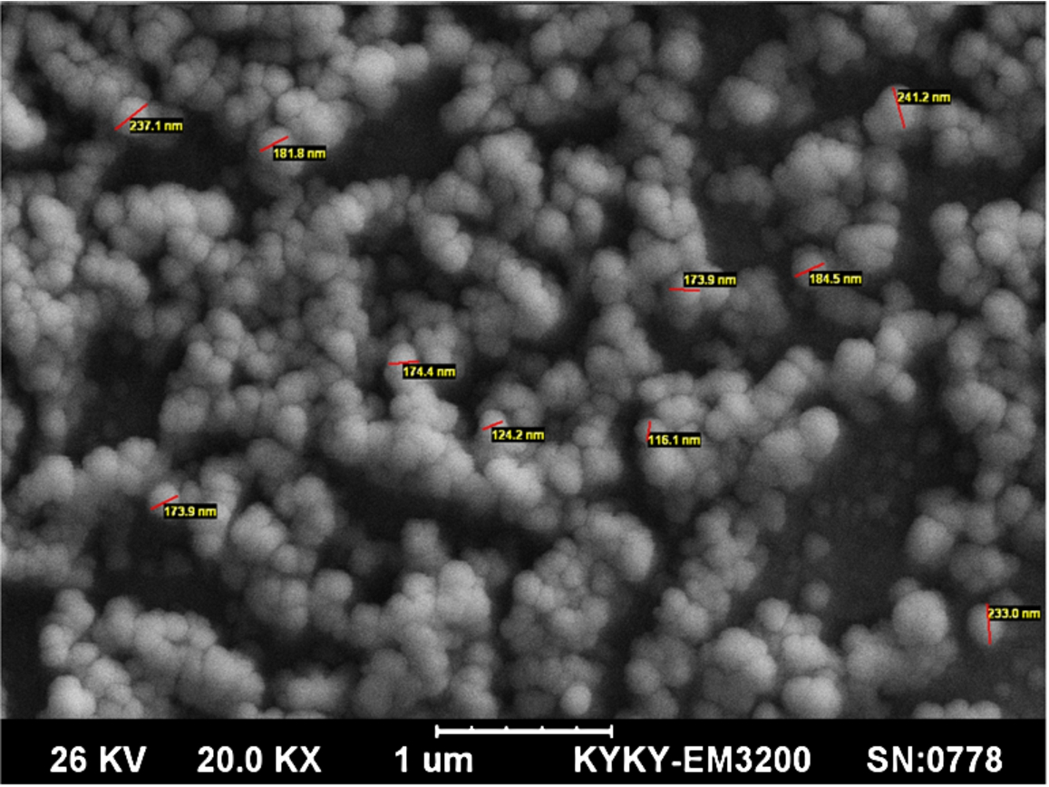
The SEM images revealed spherical and regular shape for PLGA nanoparticles.
In indirect methods, the EE% for BCA, fluorescent and radioactive methods were respectively as 81.46% ± 1.92, 88.23% ± 1.15 and 89.6% ± 1.9 (Table 1). In the indirect methods, the calculated EE% by BCA method was significantly lower than the fluorescent and radioactive methods (
Encapsulation efficiencies determined by indirect methods
Encapsulation efficiencies determined by indirect methods
Despite similarity of the results calculated by indirect methods, these methods suffer from several limitations: (1) These methods need to supernatant of preparation medium which is only available in the preparation step; (2) the low concentration of the protein in the supernatant could impose some limitations in detection methods; (3) the presence of interfering substances in the supernatant may affect the results. To analyze the EE% in the freeze-dried PLGA nanoparticles, direct methods are required. The EE% calculated by DCM, acetonitrile and modified acetonitrile methods were 12.62% ± 1.9, 17.43% ± 2.5 and 64.69% ± 4.31, respectively (Table 2). In the studies performed by Zheng et al. and Castellanos et al. the aggregation of non-soluble protein during the preparation procedure was the main drawback of these methods [1,22]. In the direct methods, after dissolving the nanoparticles, the precipitated protein could be first dissolved in the NaOH 0.1 N. This step was improved the results, however, the results were still away from the correct EE%. In a previous study demonstrated that the effect of the NaOH, as a hydrolyzer, was depended on hydrolysis time and volume of NaoH [1]. At the present study, the EE% results obtained by NaOH 1N and radioactive methods were 86.36 ± 2.2 and 90.15 ± 1.8, respectively (Table 2). The results indicated that by increasing the incubation time with NaOH 1N to overnight, the acquired results were improved [17].
Encapsulation efficiencies determined by direct methods
The direct methods for determination of encapsulation efficiencies are superior to the indirect methods, however, among the 5 methods tested, very different results were obtained and just the NaOH and radioactive methods seemed reliable. Among the results obtained by 3 indirect methods, radioactive and fluorescent methods showed more reliable results than BCA.
Footnotes
Acknowledgements
The current study was from a PhD thesis presented to the Mashhad University of Medical Sciences, Mashhad, Iran. This study was supported by Mashhad University of Medical Sciences, Mashhad, Iran (Grant No. 930461).
Conflict of interest
The authors declare that they have no conflict of interest. The authors alone are responsible for the content and writing of this article.