
Abstract
Background:
The periodontal ligament (PDL), which maintains homeostasis in the periodontium, is a group of specialized connective tissue fibers attached to both the cementum and alveolar bone. Regeneration of periodontium with PDL cells has been investigated, and the chemical and molecular structures of scaffolds control the adhesion and differentiation of cells. Therefore, the development of adequate materials for PDL-derived cells is essential to regenerate the periodontium.
Objective:
We evaluated the suitable passage time for PDL-derived cells and investigated the behaviors of PDL-derived cells grown on hydroxyapatite (HAp) scaffolds coated with type I and type III collagen.
Methods:
PDL-derived cells were isolated with enzyme from the upper molars of male Wister rats. After characterization of HAp, type I collagen, and type III collagen, PDL-derived cells at passage 2 were seeded onto collagen-coated HAp. Cell adhesion, proliferative potential, and osteoconductivity were analyzed with immunostaining, 3-(4,5-dimethylthiazol-2-yl)-2,5-diphenyltetrazolium bromide assays, Alizarin S staining, and real-time polymerase chain reaction.
Results:
Type I and III collagens were successfully coated on HAp. Gene expression analysis revealed that passage 2 was suitable for maintaining differentiation potential. Proliferative potential and cell adhesion were significantly higher on type III collagen than on HAp alone or type I collagen. In contrast, the osteoconductivity of type III collagen was significantly lower than those of HAp and type I collagen.
Conclusion:
PDL-derived cells on type I collagen differentiated into osteogenic cells and formed hard tissues. However, type III collagen enhanced the adhesion of PDL-derived cells and inhibited mineralization.
Introduction
Osseointegrated dental implants have recently become a major treatment method for tooth defects due to their esthetic and functional properties. However, osseointegrated implants have been shown to be associated with several problems, including infection, mechanical stress-related damage, sensory disorders, requirement for implant extraction, and poor bone growth. In the periodontium, the periodontal ligament (PDL), a connective tissue surrounded by two types of hard tissues, i.e., the alveolar bone and cementum, plays an important role against these external stimuli and issues and maintains the homeostasis of the periodontium [1]. PDL supports the teeth in their sockets and permits the teeth to withstand the considerable forces of mastication [1]. Furthermore, the PDL acts as a sensory receptor, which is necessary for the proper positioning of the jaws during normal function [1]. For that reason, periodontio-integrated dental implants are a major strategy used to improve the durability and function of dental implants and may be more effective than osseointegrated dental implants [2].
Recent advances in tissue engineering technology have established periodontio-integrated dental implants using hierarchical biomaterials in order to generate the complex structure of the periodontium. Inorganic biomaterials, such as hydroxyapatite (HAp) and beta-tricalcium phosphate, have been studied extensively, similar to hard tissue substitutes, and polycaprolactone and extracellular matrix (ECM) gel have been applied for soft tissues generation [3]. Moreover, hybrid materials consisting of cultured cells and biomaterials have been investigated to enhance tissue generation. Although these hybrid materials can generate periodontal tissue around dental implants in vivo, due to ankylosis, the success rate is lower than that of conventional osseointegrated implants [2,4]. Dental ankylosis, which results from fusion of the cementum and alveolar bone, is caused by disruption of PDL cell function. The main cellular component of the PDL is PDL fibroblasts, which represent a heterogeneous population of cells and exhibit cementoblast-like and/or osteoblast-like properties, including the capacity to express bone-associated markers and form mineralized nodules in vitro and in vivo [5]. Because cell proliferation, differentiation, and maturation depend on the ECM via integrin interaction [6–8], optimal ECM scaffolds are required to inhibit the mineralization of PDL and maintain PDL function [9].
The ECM of PDL is composed of two types of fibrillar collagens, i.e., type I collagen (
Interestingly, the rate of type III collagen in PDL is
However, the behaviors of PDL-derived cells attached to type III collagen, which is abundant in PDL, have not yet been clarified. Accordingly, the aim of this study was to investigate the behaviors of PDL-derived cells in the context of different types of scaffolds. The proliferative potential and osteoconductivity of PDL-derived cells attached to HAp, type I collagen, and type III collagen were investigated.
Materials and methods
Synthesis of HAp
HAp powders were synthesized by a wet method as described previously [15] using H3PO4 and Ca(OH)2, with a Ca/P molar ratio of 1.67. After the reagents were mixed and stirred for 24 h, samples were matured for 24 h. Samples were then washed in pure water and dried at 60°C for 5 days. Powders were uniaxiallly pressed at 120 MPa and sintered at 1250°C with a vaporized atmosphere to avoid dehydrating the lattice
Preparation of samples
HAp pellets were washed with acetone, ethanol, and pure water. Cellmatrix Type I-C (Nitta Gelatin Inc., Osaka, Japan) and Cellmatrix Type III (Nitta Gelatin Inc.) solutions were applied to HAp as coating materials (designated Type I and Type III solutions, respectively). Type I and Type III solutions were reconstituted using the corresponding Cellmatrix solution,
PDL-derived cell culture
Eight Wister rats (6 weeks old, male) were sacrificed by intraperitoreal injection of 1 mL pentobarbital sodium salt (Somnopentyl; Kyoritsu Seiyaku, Tokyo, Japan). After extraction of the upper first and second molars, the teeth were washed with Hanks’ Balanced Salt Solution (HBSS; Gibco, Life Technology, USA). PDL-derived cells were separated and dispersed with 0.25% trypsin (Gibco, Life Technology) and 0.5 mg/mL Liberase DL (Roche Diagnostics, Germany) for 60 min at 37°C. Single-cell suspensions were obtained with a 40-μm strainer (Falcon, BD Labware, Franklin Lakes, NJ, USA). PDL-derived cells were cultured in alpha modification of minimum essential medium Eagle (α-MEM; Wako Pure Chemical Industries, Ltd., Japan) supplemented with 20% fetal bovine serum (FBS; Gibco, Life Technology), 1% penicillin-streptomycin solution (Wako Pure Chemical Industries, Ltd.), and 1% amphotericin B solution (Sigma-Aldrich, MO, USA) at 37°C in a humidified atmosphere of 5% CO2. Floating cells were carefully removed every time the cultures were washed after cells adhered to the plates. The experiment was approved by the animal experiment ethics committee of Tokyo Medical and Dental University (grant no. 0170027A).
Osteogenetic and periodontal markers in PDL-derived cells
PDL-derived cells were subcultured until passage 4. Total RNA of PDL-derived cells from each passage was isolated with an RNeasy Mini Kit (Qiagen, Germany), and the RNA concentration was determined. cDNA was synthesized from RNA with a SuperScript VILO cDNA Synthesis Kit (Invitrogen, USA), according to the manufacturer’s instructions. Real-time polymerase chain reaction (PCR) was performed in duplicate to evaluate the expression of osteogenic and periodontal markers in PDL-derived cells using a LightCycler 480 (Roche Diagnostics) as previously described [16]. The specific primers for osteoblasts, cementoblasts, and PDL cells targeted Runt-related transcription factor 2 (RUNX2; forward: CCGTGTCAGCAAAACTTCTTT, reverse: CTCACGTCGCTCATCTTGC), osteopontin (OPN; forward: GGCTGAGTTTGGCAGCTC, reverse: TCTGCTTCTGAGATGGGTCA), F-spondin (forward: CAGCCCATCGATCCAGAA, reverse: CTCTGACCAGGCTGTCCAC), periostin (forward: TCGTGGAACCAAAAATTAAAGTC, reverse: CTTCGTCATTGCAGGTCCTT), S100A4 (forward: AGCTACTGACCAGGGAGCTG, reverse: CTGGAATGCAGCTTCGTCT), and glyceraldehyde 3-phosphate dehydrogenase (GAPDH; forward: AATGTATCCGTTGTGGATCTGA, reverse: GCTTCACCACCTTCTTGATGT) (17–21).
Cell adhesion assay
Pellets of HAp, Type I, and Type III (
Cell proliferation assay
Pellets of HAp, Type I, and Type III were separately plated onto 48-well plates, and PDL-derived cell suspensions were seeded on the samples at a density of
Mineralization assays
PDL-derived cells at passage 2 were seeded on HAp, Type I, and Type III. The medium was replaced with osteoinductive medium containing 50 μg/mL
Osteogenetic markers
Three groups of samples (HAp, Type I, and Type III) were prepared in 48-well plates. PDL-derived cells were seeded on samples at a density of
Statistical analysis
Experimental data were statistically analyzed using one-way analysis of variance, Mann-Whitney U tests with Bonferroni adjustment, Dunnet’s T3 tests, Bonferroni tests, and Tukey HSD tests for surface roughness, cell adhesion area, MTT assays, Alizarin Red S staining, and real-time PCR, respectively. Differences with P values of less than 0.05 were considered statistically significant.
Results
Characterization of HAp
The crystal structure of sintered HAp was analyzed by XRD (Fig. 1(a)). The XRD pattern corresponded to the published HAp data for ICDD; thus, HAp consisted of a single phase of hexagonal HAp. Additionally, components of the sintered HAp were analyzed by FT-IR spectroscopy with intervals of 400–1200 cm−1 and 3400–3800 cm−1 (Fig. 1(b)). The peaks coincided with PO4 and OH ions of HAp. PO4 ions were found at 1000 cm−1 (stretch), 1100 cm−1 (stretch), and 600 cm−1 (bend). OH ions were found at 3500 cm−1 (stretch) and 630 cm−1 (liberation). Therefore, XRD and FT-IR confirmed the synthesis of HAp.

Characterization of hydroxyapatite (HAp). (a) X-ray diffraction profile of HAp. Circles indicate the peaks of HAp based on ICDD 09-0432. (b) FT-IR spectrum of sintered HAp in regions of 400–1200 and 3400–3800 cm−1.
SEM micrographs of HAp (Fig. 2(a)) showed dense particles, whereas Type I (Fig. 2(b)) and Type III (Fig. 2(c)) showed fibrous structures on the surface. Moreover, FT-IR was used to analyze the surface composition (Fig. 3). Samples coated by Type I and Type III showed peaks of amide I (1600–1700 cm−1), amide II (1500–1600 cm−1), and amide III (1150–1250 cm−1), which were distinctive characteristics of collagens. Moreover, peaks of δ (CH2) and δ (CH3) were observed between 1500 and 1300 cm−1 (18). These results suggested that HAp was appropriately coated with Type I and Type III collagens. The Sa values of HAp, Type I collagen, and Type III collagen were
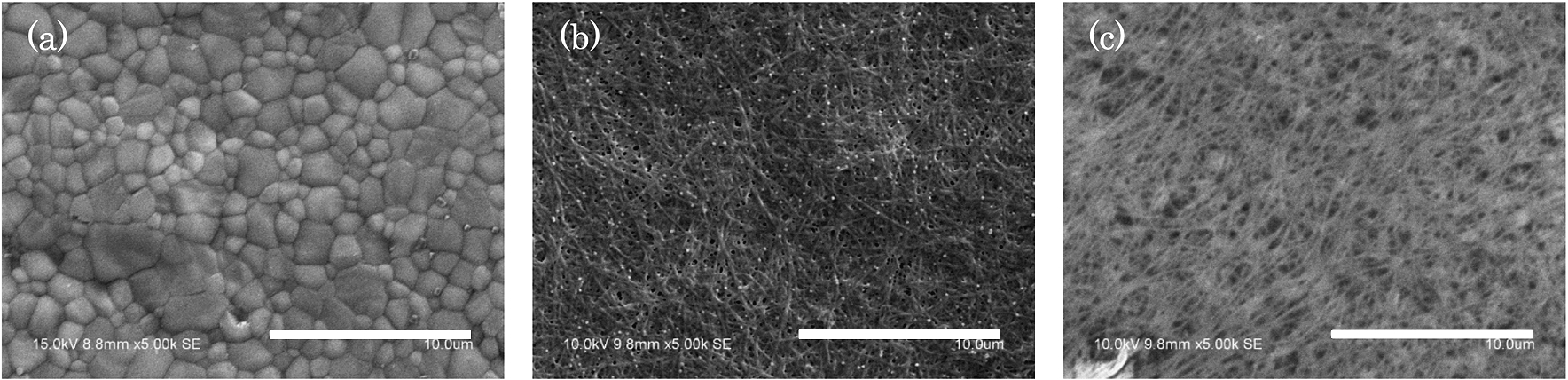
SEM micrographs of HAp (a) showing dense particles on the surface. SEM micrographs of Type I (b) and Type III (c) showing fibrous structures on the HAp surface. Scale bars, 10 μm.

ATR spectra of HAp (a), Type I (b), and Type III (c) in the region of 1800–900cm−1.
Next, the expression of osteoblastic, cementoblastic, and periodontal markers was analyzed with real-time PCR (Fig. 4). Although both markers showed gradual changes through passages 1–4, the expression levels of the osteoblastic markers RUNX2, osteopontin, and F-spondin were dramatically decreased after passage 3. Periodontal markers, such as periostin and S100A4, were markedly changed at passage 3.

Effects of scaffolds on PDL-derived cell potential with increasing passage numbers. Cells were cultured for 5 days for each passage. The expression levels of osteoblastic, cementoblastic, and periodontal markers were analyzed with real-time PCR.
PDL-derived cells attached on the samples showed different shapes related to the composition of the substrates. Cells on HAp showed a round shape at up to 6 h of incubation, after which wide spreading and extended pseudopodia were observed, whereas cells on type I and type III collagens showed wide-spread amoeboid-like shapes (Fig. 5(a)). Measurement of the cell area by fluorescent staining of F-actin is shown in Fig. 5(b). Cells attached on type III collagen showed the largest cell area after 6 h of cultivation. Cells on type I collagen were larger than those on HAp surfaces throughout the cultivation period.

Initial adhesion area of PDL-derived cells on HAp, Type I, and Type III. (a) PDL-derived cells with fluorescence staining at 3, 6, 18, and 24 h after seeding (red: rhodamine phalloidin, blue: DAPI). Scale bars, 100 μm. (b) The footprint area of PDL-derived cells on HAp, Type I, and Type III after 3 h. *:
One and 3 days after seeding, MTT assays were performed to evaluate the proliferative potential of PDL-derived cells on HAp, Type I, and Type III (Fig. 6). As a result, proliferation potential was significantly higher in Type III compared with those of HAp and Type I.

Proliferation ability of PDL-derived cells on HAp, Type I, and Type III (
Alizarin Red S staining and real-time PCR were performed in order to evaluate the mineralization of PDL-derived cells. Seven, 10, and 14 days after the start of culture, stained calcium compounds were observed by optical microscopy. On day 14, PDL-derived cells grown on HAp and Type I formed red-stained calcium compounds three-dimensionally, whereas Type III resulted in mottled pale pink deposits on the surfaces. For all culture periods, the mineralization ability of PDL-derived cells was lower on Type III than on HAp and Type I. The absorbance of dissolved calcium compounds on each sample was then read at 415 nm to quantify the osteoconductivity of PDL-derived cells (Fig. 7). The absorbance increased in HAp, Type I, and Type III over time. At 10 and 14 days, the absorbance of Type III was significantly lower than those of HAp and Type I.

Calcification ability of PDL-derived cells. HAp (a) and Type I (b) formed rough surfaces of calcium compounds. Type III (c) merely showed a mottled pale red color with smooth surfaces. Scale bar, 8 mm. (d) Absorbance after 10 and 14 days of culture under osteogenic conditions (
Furthermore, real-time PCR showed that PDL-derived cells expressed known osteoblast-specific markers, i.e., ALP and Col1a1, on day 7 (Fig. 8). ALP and Col1a1 gene expression levels were enhanced on HAp and Type I, whereas Type III inhibited osteoblast-specific gene expression.

Osteogenic markers of PDL-derived cells according to collagen type. Cells were cultured for 7 days in osteoinductive medium, and the expression of alkaline phosphatase and collagen type I alpha I genes was evaluated. The ratios are relative to the GAPDH value. *:
In our study, primary cells used for evaluation of scaffolds were isolated from rat molars using an enzyme-based method. Izumi et al reported that a high proliferation rate and mesenchymal stem cell-like properties were observed after isolation of cells using an enzyme-based method [17]. Furthermore, increasing the number of passages has been shown to cause gradual loss of mesenchymal stem cell-like potential [18]. Itaya et al also demonstrated that PDL-derived cells at passage 3 showed decreased levels of tendo/ligamentogenesis-related genes [19]. In our study, the expression of osteogenetic genes (RUNX2, osteopontin, and F-spondin) decreased at passage 3, whereas markers for PDL cells (periostin and S100A4) increased at passage 3. The second passage, which did not show major differences compared with primary cells, was used to prevent PDL-derived cells from unnecessary passaging. The properties of PDL-derived cells dramatically changed after passage 3; therefore, the passage time must be limited to two passages in order to maintain stem cell-like properties.
MTT assays showed that type III collagen had significantly higher initial adhesive and proliferative abilities than HAp and type I collagen, indicating that type III collagen could contribute to the generation of new tissues requiring rapid wound healing. The turnover of collagen in PDL is five times faster than in gingiva and 15 times faster than in skin [20]. This rapid turnover would prevent type III collagen from being replaced with type I collagen, which results in a high proliferation rate in PDL-derived cells. Hence, PDL-derived cells maintained a high proliferative potential on type III collagen, implying that type III collagen played an important role in the homeostasis of PDL tissue.
In mineralization assays, HAp and type I collagen, as the main components of the bone and cementum, enhanced the mineralization ability of PDL-derived cells. Real-time PCR analysis showed that the expression levels of the ALP and Col1a1 genes as markers of bone formation were significantly higher on HAp and Type I compared with those on Type III. Type III collagen apparently colocalizes with type I collagen within the same fibrils as Sharpey’s fibers in the periodointium [21]. Since Sharpey’s fibers are embedded and fastened to both hard tissues without mineralization, type III collagen is thought to regulate mineralization in the PDL [22,23]. Therefore, type III collagen appeared to prevent mineralization of PDL via cell differentiation and Sharpey’s fibers and contribute to the maintenance of homeostasis in the PDL.
The cell morphology and spread area were controlled by HAp, type I collagen, and type III collagen used in this study. The adhesion motif of the ECM affects cell behavior via the integrin superfamily [24]. Type I and III collagen have been reported to bind integrin
Conclusions
In summary, our findings showed that PDL-derived cells attached to type I collagen and differentiated into osteogenic cells to form hard tissues, whereas type III collagen enhanced the adhesion of PDL-derived cells and maintained a state of nondifferentiation in order to inhibit mineralization of PDL tissue. Although the mechanisms mediating the interactions between the ECM and PDL-derived cells are still unknown, our experiments strongly indicated the important and predominant roles of type III collagen as an inhibitor of bone formation and in the maintenance of the homeostasis of the periodontium. Although the interaction between the ECM and integrin may trigger these behaviors in PDL-derived cells, further experiments are needed to elucidate the intracellular signaling cascade.
Footnotes
Acknowledgements
This study was partially supported by a JSPS Grant-in-Aid for Scientific Research (C) (grant no. 26462964 and 17K11796) and Creation of Life Innovation Materials for Interdisciplinary and International Researcher Development, MEXT Japan.
Conflict of interest
The authors have no conflict of interest to report.