
Abstract
Background:
Recently, polyethylene glycol (PEG) modified gold nanoparticles have been studied to maintaining long-term stability in biological fluids. Its biodistribution was also reported, however, comparison of bare gold nanoparticles and PEGylated gold nanoparticles with equal particle size is not sufficient.
Objective:
We prepared bare gold nanoparticles and PEGylated gold nanoparticles with diameters of 20-30-nm or 50-nm to avoid the influence of particle diameter, and studied their biodistribution in the mouse.
Methods:
Gold concentrations in brain, heart, lungs, liver, stomach, pancreas, spleen, kidneys, blood, urine, and feces were measured at 0.5, 1, 2, 3, 6, 12, 18, 24, and 48 h after administration of gold nanoparticles using inductively coupled plasma atomic emission spectrometry.
Results:
At 48 h after intravenous administration, accumulation in the liver and spleen was significantly reduced by PEGylation, and the gold amounts of PEGylated gold nanoparticles with diameters of 20-30 nm and 50-nm in the brain were 3.6 times and 2.7 times higher than those of bare gold nanoparticles, respectively.
Conclusions:
These results indicated that the usefulness of PEGylated gold nanoparticles with small particle size for a drug carrier.
Introduction
Studies on fine particles have drawn attention, and micro- and nanoparticles are used as an effective drug delivery device. Many organic carriers are being studied in this field [16,23,25], and colloidal gold nanoparticles are also investigated. Inorganic gold nanoparticles of various size range provided several opportunities in targeted drug delivery, imaging and as a diagnosis [1,4,19,22]. Similar to silver nanoparticles, gold nanoparticles have unique optical properties due to surface plasmon resonance. This property makes gold NPs useful for many applications such as spectroscopy, Raman sensors and bright optical tags for molecular-specific biological imaging [8,27,29]. Many toxicity studies on gold nanoparticles have been conducted [10,15,18]. Besides toxicity tests, biokinetics studies are a fundamental part of investigations to evaluate a safe and sustainable use of nanoparticles [11]. In previous studies, we reported the synthesis of gold nanoparticles with different particle sizes mainly 15-, 50-, 100-, and 200-nm. Their biodistribution data at 24 h after administration revealed particle size dependence. Gold nanoparticles with a diameter of 15 nm showed a higher distribution in tissues compared to the larger gold nanoparticles and mainly accumulated in the liver followed by lungs and kidneys. The Smaller percentage was also observed in spleen, brain, heart, blood, and stomach [20]. Recently, polyethylene glycol (PEG) modified gold nanoparticles have been studied to maintaining long-term stability in biological fluids [17]. Functional groups such as mercapto (-SH) and amino (-NH2) groups are known to have a high affinity for gold, and polymers having such functional groups are expected to be useful stabilizers for gold nanoparticles. PEG is known to prolong the circulation time of biomedicines in the bloodstream, by reducing the non-specific binding of proteins as well as their cytotoxicity [3,12,13,17]. The effects of PEG molecular weight, type of anchoring ligand, and particle size on the assembly properties and colloidal stability of PEG-coated gold nanoparticles were reported. This paper showed that gold nanoparticles coated with high molecular weight PEG were more stable than gold nanoparticles coated with low molecular weight PEG. In addition, among the 20-, 40- and 80-nm gold nanoparticles coated with PEG, 20-nm gold nanoparticles exhibited the lowest uptake by reticuloendothelial cells and the slowest clearance from the body [30]. Investigations on the biokinetics of PEGylated gold nanoparticles after intravenous and intratracheal application compared to bare gold nanoparticles have also been reported. Gold nanoparticles with an inorganic core diameter of 5 nm were applied to rats and the biodistribution was measured after 1 and 24 h. PEGylated gold nanoparticles showed a prolonged blood circulation time, and non-PEGylated gold nanoparticles accumulated mostly in liver and spleen [11]. Although these studies provide valuable information, comparison of bare gold nanoparticles and PEGylated gold nanoparticles with equal particle size is not sufficient.
The main aim of the present study was to prepared bare gold nanoparticles and PEGylated gold nanoparticles with equal particle size and observed detailed biodistribution profiles in mice after intravenous injection to evaluate their in vivo kinetics. We prepared bare gold nanoparticles and PEGylated gold nanoparticles with diameters of 20–30-nm or 50-nm to avoid the influence of particle diameter.
Materials and methods
Materials
Hydrogen tetrachloroaurate(III) tetrahydrate (HAuCl4·4H2O, purity ⩾ 99%), trisodium citrate dihydrate (C6H5Na3O7·2H2O, purity ⩾ 99%), polyoxyethylene (20) sorbitan monooleate (polysorbate 80), nitric acid (HNO3, 60–61%), hydrochloric acid (HCl, 35–37%), gold standard solution (1000 ppm, for atomic absorption spectrochemical analysis) and d(+)-glucose (C6H12O6, purity ⩾ 98%) were purchased from Wako Pure Chemical Industries, Ltd. (Osaka, Japan). Methoxy poly(ethylene glycol) thiol (mPEG-thiol) with molecular weights of 2000 and 5000 were purchased from Laysan Bio Inc. (Alabama, USA). Isoflurane for the animal was purchased from Mylan Inc. (Pittsburgh, PA, USA). All other chemicals were of the highest grade commercially available.
Synthesis conditions of gold nanoparticles (GNPs)
Synthesis conditions of gold nanoparticles (GNPs)
PEGylation conditions of gold nanoparticles (GNPs)
Gold nanoparticles with average diameters of 10, 20, and 50 nm were synthesized by using the method previously described by Turkevich et al. [13,21,24,30]. As shown in Table 1, four different sizes of gold nanoparticles were synthesized from hydrogen tetrachloroaurate(III) solution and sodium citrate solution, which were prepared by dissolving 10 mg/ml of hydrogen tetrachloroaurate(III) tetrahydrate and trisodium citrate dihydrate in purified water, respectively. Hydrogen tetrachloroaurate (III) solution was added to purified water, and then subsequently added to three-necked round bottom flask equipped with a reflux condenser. The solution was heated at 100°C in an oil bath. Sodium citrate solution was added to the prepared solution, and it was heated and reflexed. After that, the prepared gold nanoparticulate suspension was cooled to room temperature using an ice bath. To disperse the 20-nm and 50-nm gold nanoparticles, polysorbate 80 was added to the suspension at 3.0 g/l. Then, the sample suspensions were centrifuged under the conditions in Table 2 (Himac 80WX, Hitachi Koki Co. Ltd., Tokyo, Japan). Following centrifugation, the precipitated gold nanoparticles were rinsed with polysorbate 80 solution (3.0 g/l) to remove foreign substances. Centrifugation and rinsing were repeated for a total of three cycles. As shown in Table 2, mPEG-thiol with molecular weights of 2000 or 5000 was added to the suspension containing 10-nm gold nanoparticles. To PEGylate the bare 10-nm gold nanoparticles, it was stirred with mPEG-thiol for 24 h at 10°C. Then, the sample suspension was centrifuged (Himac 80WX) and rinsed with purified water to remove foreign substances. Centrifugation and rinsing were repeated for a total of three cycles. Finally, the synthesized bare and PEGylated gold nanoparticles were suspended in 5% (w/v) d(+)-glucose solution and then filtered through a 0.22-μm membrane filter (Toyo Roshi Kaisha, Ltd., Tokyo, Japan).
Characterization of bare and PEGylated gold nanoparticles
The mean volume diameter and polydispersity index values of bare and PEGylated gold nanoparticles were determined using a particle size analyzer (ELSZ-1000ZS, Otsuka Electronics Co., Ltd., Osaka, Japan) which is one of the dynamic light scattering systems. Samples were dispersed in purified water and measured at 25°C. Also, the zeta potential of the samples was determined at 37°C, following resuspended in phosphate buffer saline (pH: 7.4, ionic strength: 0.154 M). Surface properties of the gold particles were observed using a scanning electron microscope (SEM, JSM-6060LA, JEOL Ltd., Akishima, Japan).
In order to determine contained an amount of gold in the samples, 50 μl of sample suspension was added to 950 μl of aqua regia to dissolve gold. Subsequently, 9 ml of purified water was added, followed by vortexing. Ten milliliters of the sample solution was directly analyzed using an inductively coupled plasma atomic emission spectrometry (ICPE-9000, Shimadzu Co., Kyoto, Japan). The gold content was determined by previously reported method [20,21], by comparing the value of the calibration curve of the gold standard solution.
Biodistribution study
Animals
Male mice (ddY, 7–8 weeks old) were housed in stainless steel cages under standard environmental conditions (23 ± 1°C, 55 ± 5% humidity and a 12/12 h light/dark cycle) and maintained with free access to water and a standard laboratory diet (carbohydrates 30%; proteins 22%; lipids 12%; vitamins 3%, Nihon Nosan Kogyo Co., Yokohama, Japan). They were used in accordance with the Guidelines for Animal Experimentation of Tokyo University of Science, which are based on the Guidelines for Animal Experimentation of the Japanese Association for Laboratory Animal Science.
Experimental design
On the day of experiments, mice were housed in metabolic cage 3600M021 (Tecniplast Japan Co., Ltd., Tokyo, Japan) and divided into three groups (
Results and discussion
Preparation and characterization of bare and PEGylated gold nanoparticles
The gold nanoparticles were synthesized using hydrogen tetrachloroaurate(III) solution and sodium citrate solution. We changed the concentration of tetrachloroauric acid and sodium citrate to obtain gold nanoparticles of the required particle size. In particular, it was assumed that the concentration of sodium citrate was important for the preparation of gold nanoparticles and for controlling the particle size [9]. A lower concentration of sodium citrate solution resulted in an increase in the particle size of gold nanoparticles due to aggregation of more gold atoms into the nanoparticles [6]. Mean volume diameters, polydispersity indexes, and zeta potentials of the gold nanoparticles are shown in Table 3. As a result of the t-test, there were no significant differences in particle size between 20-nm bare gold nanoparticles and 10-nm PEGylated gold nanoparticles (Mw: 2000), and 10-nm PEGylated gold nanoparticles (Mw: 5000) and 50-nm bare gold nanoparticles. Their particles size distributions are shown in Fig. 1. The value of zeta potential was reduced by PEGylation. Also, it tended to decrease as the PEG chain was longer. It has been reported that the surface charge of gold nanoparticles changes from negative charge to positive charge by PEGylating thiol-terminated poly(ethylene glycol), having a positive charge at the terminal, to gold nanoparticles [2]. In this study, we considered that the charge on gold nanoparticle surface was covered by PEGylation and the absolute value of the zeta potential was decreased. Figure 2 displays SEM images of the nanoparticles, showing spherical dispersed particles. From these findings, we confirmed that bare and PEGylated gold nanoparticles were prepared successfully.
Particle size, polydispersity index, and zeta potential of gold nanoparticles (GNPs,
, mean ± SD)
Particle size, polydispersity index, and zeta potential of gold nanoparticles (GNPs,
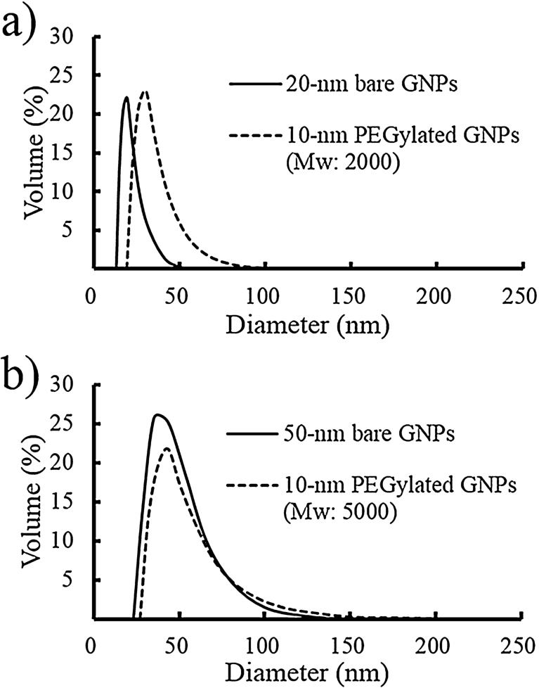
Particle size distribution. (a) 20-nm bare gold nanoparticles (GNPs) and 10-nm PEGylated GNPs (Mw: 2000). (b) 50-nm bare GNPs and 10-nm PEGylated GNPs (Mw: 5000).

Scanning electron microscopy images of bare and PEGylated gold nanoparticles taken at an accelerating voltage of 15 kV (magnification: 90,000–110,000×). (a) Twenty-nm bare gold nanoparticles. (b) Ten-nm PEGylated gold nanoparticles (Mw: 2000). (c) Fifty-nm bare gold nanoparticles. (d) Ten-nm PEGylated gold nanoparticles (Mw: 5000). (e) Forty-nm PEGylated gold nanoparticles (Mw: 2000). (f) Forty-nm PEGylated gold nanoparticles (Mw: 5000).
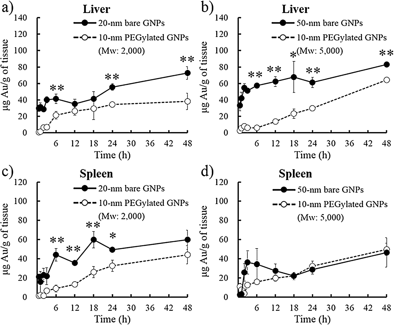
Gold concentration of various gold nanoparticles (GNPs) in mouse liver and spleen (mean ± SD,

Gold concentration of various gold nanoparticles (GNPs) in mouse pancreas and stomach (mean ± SD,
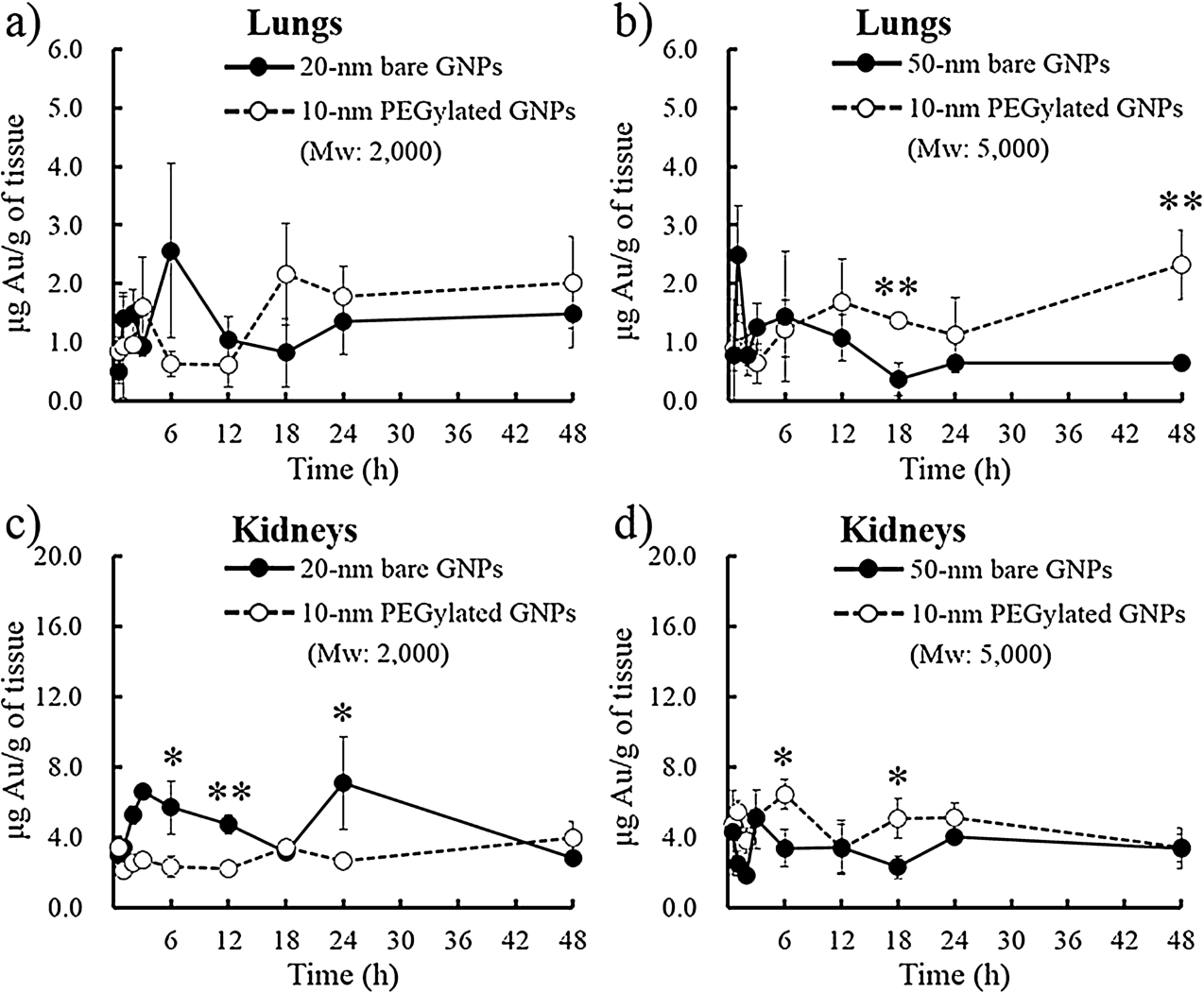
Gold concentration of various gold nanoparticles (GNPs) in mouse lungs and kidneys (mean ± SD,
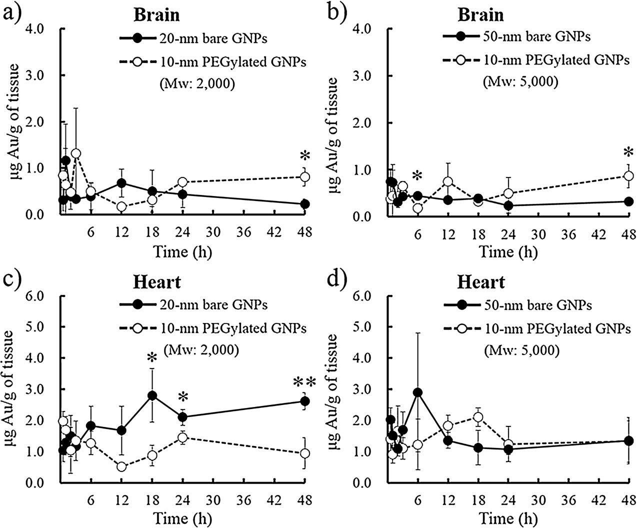
Gold concentration of various gold nanoparticles (GNPs) in mouse brain and heart (mean ± SD,
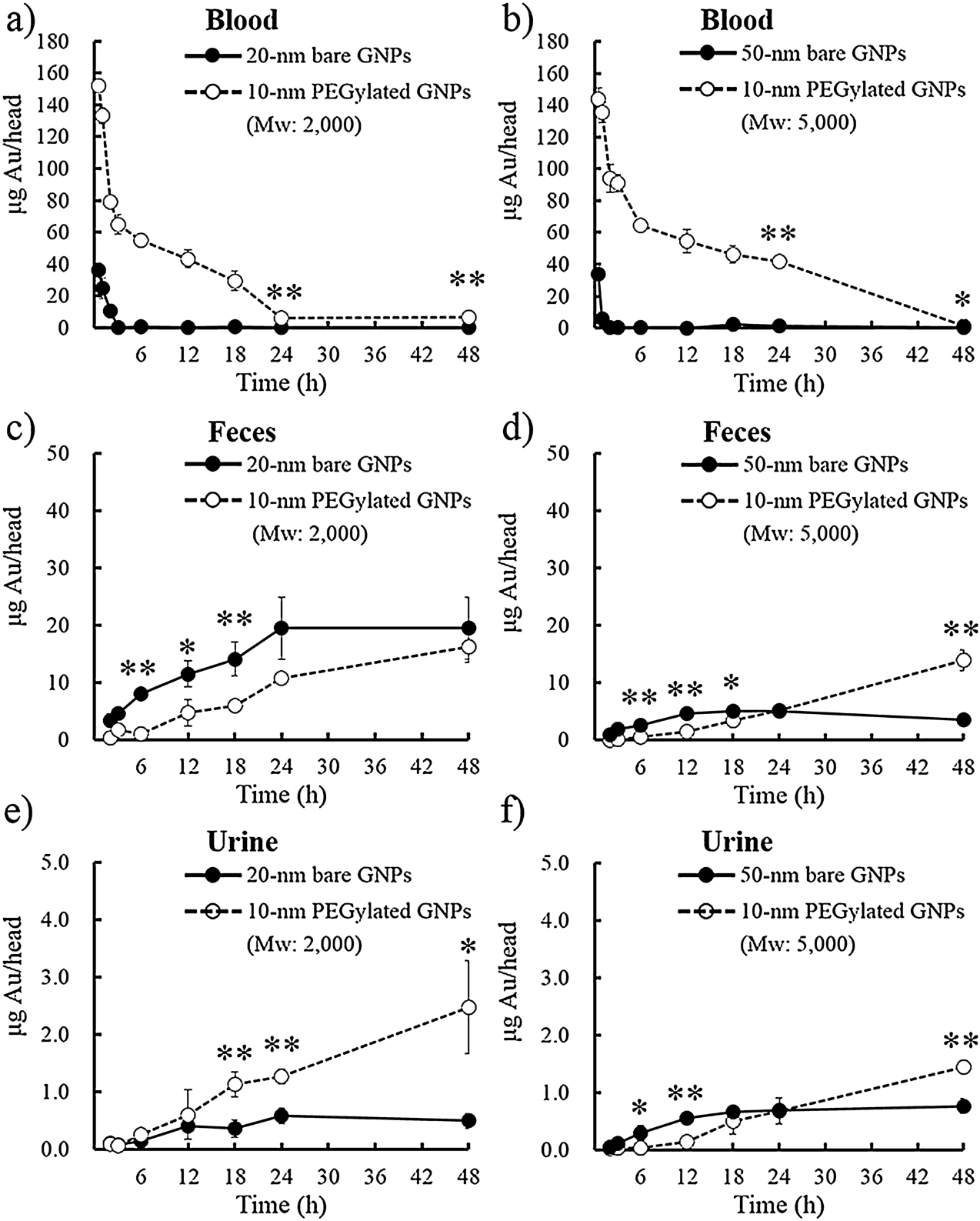
Gold amounts of various gold nanoparticles (GNPs) in mouse blood, feces, and urine (mean ± SD,
Figures 3–7 shows the gold concentration of blood, feces, urine, and all organs (lung, kidneys, liver, spleen, brain, heart, pancreas, and stomach). As shown in Figs 3 and 4, in gold nanoparticles with a particle size of 20-30 nm, accumulation of gold nanoparticles in the liver, spleen, and pancreas was significantly reduced by PEGylation. Detection of high concentration of gold in the liver and spleen, and a significant reduction of liver accumulation of gold nanoparticle by PEGylation were also observed in 50-nm gold nanoparticles. These results indicated that side effects could be reduced by PEGylation. This will be important, especially in the liver, because the accumulation of gold nanoparticles was large in this RES organ [20,30]. At 48 h after administration, the gold amount of PEGylated gold nanoparticles with a particle size of 20-30 nm and 50-nm accumulated in the brain was 3.6 times and 2.7 times higher than that of bare gold nanoparticles, respectively (Fig. 6). As shown in Figs 7(a) and 7(b), PEGylation prolonged a retention time of gold nanoparticles in the blood circulation and thus, it was suggested that more gold nanoparticles cross the blood-brain barrier through the 20 nm gap between astrocytic end-feet basement membrane and capillary endothelium [5]. The previous study showed that modifying 5-nm gold nanoparticles with PEG having a molecular weight of 10,000 improves the retention of gold nanoparticles in the bloodstream [11]. In this study, it was shown that even when shorter PEG chains were modified, the retention of gold nanoparticles in the blood was improved. This result suggested the influence of the PEG configuration on the particle surface. The low surface coverage of PEG chains leads to the configuration in which most of the chains are located closer to the particles surface, and the high surface coverage and lack of mobility of the PEG chains leads to the configuration in which most of the chains are extended from the surface [14]. In the PEGylated gold nanoparticles prepared in this study, we considered that the PEG density on the particle surface was high and a hydration layer with a sufficient thickness was formed on the particle surface because the PEG chain was short and the core size was large. As shown in Figs 7(c)–(f), the main excretion route of gold nanoparticles was feces, and it was found that excretion was faster in smaller particles. Excretion of 10-nm PEGylated gold nanoparticles to feces was significantly faster than 50-nm bare gold nanoparticles at 48 h, however, increase in excretion amount of gold nanoparticles by PEGylation was not observed in Fig. 7(c). Therefore, it was suggested that the inorganic core diameter of gold nanoparticles has a great influence on the excretion rate of gold nanoparticles into feces. The urinary excretion of gold nanoparticles increased by PEGylation (Figs 7(e) and 7(f)). This result may be due to a change in retention property of the gold nanoparticles in the bloodstream by PEGylation. However, it was reported that gold nanoparticles were taken up into cells by endocytosis, and the gold nanoparticles observed inside the urothelial cells exfoliated into the urine together with urothelial cells [7]. Therefore, further investigation is necessary for urinary excretion of gold nanoparticles.
Conclusions
We confirmed that accumulation in the liver and spleen was significantly reduced by PEGylating gold nanoparticles. In addition, PEGylation improved the delivery of gold nanoparticles to the brain. These results were more remarkable in smaller particles. As the size of the core gold nanoparticles increased, the amount of gold nanoparticles excreted into the feces decreased. From these results, it was suggested that PEGylated gold nanoparticles with small particle size are useful when gold nanoparticles are used as drug carriers.
Footnotes
Acknowledgements
We gratefully acknowledge the work of past and present members of our laboratory.
Conflict of interest
The authors have no conflict of interest to report.