
Abstract
BACKGROUND:
The development of biomaterial scaffolds and implementation of tissue engineering techniques are necessary. Therefore, Polycaprolactone/Sodium Hyaluronate/Multiwalled Carbon Nanotubes/Extract of Mimosa tenuiflora composites have been produced by a thermally-induced phase separation method.
OBJECTIVE:
The objective of this research was to evaluate the in vitro bioactivity and in vitro biocompatibility of the composites.
METHODS:
The in vitro bioactivity of the composites was assessed by soaking them in simulated body fluid for 7, 14, 21, and 28 days. The structure and composition of the composites were analyzed using scanning electron microscopy coupled with energy dispersive spectroscopy and Fourier transform infrared spectroscopy. Also, the in vitro biocompatibility of the composites was evaluated by means of alkaline phosphatase activity of the osteoblasts and by measuring the metabolic activity of the cells using MTT assay.
RESULTS:
The results show a porous and interconnected morphology with enhanced bioactivity. It was observed that the incorporation of Mimosa tenuiflora in the composites promotes increased viability of osteoblasts in the scaffolds.
CONCLUSIONS:
The results show the efficiency of bioactive and biocompatible composites and their potential as candidates for tissue engineering applications.
Introduction
Bone tissue plays an important role in the body since it acts as a support for mechanical stress and maintains ionic balance [1]. Nevertheless, bone tissue defects are significant problems that are mainly observed in the median age of population [2]. The best integration results are obtained using autografts to repair or restore critical bone defects [3]. However, some of their disadvantages include pain and morbidity in the site and limited quantity and availability of transplantable bone [4,5]. In order to get an ideal composite for this application, there is a significant interest in developing bioactive scaffolds [6]. Therefore, the development of biomaterial scaffolds and implementation of tissue engineering techniques are necessary [7]. The scaffold plays a vital role in maintaining the cell functions and developing new cells or tissues [6].
Polycaprolactone (PCL) is a synthetic, biocompatible, semicrystalline, and hydrophobic material [8]. It has gained much interest in the biomaterials field due to its good solubility [9]. It is an aliphatic polyester prepared by ring opening polymerization of 𝜀-caprolactone, with a degradation rate of approximately 24 months [10]. This makes it a good candidate for the manufacturing of composite materials with appropriate degradation kinetics [11]. Furthermore, it has not showed cytotoxicity in osteoblast cultures [9].
On the other hand, hyaluronic acid (HA) is a glycosaminoglycan polysaccharide found in the extracellular matrix of all living creatures [12]. HA is hydrophilic and has negatively charged linear D-glucuronic acid and N-acetyl-D-glucosamine copolymer [6]. It has an important role in the maintenance of joint lubrication of tissues [13]. HA interacts with extracellular molecules to form the extracellular matrix and is associated with the migration and differentiation of cells [14]. One of its most important characteristics is its great capacity to retain water, providing the tissues pressure resistance to mechanical stresses, lubrication, and cell differentiation [15]. Nevertheless, its poor mechanical properties limit its uses as a biomaterial [16]. It has been used for medical applications including viscosupplementation, drug delivery, eye surgery, tissue regeneration, and embryo protection [14].
Multiwalled carbon nanotubes (MWCNT) have been used to enhance the mechanical properties of biodegradable materials [17]. Recent studies have shown that they can improve the osteogenic process, making them ideal candidates for the development of nanocomposites for tissue engineering application [18]. They have shown high-binding biomolecules of the extracellular matrix [19]. MWCNTs have a surface area similar to collagen fiber in natural bone and were found suitable to promote bone ingrowth and enhance different osteoblast in vitro activities [20]. Also, CNTs exhibit a wide range of electrical properties that can be used for cell electrical stimulation applications for accelerating bone formation and regeneration [21].
Mimosa tenuiflora is a tree distributed in the tropical forests from the southeastern regions of Mexico to northern Brazil and Venezuela [22]. In recent years, it has been discovered that the cortex of M. tenuiflora contains high contents of saponins and tannins [23]. Furthermore, it has also been shown that M. tenuiflora composites have good bioactivity characteristics and are, thus, a good candidate for use in bone regeneration [24].
Based on these characteristics, it could be reasonable to believe in the bioactivity of PCL/HA/MWCNT/Extract of M. tenuiflora composites. So, in the present work, the scaffolds of these substances were prepared by thermally-induced phase separation method. Then, they were used to evaluate the in vitro bioactivity and the in vitro biocompatibility of the composites.
Materials and methods
Materials
PCL was purchased from Sigma-Aldrich (United States). The bark of M. tenuiflora was obtained in the region of Jiquipilas Chiapas. Sodium Hyaluronate was provided by Lifecore Biomedical (United States). MWCNTs were purchased from Sigma-Aldrich (United States). Glacial Acetic Acid (Mallinckrodt, United States) was used as solvent. SBF was prepared in our laboratory according to a previously published method [25].
Preparation of composites
Extract of M. tenuiflora was obtained according to a previously proposed method [26]. It was prepared using a mixture of PCL/HA/MWCNT (96/2/2% wt). The extract from M. tenuiflora was added in a relation of 20% wt to the prepared mixture. The composites were prepared using the thermally-induced phase separation technique. Briefly, PCL was dissolved in concentrated acetic acid at 40 °C with constant stirring; once dissolved, the mixture was added to HA that was dissolved previously in deionized water at room temperature. On the other hand, MWCNTs were placed in 1 ml of concentrated acetic acid in ultrasound bath for 25 min, and then the solution was added to the previous mixture of PCL and HA. Finally, the extract from M. tenuiflora was added to the PCL/HA/MWCNT mixture and stirred until a homogeneous mixture was obtained (approximately 1 h). The solution was placed in an ultrasound bath for 10 s and then placed in the freezer at −40 °C for 24 h. Then, the sample was removed from the freezer and lyophilized under a vacuum of 0.01 mbar and −80 °C for 72 h.
The composites were neutralized by immersion in 0.05 M NaOH for 24 h at room temperature and rinsed in distilled water and immersed in phosphate-buffered saline (PBS) at room temperature for 6 h and dried.
FTIR spectroscopy
To evaluate the chemical composition of PCL/HA/MWCNT/M. tenuiflora composites, FTIR spectra were recorded using a transmission mode in an IR spectrometer (Nicolet 6700, Thermo Scientific, United States). For each spectrum, 100 scans at 16 cm−1 using the Scandium Universal SEM Imaging Platform software resolution were averaged.
Mechanical properties
The compression tests were carried out at room temperature using a Sintech 20/D universal machine. Tests were run at a strain rate of 1 mm/min using a 5000 kN load cell; three specimens were tested for each sample to elaborate a stress-strain curve.
Characterization of morphology and in vitro bioactivity
For analysis of in vitro bioactivity study, the samples were tested using a previously reported method [24,25]. Briefly, the samples were placed in 5 mL of SBF (1.5X) at pH 7.4 in an incubator at 37 °C for a period of 7, 14, 21, and 28 days. The medium (SBF) was changed every 48 h. After the incubation period, the samples were rinsed with distilled water and finally dried in a vacuum oven at room temperature. Morphological characterization and detection of calcium and phosphorous were done using the Field Emission Scanning Electron Microscopy (FE-SEM, JEOL JSM-7000F) coupled with an energy dispersive system (INCA 7557 de Oxford Instruments). The average pore size and the pore size distribution were measured using the Scandium Universal SEM Imaging 95 Platform software (Soft Imaging 96 System by Olympus) from the SEM micrographs in the original magnification. Three different cross-sections of each scaffold were used to estimate the pore size, and at least 100 pores were used to estimate the distribution.
Alkaline phosphatase activity expression and cytotoxicity assay
Alkaline phosphatase (ALP) activity of the cells on the scaffolds was determined using an APL substrate assay kit (Pierce Biotechnology, Monterrey, Mexico). As a control sample, osteoblasts were seeded directly on the well with the medium but without scaffold. Culture test was prepared using scaffolds with a density of 5 ×10³ cells/cm2 immersed in a prepared solution of alpha-MEM supplemented with 10% fetal bovine, 1% antibiotics, 2.1 mg/ml betha-glycerophosphate, and 50.1 g/ml ascorbic acid. The treated materials were located in an incubator at 37 °C with 5% CO2. Sets of control and test cultures were harvested on days 7, 14 and 21 and then washed twice with PBS. The cells were lysed with 3 ml of 1% Triton X-100 in DEPC-treated water and three freeze-thaw cycles at −70 °C. The ALP activity was measured using a spectrophotometry at 405 nm (Microplate spectrophotometer Bench mark plus, BIORAD).
Cytotoxicity assay was evaluated using the tetrazolium colorimetric MTT assay (SIGMA). Fibroblast cells (3T3 ATCC®) were seeded in samples immersed at a density of 5 ×10³ and incubated in DMEM supplemented with fetal bovine serum at 37 °C for 7, 14 and 21 days. The control sample consisted of cells immersed directly on the medium without scaffolds. The medium was carefully removed, and the purple formazan crystals were dissolved in dimethyl sulfoxide (DMSO) after a few minutes of incubation at 25 °C. Absorbance was measured at 570 nm by a microplate reader (Benchmark Plus, microplate spectrophotometer BioRad), and the results were normalized to the non-treated control cells to determine relative cell viability. Statistical significance was determined by Student’s t-test that assesses statistically significant differences of data between two groups (control sample and each sample). Significance was assigned at p < 0.05.
Results and discussion
Synthesis and characterization of morphology
PCL is a biodegradable polyester that has been tested as scaffold material. It is a semi-crystalline material with good mechanical properties that degrades much more slowly than other polyesters. This material supports cell attachment, proliferation, and matrix production for a variety of cell types, including chondrocytes, osteoblasts, and mesenchymal stem cells [27]. Due to the degradation properties of PCL, it can be used as the main structure of scaffolds.
The modification of PCL scaffolds with an hydrophilic component such as HA can furthermore be utilized to decrease the inherent hydrophobic property of PCL [28]. HA is an important component of ECM and disperses in all tissues. It is a unique, linear, unmodified glycosaminoglycan (GAG) consisting of a repeating disaccharide unit. In long bones, HA amounts to 3% of the total GAG [29]. In 2014, Lebourg et al. [30] used 2% HA concentration to produce macropores for cell colonization without any significant change in mechanical properties. They concluded that the presence of HA improves the quality and speed of regeneration. So, during our experiment, we decided to use the same quantity of HA.
Recent studies have shown that HA, SWCNTs, and HA-SWCNTs at low concentrations did not exhibit cytotoxic effects on primary osteoblasts. Moreover, these biocomposites increased the deposition of inorganic crystals in osteoblast cultures [31]. Previous composites with CNTs (from 2% to 10% in wt) showed that the presence of CNTs affects the mechanical properties of the composites [32,33]. Therefore, we decided to use 2% of MWCNT to evaluate its effect on the composite.
The cytotoxicity of M. tenuiflora cortex in combination with chitosan for 3T3 fibroblast cells decreased significantly at the 80/20 chitosan/M. tenuiflora films, and it was similar for 90/10 chitosan/M. tenuiflora films. At high concentrations of M. tenuiflora (70/30) and chitosan (100/0) films, the cytotoxicity increased [34]. Thus, this present research also considered 20% of M. tenuiflora for the composites.
Based on the above-mentioned reasons, PCL/HA/MWCNT/M. tenuiflora composites were produced by freeze-drying technique in the form of porous materials with a well-interconnected network of pores. The structural properties of bone tissue scaffolds such as pore interconnectivity, pore size and porosity should be considered to ensure a biological environment to cell attachment and proliferation to promote new bone formation [35]. Since the biomaterial surface is the medium in which the interactions occur in the biological environment, a porous surface has been described as an ideal surface medium [36]. Cells tend to adhere and proliferate in porous constructs [37]. In order to evaluate the structural characteristics and the surface morphology, an SEM was used. Composites give a homogenous, porous and interconnected structures (Fig. 1 and Table 1) with desired pore sizes (Table 1) for bone regeneration [38]. The addition of MWCNT increased the pore size, as shown previously [21]. Also, the addition of M. tenuiflora to the ternary biopolymeric blends leads to the increase of the pore size, which is related with an improvement of the adhesion of the osteoblasts due to cell size, migration requirements, and transport [39]. It was previously shown that the use of ternary polymers mixture to get a composite material improves the porosity of the material in comparison with scaffolds produced from binary blends only [40]. In Fig. 1, the morphology of the composites is shown where a porous structure can be appreciated. The composites present a highly interconnected porous structure (<50 μm porous), and at least 20% of the pores were smaller than 20 μm so as to provide a structure in which the cells can interact with proteins and show bioactivity properties [2]. Here, it should be noted that bone is a tissue with 50–90% porosity [40].
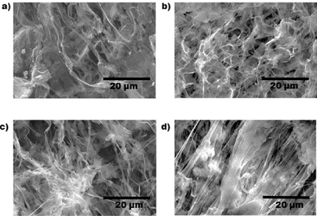
SEM images of (a) PCL, (b) PCL/HA, (c) PCL/HA/MWCNT, and (d) PCL/HA/MWCNT/MT composites.
Pore size distribution of PCL, PCL/HA, PCL/HA/MWCNT, PCL/HA/MWCNT/MT composites
The porous structure of the composite materials changes with the incorporation of MWCNT and HA. In the presence of PLC, the porosity is heterogeneous. However, when HA is added, the homogenous pores tend to reduce their size when MWCNT are added. Finally, once the M. tenuiflora is incorporated, the pores can be seen covered by small particles that can be associated with precipitations of the M. tenuiflora bark and the formation of more elongated pores covered by small fibers.
For this analysis, a universal machine was used. The mechanical strength affects the ability of the scaffold to be used for cell attachment, migration and differentiation, and it is considered a substantial factor in tissue engineering [41,42]. So, during this study, we evaluated the compression strength of the samples. Figure 2 exhibits the strain-stress deformation curves for PCL and PCL/HA/MWCNT/M. tenuiflora composites, and Table 1 shows the mechanical properties of the composites. The PCL/HA/MWCNTs composites exhibited the biggest module, indicating that it is a more resistant material than the other samples tested. PCL is a synthetic polymer known by its mechanical strength [37], so interactions between PCL/HA and MWCNT are favorable to the cohesion of the structure. It has been reported that scaffolds prepared with natural polymers exhibit weak mechanical properties. However, PCL show an enhancement of mechanical properties of scaffolds [43].
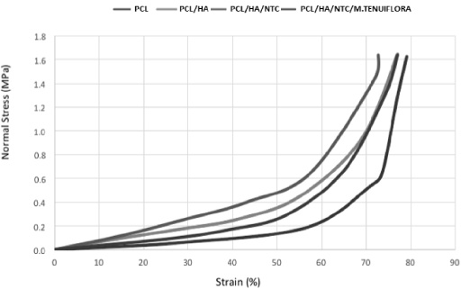
Compressive stress vs strain plot of the PCL, PCL/HA, PCL/HA/MWCNT/ and PCL/HA/MWCNT/MT composites.
The addition of M. tenuiflora caused a reduction in the modulus, possibly because of heterogeneous dispersion of the nanotubes and the poor mechanical properties of M. tenuiflora [44,45]. The PCL/HA composite showed less strain (Table 2) than PCL/HA/MWCNTs/M. tenuiflora composite, possibly due to the accommodation of the material into polymer matrix.
Compressive properties of PCL, PCL/HA, PCL/HA/MWCNTs, PCL/HA/MWCNTs/Mimosa tenuiflora composites
These results exhibited improved mechanical properties for the composites for use in tissue engineering. It is known that mechanical performance of the scaffolds is related with a number of factors including composition and morphology [42].
The infrared spectra for the various samples after SBF treatment showed the presence of the bands assigned to the phosphate bending vibration at 1079, 670, 609 and 570 cm−1 and carbonate group out of plane and stretching mode at 858 and 1454 cm−1 (Fig. 3) [24]. This suggests that carbonated apatite was formed in all compositions. Nevertheless, it is important to mention that the band corresponding to the phosphate bending vibration was not so evident in the scaffold only by PCL. This suggests that the incorporation of MWCNT, HA and M. tenuiflora could improve the bioactivity properties of the composites.
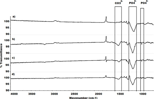
FTIR of (a) PCL, (b) PCL/HA, (c) PCL/HA/MWCNT, and (d) PCL/HA/MWCNT/MT composites after 28 days immersed of SBF solution.
In biomaterials used as scaffolds, the physical properties and chemical composition are essential and may modulate the condition of the cells [19]. According to the SEM images (Fig. 4), as the time exposition to SBF is increased, the deposition of apatite was higher. With the extension of mineralization time, a large quantity of spherical crystals was observed at the surface of the scaffolds. After 28 days of immersion, a layer of apatite was evident in all the coating surfaces. The apatite formation followed the texture of sub-micron pores present on its flake surfaces. The composites showed typical hydroxyapatite structures [25], with spherical morphology of apatite particles that were irregularly shaped, forming a continuous uniform layer. On this layer, some particles are agglomerated, suggesting the existence of secondary nucleation sites. The densification for individual layers increased with the addition of M. tenuiflora to the composite. The CA/P atomic ratio calculated at 28 days of immersion are 1.84, 1.76, 1.94, and 1.84 for the PCL, PCL/HA, PCL/HA/MWCNT, and PCL/HA/MWCNT/M. tenuiflora composites, respectively, which are near to the stoichiometric value of 1.67 [46]. The ternary composites of PCL/HA/MWCNT showed evidence of apatite crystals that do not form agglomeration. It is also evident that the material that incorporates M. tenuiflora shows a surface completely covered by these agglomerations of apatite particles, forming even multiple layers that form small calcified clusters. It was possible to find that CNTs do not affect the bioactivity of the scaffolds, as shown in other researches [47]. Furthermore, in this study, it was revealed that the nucleation, shape and growth of apatite formation depended on the surface characteristics, improving with the presence of M. tenuiflora.
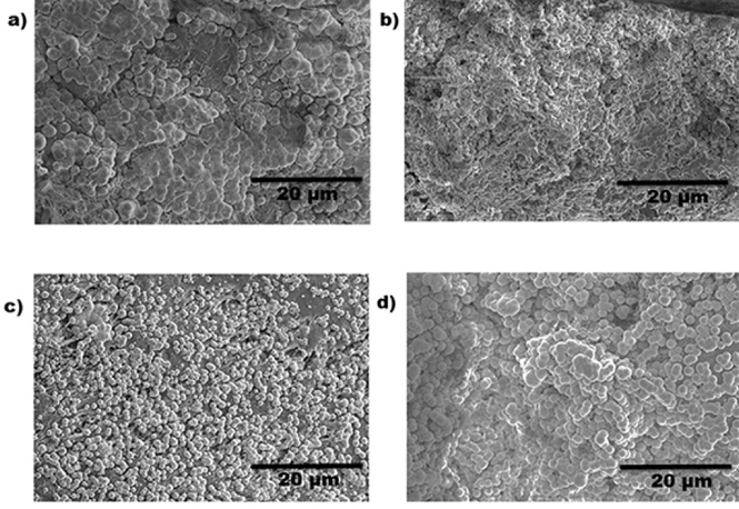
SEM of PCL, PCL/HA, PCL/HA/MWCNT, and PCL/HA/MWCNT/MT at 28 days.
Materials used for bone tissue engineering should be able to support the organic and inorganic constituents of the natural bone tissue [35]. It has been previously reported that PCL composites are bioactive and exhibit cell adhesion and proliferation in osteoblast culture [36]. Alkaline phosphatase activity is an important osteoblast differentiation marker having a stimulatory effect for matrix calcification in bone tissue [31]. An increased level of the ALP is a sign of the metabolic activity of the osteoblasts.
The bar graph in Fig. 5a illustrates the results of the ALP activity of the cells in the scaffolds. All composites showed ALP expression, indicating that the osteoblasts could begin the differentiation process. The behavior of ALP activity for the osteoblast is typical during the early stages of differentiation, showing the maximum level of expression at day 21 [30]. Previous studies had found that on increasing the CNT content, the ALP activity also increased [21,48]. Nevertheless, in this study, a significant difference (p < 0.05) was not observed among the scaffolds, but a difference was observed among the different periods. Furthermore, it is important to mention that the incorporation of MWCNT does not affect biocompatibility and bioactivity as shown previously [48].
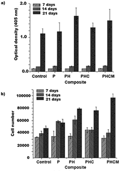
(a) Optical density of the ALP activity of the cells in the scaffolds at 405 nm and (b) cell viability in the scaffold measured using MTT assay. The results are reported as mean ± SD and the differences observed between composites results were considered significant when p < 0.05.
On the other hand, cell viability was characterized by MTT assay. The metabolic conversion of MTT, a yellow tetrazole salt, to purple formazan crystals occurred in living cells, and this process is proportional to the number of viable cells [37]. Metabolic activity from MTT assay data (Fig. 5b) showed that, on day 21, the composite PCL/HA/MWCNT/M. tenuiflora showed a significantly higher number of cells (p < 0.05) than the rest of the compositions. These results suggest that the incorporation of M. tenuiflora in the composites promotes an increased viability in the scaffolds. This result matches with the in vitro bioactivity test, where apatite formation all around the surface was evident. Furthermore, it is also evident that the incorporation of the MWCNT does not affect the cellular proliferation in the scaffolds, showing even more cells (p < 0.05) with respect to the control at 21 days, without any visible cytotoxicity. These results are in agreement with those of other researches [21]. Previous studies have showed that MWCNT may interfere with proliferation assays [17]. Also, it is difficult to confirm that cell viability increases with the presence of HA, as it previously reported by other research works [13].
Biodegradable PCL/HA/MWCNT/M. tenuiflora composites have been successfully prepared by a thermally-induced phase separation method. The scaffolds mimic both the structure and composition of natural bone. There are many amine carbonyl groups on the composite scaffolds that could act as nucleation sites. As a result, apatite could be formed more efficiently on the composite scaffolds than on the PCL scaffold.
Also, a porous and interconnected morphology with enhanced bioactivity that could be appropriate for tissue engineering has been found. The ability of the composites to form in their surface a mineralized layer was evident through the cumulative results obtained from IR spectra and SEM-EDS analysis. Also, it was observed that the incorporation of M. tenuiflora in the composites promotes increased viability of osteoblasts in the scaffolds.
Footnotes
Acknowledgements
The authors acknowledge the financial support of the Mexican Public Education Secretary and the Mexican National Council for Science and Technology (CONACyT) through project CB-2015-01-252439 and the CONACyT Pharmachemicals Net.
Conflict of interest
None to report.