
Abstract
Background:
Novel pectin-honey hydrogels have been developed and characterized as medical device. Ideally, a wound dressing should maintain optimal fluid affinity, permit moisture evaporation, protect the wound from microbes, and have shape-conformability, biocompatibility, and antibacterial activity.
Objective:
A novel, simple and fast method to produce pectin-honey wound dressings is described.
Methods:
The properties of these pectin-honey hydrogels were investigated, including swelling ability, water vapour transmission rate, hydrogen peroxide production, methylglyoxal content and antibacterial activity. Biocompatibility was assessed by proliferation assays using cultured fibroblast cells and by in vivo study with subcutaneous and intraperitoneal implantation in rats.
Results:
Hydrogel showed a good water vapour transmission rate, fluid uptake and were not cytotoxic for fibroblasts. The hydrogel demonstrated good antibacterial activity toward clinically relevant pathogens, including S. aureus and E. coli. Biocompatibility was confirmed by the measurement of plasma levels of interleukin (IL)1 beta, IL-6, tumour necrosis factor (TNF) alpha, and prostaglandin (PG)E2. No histological changes were observed.
Conclusions:
The presence of a natural active component, conformability, and complete resorbability are the main characteristics of this new biocompatible biomaterial that is well tolerated by the body, possibly improves healing, may be used for surgical complications prevention, with a simple and inexpensive production process.
Introduction
Healing wounds are sites that are easily attacked by bacteria, which leads to the formation of a biofilm that devitalises tissues [1]. The development of a new wound dressing is a focus area for many researchers since the dressing can create a barrier against infection while maintaining a physiological environment in contact with the wound [2,3]. These dressings help to maintain a moist environment at the wound site, promote tissue healing, and reduce infection, pain, and costs [1,4,5]. Antimicrobial agents play an important role in reducing bacterial contamination, but the resistance of pathogens to these substances has led to a decrease in the efficacy of antibiotics. Researchers have therefore advised against the use of systemic antimicrobial agents for the treatment of wounds [1,6].
Various wound dressings consisting of vegetable fibres, protective films, hydrogels, and hydrogel enriched with nitrogen oxides are available commercially [1,2,5,7]. Hydrogels are reported to be suitable for use in healing wounds but they have limitations in current use, which include a requirement for high frequency of application, inactivation by wound fluids, and formation of eschars [5]. The requirement for a new antimicrobial substance led to revaluation of ancient remedies, including the use of honey [8,9].
Honey has been used alone or in combination with other compounds for medical use since ancient times. Honey is a heterogeneous substance, has antimicrobial activity, anti-inflammatory effects and increase the healing process following skin or peritoneal damage [8,10]. It contains high levels of glycine, methionine and proline which are all fundamental for collagen formation and fibroblast deposition, which are the main factors for wound healing [11].
Manuka honey, produced in New Zealand, is the most studied honey having antibacterial properties against major aerobic and anaerobic bacteria species [12,13].
Several components are known to contribute antibacterial activity of honey. The osmotic effect of the sugars in honey and its characteristically acidic pH are known factors hindering bacterial multiplication. Recently, two components of honey, hydrogen peroxide (H2O2) and methylglyoxal (MGO), have been identified as effective antibacterials [14–16].
An ideal wound dressing is yet to be developed. Ideally, a wound dressing should maintain optimal fluid affinity, permit moisture evaporation, protect the wound from foreign microbes, and have shape-conformability, biocompatibility, and antibacterial activity [2,4,5,17].
Based on these premises, the aim of the present study was to describe and characterize the properties of pectin-honey hydrogels (PHHs) for wound healing and to assess their biocompatibility through an in vitro and in vivo assay.
Material and methods
Materials
Honey was purchased from Manuka Health (66 Weona Court,Te Awamutu 3800, New Zealand) and pectin from Ardet s.r.l. (Torino, Italy). Culture media, that is, tryptone soy agar (TSA), tryptone soy broth (TSB), peptone water, and 5% sheep blood agar, were purchased from Oxoid (Milan, Italy). Escherichia coli (Turin strain) and Staphylococcus aureus (Turin strain) isolated from canine wounds were used for this work.
Preparation of PHHs
The preparation method was modified after the procedure described by Walker (Walker, 1942) [18]. Briefly, the pectin-honey hydrogels were prepared starting from a solution (1:1 v/v) of liquid honey (Manuka Health, New Zealand) and sterile deionized water. The same volume of pectin powder (ARDET s.r.l., Italy) was then added and with continuous stirring until the mixture was homogeneous. The resulting gel was spread into 2 mm thick films and hot air dried at
Fluid uptake test (swelling test)
To investigate the fluid swelling ratio of PHHs, samples were cut into disks with a diameter of approximately 25 mm. The dry weights (Wdry) of the membranes were measured and recorded. Afterwards, pre-weighed dry samples were immersed in PBS solution, pH 7.4, at 37°C. The weights of the swollen PHHs were determined every 5 min subsequently by sandwiching the membranes between two paper towels to remove excess water on the surface, and then wet weights (Wwet) were measured. All experiments were performed in triplicate. The swelling ratios were calculated as the average value according to the following formula:
Water vapour transmission rate (WVTR)
The moisture permeability of the PHH was determined by measuring the WVTR. A piece of the specimen was fixed over the top of a tube (diameter, 34 mm) containing 10 mL PBS. The tube was then placed in an incubator at 37°C and 35% relative humidity. The membranes were weighed at regular intervals of time and the weight loss was recorded and plotted on a graph versus time. The WVTR was calculated from the slope of the graph by the following formula:
H2O2 analysis
The analysis of H2O2 from honey and PHHs was carried out according to the method reported by Long (1999) [20] (ferrous ion oxidation-xylenol orange [FOX] assay), with minor modifications. The stock reagents were as follows: reagent 1, 4.4 mM butylated hydroxy toluene (BHT) in HPLC-grade methanol; reagent 2, 1 mM xylenol orange and 2.56 mM ammonium ferrous sulphate in 250 mM H2SO4; working reagent: one volume of reagent 2 added to nine volumes of reagent 1. Approximately 200 mg of honey were diluted with deionized water to the ratio 20% w/w and immediately kept in a thermostat at
To reduce the processing time required to extract the water-soluble components from PHHs, their size was reduced prior to incubation by cutting the specimens into small pieces with the aid of a small and clean knife. Aliquots of 100 μL of the aqueous layer of the final mixture (40% w/w) were subjected to FOX analysis as described above for honey. Monitoring of H2O2 was carried out at 60 min, 90 min, 24 h, and 48 h. The FOX assay was calibrated using standard H2O2 (molar extinction coefficient, 43 M–1 cm–1; absorbance wave length of H2O2
Methylglyoxal (MGO) analysis
MGO was evaluated by the method proposed by Wild (2012) [21] with slight modifications. The method is based on the reaction between N-acetyl-L-cysteine (Sigma Aldrich) and MGO at room temperature. Samples were diluted in water (340 mg/ml) and the reaction was performed in 100 mM sodium dihydrogen phosphate buffer (adjusted to pH 7.0 with 10 M NaOH) at 22°C. For the standard curve of the reaction, different concentrations of MGO (0.5, 1, 2, and 5 mM) were used. MGO solutions (Sigma Aldrich) equivalent to 0.5, 2, and 5 mM and 10 μl of each honey solution (170 mg HBM/ml water) were added up to a volume of 980 μL with sodium dihydrogen phosphate. The reaction was started by adding 20 μL 500 mM N-acetyl-L-cysteine, and the absorption was recorded after 7 min. The condensation product, N-α-acetyl-S-(1-hydroxy-2-oxo-prop-1-yl) cysteine, was determined by recording the absorption at 288 nm (UVIKON 923, Bio-Tek Instrument). Results are given in μmol/mg of honey.
Protein content analysis of honeys
Honey samples were diluted in water and the protein content was determined using the BCA protein assay kit according to the manufacturer’s instructions (Pierce, BCA Protein Assay).
Microbiological analysis
The success of sterilization was verified by the absence of bacterial growth on solid medium (5% sheep blood agar) at 37°C for 24 h, both in aerobiosis and anaerobiosis. Subsequently, the antibacterial activity of PHHs was determined against S. aureus and E. coli, which had been previously isolated from canine wound infections. The agar good diffusion method was used to screen the antimicrobial activity of the Manuka PHHs [22]. Clinical strains were grown overnight in TSB at 37°C and adjusted to 0.5 McFarland standard. Each culture was inoculated on the surface of Petri plates. Subsequently, wells with 6-mm diameter were bored into the surface of the agar. The wells were filled with 6 mm of a Manuka honey-based patch, 80 μl of a Manuka honey sample 100% v/v (as positive control), 80 μl of a Manuka honey sample 50% v/v (as a positive control that resembles the concentration of honey present in PHHs), and 6 mm of pectin (as negative control). Plates were incubated at 37°C, and after 24 h, the diameters of the inhibition zones were measured. Each assay was carried out in triplicate.
In vitro cytotoxicity assay
The cytocompatibility of PHHs was evaluated using L929 cells (mice fibroblasts) (ECACC Cell Lines-Sigma Aldrich, Milan, Italy) that were cultured in 75 mL flasks containing Modified Eagles Medium (MEM; Sigma Aldrich, Milan, Italy), 10% fetal bovine serum (FBS; Sigma Aldrich, Milan, Italy), 2% L-glutamine (Sigma Aldrich, Milan, Italy), and 2% penicillin-streptomicin-amphotericin B solution (Sigma Aldrich) at 37°C with 5% CO2, 95% air, and complete humidity. When a confluence of 80% was reached, cells were detached using 0.1% trypsin/EDTA solution (Sigma Aldrich, Milan, Italy), centrifuged, and counted using Trypan Blue solution (Sigma Aldrich) with a Burker chamber. Cells were either resuspended at a concentration of 5 × 10⁵ cells/mL or stored at
Cells (
In vivo biocompatibility study
All procedures were approved by the Bioethical Committee of the University of Turin and by the Italian Ministry of Health (protocol number 304/2015-PR, 20/04/2015).
A total of 39 adult male Sprague-Dawley rats, weighing 225–250 grams, were purchased by Harlan Laboratories (Italy). All rats were housed in single cages for 7 days prior to the beginning of the experiment. The room temperature was set at 23°C throughout the duration of the experiment and cages were cleaned daily. Animals were fed with a commercial diet and water was given ad libitum. Anaesthesia was induced by administering 5 mg/kg of xylazine (Rompum®, Bayer Animal Health, Italy) and 50 mg/kg of tiletamine and zolazepam (Zoletil 50, Virbac, Italy) intramuscularly. Anaesthesia lasted for approximately 1 hour. Under anaesthesia, blood samples were collected from the caudal vein to perform biochemical analysis. Soon after, the ventral hair was shaved and the skin was prepared using a 3-step iodopovidone–chlorhexidine scrub. The animals were randomly assigned to the treatment or control groups. A 4-cm midline incision was made in the abdominal wall. In the treatment group, subcutaneous and intraperitoneal implantation of PHH was performed as previously described [24]. Briefly, one square each of PHH measuring 1 × 1 cm was implanted intraperitoneally under the left abdominal wall and subcutaneously at ∼1 cm left to the midline between the muscle and skin. In the control group, only the surgical procedure was performed without PHH implantation. In both groups, the midline incision was closed in two layers, with 3–0 USP glycomer 631 for the fascia, and with 3–0 USP nylon for the skin. Each surgical procedure lasted about ∼20 minutes.
Blood samples for the biochemical analysis were collected from the caudal vein. The blood was collected into tubes before surgery and at 6 h (T6), 24 h (T24), and 72 h (T72) post-surgery and at the time of euthanasia. Plasma levels of IL-1β, IL-6, TNF-α and PG(E2) were measured by using a commercial ELISA kit (Rat IL1 beta ELISA kit, Booster Biological Technology; Rat Il-6 ELISA, AB Frontier; Rat TNF alpha ELISA, AB Frontier; Prostaglandin E2 Express EIA kit, Cayman Chemical).
At 0, 6, 24, 72 hour’s post-surgery, three rats from each group were euthanized and target organs (liver, kidney, left abdominal wall) were collected and fixed in 4% formaldehyde. These tissues were sectioned, stained in H&E, and observed by two pathologists in a blinded manner [24].
Statistical analysis
Normality of the data was evaluated using the Shapiro–Wilk normality test. For the in vitro cytotoxicity assay, all experiments were performed in triplicate, and the data are representative of at least three independent experiments. The results, expressed as mean ± SEM values, were analysed using the Kruskal–Wallis test and a Dunn’s post-test.
For plasma cytokine levels, all experiments were performed in duplicate. For IL-1β, IL-6, and TNF-α, the results have been expressed as median (95% IC) values and were analysed using the one-way ANOVA test. For PG(E2), the results are expressed as median (95% IC) and were analysed using the Friedman test. Statistical analysis was performed with the GraphPad Prism 6.01 software. Values with
Results
Fluid uptake test (swelling test)
The weight of PHHs immersed in PBS solution under physiological mimicking conditions (pH 7.4, 37°C). The fluid content increased to about 150% after 180 minutes. The results from the fluid uptake experiment revealed that the dressing has wide capacity to prevent fluid accumulation if used on wound.
WVTR
The transmission of water vapour through the membranes is an important parameter for the evaluation of their effectiveness as a hydration factor when placed on a wound. The WVTR recommended for wound dressing is 2000–2500 g/m2/day in order to ensure proper wound moisture without risk of dehydration or excessive production of exudates [1,4]. A good WVTR facilitates the healing process because it improves cell migration and promotes re-epithelialization.
The water loss from a fully hydrated dressing on exposure to air was evaluated. The mean evaporative water loss from PHHs was 2689.8 ± 158.5 g/m2/day.
H2O2-producing activity
All the honey samples, before inclusion in the membranes, were able to produce significant amounts of H2O2 while no H2O2 development was observed in the case of corresponding PHHs (data not shown). In Manuka honey, at shorter incubation times up to 90 min of incubation, the concentration of H2O2 generated by honey glucose oxidase was in the range reported in the literature for other types of honey with different methods of analyses (1–2 mM at 30 min of incubation) [16,25–27]. By contrast, Manuka honey showed the lowest H2O2 production at all incubation times, probably because of its high content of MGO, which has previously been suggested to be a glucose oxidase-inhibitory agent [16,25]. At longer incubation times (24 and 48 h), the tested honey samples displayed a significantly different behaviour: The dramatic loss of H2O2 producing activity found for PHHs indicated that their production procedure, which included a heating step at 80°C and exposure to γ-rays for final sterilization, induced complete loss of glucose oxidase activity. This demonstrated that the preserved antibacterial activity in the microbiological testing was generated solely by the action of nonperoxide agents.
MGO analysis
Dihydroxyacetone (DHA) is a direct precursor of MG in Manuka honey [15]. The MGO concentration in PHHs, determined by the N-acetyl-L-cysteine assay, was 0.26 ± 0.07 μmol/mg of proteins. The PHHs had higher MG concentration than bulk honey. MGO content is important because it can serve as a suitable quality and cost parameter for Manuka honey. The H2O2 and MG content is responsible for the antibacterial activity of honey [28] and PHHs maintain antibacterial activity similar to that of bulk honey.
Microbiological analysis
Manuka membranes did not show bacterial contamination after sterilization by gamma-irradiation. Table 1 outlines the antibacterial activity based on the clear zone that was produced.
Mean zones of inhibition (diameter [including that of the well], 6 mm)
Mean zones of inhibition (diameter [including that of the well], 6 mm)
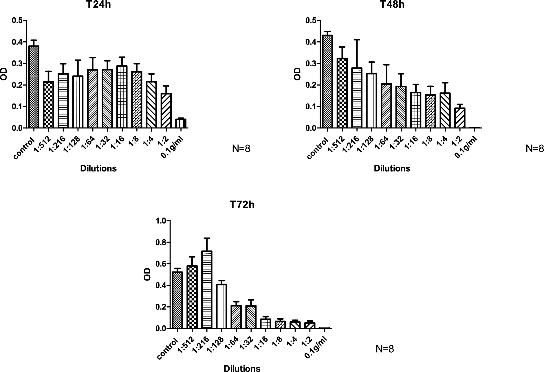
Results concerning the effects induced by different concentrations of dissolved honey membrane on viability of L929 cells are represented in Fig. 1.
MTT assay (
Three rats from the treated group died of ascites in the first 24 hours. The gross evaluation in 36 rats showed no wound site infection or presence of adhesions. On performing histological analysis 24 and 72 hours’ post-surgery, the tissue near the implant was found to be characterized by the presence of fibroblasts with some cellular response, including lymphocytes, macrophages, and neovascularisation. No reaction was observed in distant organs. Thus, the PHHs did not induce a foreign body reaction. The differences in the blood levels of the IL-1β, IL-6, TNF-α and PG(E2) at the 0, 6, 24, and 72 hours’ time points were not statistically significant. The results are summarized in Table 2.
Results for blood levels of the IL-1β, IL-6, TNF alpha and PG(E2)
Results for blood levels of the IL-1β, IL-6, TNF alpha and PG(E2)
The new membranes may be used as wound dressings as they have a good WVTR and fluid uptake and show no cytotoxicity to fibroblasts; they also have good swelling capability, which is an important factor for reducing the risk of wound dehydration.
The results obtained by the citoxicity assay after 24 hours from the seeding, in presence of decreasing concentrations of dissolved honey membranes, have shown a statistically significant decrease (
After 72 hours of incubation, the highest concentrations (from 1:2 to 1:64) caused an inhibition in cell growth while the lowest (1:256 and 1:512) induces a statistically significant increase in cell growth compared to the control. During the in vivo experiments, tree rats died: in authors’ opinion, this was because of the excessively large sheet of membrane implanted intraperitoneally because, initially, a dimension of
In the light of PHHs antibacterial activity [12] and since administration of systemic antibiotics does not always lead to good outcomes in terms of: wound healing, matrix penetration of the EPS biofilm and antibiotic resistance, in this study we propose the use of Manuka honey to prepare PHH for wound dressings. Interestingly, our membranes demonstrate a good antibacterial activity toward clinically relevant pathogenic microorganisms such as S. aureus and E. coli.
Honey membranes possess a wide variety of properties that can make them suitable (as for other natural materials such as chitosan hydrogels) [2,24,29], for very different uses that we can hypothesise ranging from wound healing to adhesion prevention to drug delivery. The presence of natural active components, conformability, and complete resorbability are the main characteristics of this new biocompatible biomaterial that respects the pathophysiology of tissue, is well tolerated by the body, possibly improves healing, and may be used for the prevention of surgical complications. Furthermore, the production of these devices is extremely simple and inexpensive.
Footnotes
Acknowledgements
The authors gratefully acknowledge the financial support from grant PRIN 2012.
Conflict of interest
The authors have no conflict of interest to report.