
Abstract
Fibrinogen plays a necessary role in blood clotting and wound healing. In this study, a new solvent mixture of formic acid/acetic acid with low toxicity was investigated as an alternative solvent for fibrinogen electrospinning. The nanofibers were analyzed by scanning electron microscope (SEM), simultaneous thermal analysis (STA) and attenuated total reflectance-Fourier transform infrared spectroscopy (ATR-FTIR). The results showed that when the ratio of formic acid to acetic acid was 75/25 (v/v) the finest defect-free fibres with diameters ranging from 184 ± 37 to 241 ± 70 nm were obtained. In addition, the average fibre diameters increase with increasing concentration of fibrinogen from 10wt% to 12wt%. It is concluded that solvent mixture consisting of formic acid/acetic acid can be a great solvent for electrospinning of fibrinogen and is able to produce nanofiber structures.
Introduction
Nanofibrous materials have gained much attention in biomedical fields, such as drug delivery and tissue regeneration matrices [1]. Electrospinning is a favorable technique to produce ultrafine fibers with diameters ranging from microns to nanometers [1,2]. Nanofibers can be considered as scaffolds for tissue engineering [3]. Fibrinogen scaffolds have been widely used in tissue engineering applications, because of their ability to mimic the native tissue structures and non-immunogenicity [4]. Fibrinogen, a natural plasma protein containing six chains (2 Aα, 2Bβ, and 2γ) is a major element in the coagulation process, normal hemostasis and wound healing [5,6]. Various techniques of physical and chemical methods including cryoprecipitate, ammonium sulfate, ethanol and poly(ethylene glycol) (PEG) precipitation are used to isolation and preparation of fibrinogen [7]. Cryoprecipitate is the gold standard of concentrated fibrinogen due to its frequency of use and providing the highest fibrinogen yield [8,9]. Electrospun of natural polymers including proteins and polysaccharides is very difficult as a result of their high viscosity and low solubility in general organic solvents. A limited number of solvents such as 1,1,1,3,3,3-hexafluoroisopropanol (HFIP) and trifluoroacetic acid (TFA) are available that can be used to dissolve natural polymers. However, these solvents are very toxic and expensive which limited its application as a solvent [10,11]. Some studies suggested non-toxic solvent such as acetic acid or formic acid for electrospinning of natural polymers [12]. Furthermore, electrospun of some natural polymers like gelatin and silk fibroin in acidic solvents is reported [13]. To the best of our knowledge, the solvent is used for dissolve and electrospinning of fibrinogen is a mixed solvent of 9 part HFIP and 1 part minimal essential medium (MEM). It has been reported that a volatile, fluorinated hydrocarbon solvent such as HFIP as well as MEM is needed to spin fibrinogen [14,15]. The goal of this study was to evaluate the feasibility of electrospinning of fibrinogen using formic acid/acetic acid to determine an alternative solvent for electrospinning of fibrinogen and compare their morphology and diameter with the nanofibers obtained from HFIP solvent.
Materials and methods
Preparation of fibrinogen
The cryoprecipitate was donated by blood bank unit of Imam Khomeini Hospital, Tehran, Iran. Briefly, cryoprecipitate was obtained from fresh frozen plasma (FFP) followed by thawed at 4°C and centrifuged at
SDS- PAGE electrophoresis
Sodium dodecyl sulfate-polyacrylamide gel electrophoresis (SDS PAGE) was used to confirm the polypeptide chains of the fibrinogen molecule. The electrophoresis was performed in 12% acrylamide running gel and a 4% stacking. The electrophoretic bands stained with Coomassie Brilliant Blue R-250.
Electrospinning of fibrinogen
For electrospinning, the fibrinogen solution at a concentration of 10, 11 and 12wt% was prepared in HFIP (Hangzhou Dingyan, China)/DMEM (Inoclon, Iran) 9/1 (v/v), formic acid (FA), formic acid/acetic acid (FA/AA) with ratios of 75/25, 50/50 and 25/75(v/v). The electrospinning process was performed using electrospinning equipment (Electroris, FNM, Tehran, Iran). The polymer solutions were electrospun at a high voltage of 20 kV with flow rate of 1.0 ml/h. The distance between syringe tip and collector was 12 cm.
Characterization
The morphology of the electrospun nanofibers was characterized by SEM (model Philips XL-30, Netherlands, at 25 kV). The nanofibers were sputter-coated with gold, and the average diameters of nanofibers were measured via Image J software. The surface of the nanofibers was characterized by attenuated total reflectance-Fourier transform infrared (ATR-FTIR) spectroscopy (Nicolet iS10). The spectroscopic analysis of samples was recorded over a range of 400-4000 cm−1 at a resolution of 4 cm−1. The thermal behavior of fibrinogen nanofibers were studied by simultaneously thermal analysis (TGA–DTA) with an STA 503, (BÄHR, Germany). The samples were heated to 600°C at a rate of 10°C/min under argon flow rate of 3 l/h.
Statistics analysis
All data are presented as the Mean ± standard deviation (SD). Statistical analyses were performed by using one-way analysis of variance (ANOVA) with a Games-Howell post hoc test and Wilcoxon signed rank test. The significance level was set to
Results and discussion
Preparation of fibrinogen
Several modifications on the cryoprecipitate method such as different kind of anticoagulant, various centrifugation speeds and the addition of chemical additives have been described [16]. Here we prepared fibrinogen from cryoprecipitate based on described in materials and methods, then compared with commercial product in terms of purity. It has been known that fibrinogen is composed of three pairs of different polypeptide chains with a molecular weights of 66 kDa, 54 kDa, and 48 kDa, which assigned to Aα, Bβ and γ polypeptide chains respectively [17]. Figure 1 shows SDS-PAGE of prepared fibrinogen in comparison with commercial fibrinogen (cod. F4129, Sigma-Aldrich). According to the SDS-PAGE patterns, the results are similar in terms of purity between prepared fibrinogen and the commercial product. On the basis of these results, it is concluded that the cryoprecipitate method is simple and easy way which can give good fibrinogen yield with admissible purity.

The SDS-PAGE of fibrinogen isolated compared with commercial fibrinogen. Lane 1: Fibrinogen obtained by cryoprecipitate method, Lane 2: commercial product fibrinogen (cod. F4129, Sigma-Aldrich), Lane 3: Molecular weight markers (11–180 kDa) from Avagene (Tehran, Iran).
Several parameters including solution properties (such as concentration, electrical conductivity, dielectric properties, surface tension, viscosity), processing parameters (e.g. applied voltage, flow rate, distance between the collector and the tip) and ambient parameters such as humidity and temperature have effect on the nanofibers [18,19]. In this study, the effect of the volume ratios of the FA/AA solvent and various concentrations of fibrinogen on nanofiber morphology and diameter was investigated. However, the processing parameters were optimised and fixed during electrospinning. As shown in Table 1, when HFIP was used as the solvent, the average diameters were 226 ± 146, 327 ± 171 and 537 ± 370 nm at 10%, 11% and 12wt%, respectively. It was found that fiber diameters increase with increasing fibrinogen concentration (
Average fiber diameters of fibrinogen nanofibers in HFIP and acidic solvent. The data presented as mean ± SD
Average fiber diameters of fibrinogen nanofibers in HFIP and acidic solvent. The data presented as mean ± SD
ATR-FTIR spectroscopy was performed to reveal the presence of fibrinogen. ATR-FTIR spectra of fibrinogen in acidic and HFIP solvent are presented in Fig. 3. The spectrum of fibrinogen showed the peak at 1650 cm−1 (amide I for C=O stretching) and 1538 cm−1 (amide II for –NH deformation), which are typical peaks of proteins. Similar results were obtained by other researchers [25,26]. As it shown in Fig. 3, there was no difference in ATR-FTIR spectra of fibrinogen in two solvents.
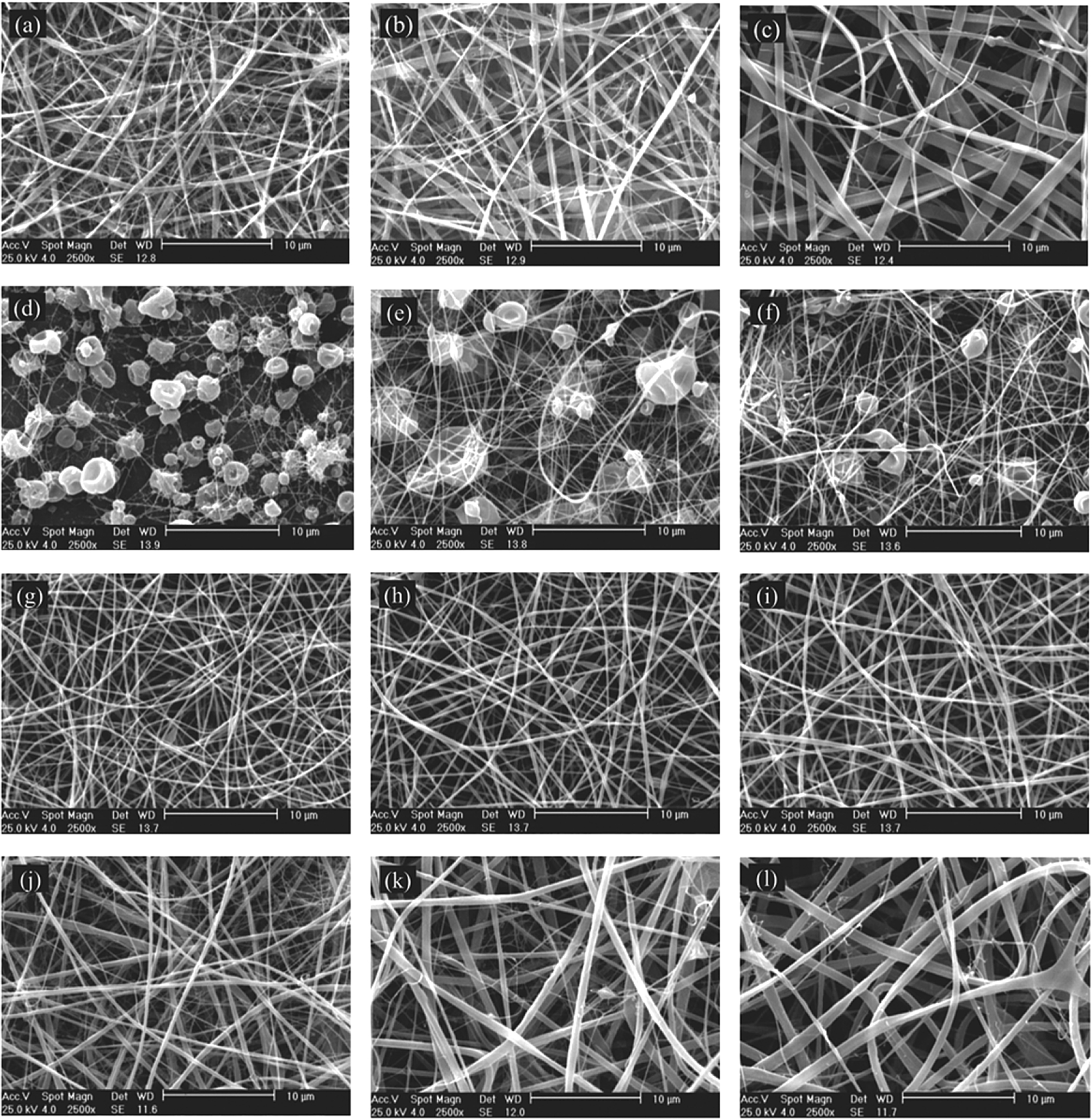
SEM of fibrinogen nanofibers at various concentrations in HFIP and acidic solvent. (a), (b), (c) are nanofibers of 10%, 11% and 12% in HFIP solvent. (d), (e), (f) are nanofibers of 10%, 11%, and 12% in formic acid. (g), (h), (i) are nanofibers of 10%, 11%, and 12% in formic acid/acetic acid (75/25) and (j), (k), (l) are nanofibers of 10%, 11%, and 12% in formic acid/acetic acid (50/50).
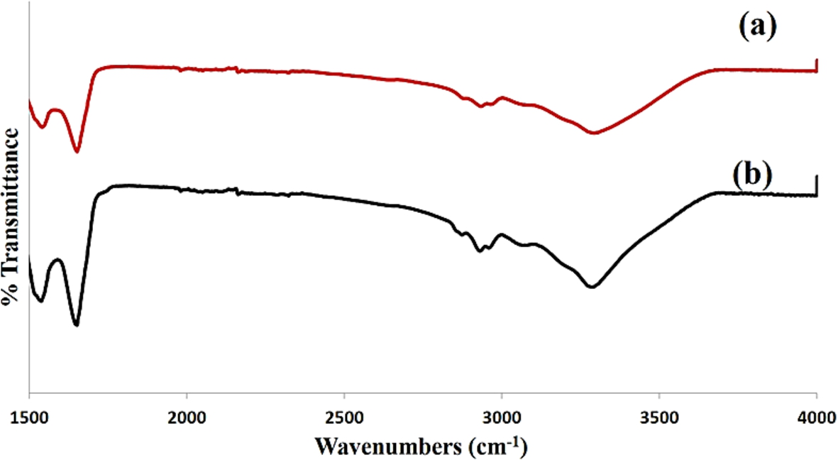
ATR-FTIR spectroscopy of fibrinogen nanofibers. (a) fibrinogen nanofibers in HFIP solvent. (b) fibrinogen nanofibers in acidic solvent.
Many studies have investigated the stability of fibrinogen structure under electrospinning conditions [15,27] but, few studies have examined the thermal properties of fibrinogen electrospun nanofibers. Thermal analyses techniques are beneficial to give essential information about a material’s properties and phase transitions [28]. For thermal stability and degradation of the fibrinogen nanofibers in HFIP and acidic solvent, the TGA and DTA analysis was performed and shown in Fig. 4. The thermogram of derivative thermal gravimetric (DTG) is prepared on the basis of the first derivative of the TG plots to show the decomposition temperature. According to the DTG curve of Fig. 4, there are two weight loss peaks. The first is at around 100°C and the second peak at 315°C, which nearly similar to the DTG results of fibrinogen scaffold obtained by Chuanglong He [26]. The first broad peak in all polymeric curves is mainly due to the evaporation of solvent or water [28–30] which can be confirmed by the DTA curve at the same temperature range. In addition Haiguang Zhao mentioned that the weight loss of fibrinogen before 260°C should be attributed to the water evaporation and dehydration between the protein molecules [31]. The second weight loss phase is likely caused by the decomposition and degradation of fibrinogen. As it shown in Fig. 4 the thermal behaviour of electrospun fibrinogen nanofibers in HFIP solvent was similar to the profile of fibrinogen nanofibers in acidic solvent. These results showed that mix solvent of formic acid/acetic acid had no influence on the structure or stability of fibrinogen nanofibers.

Thermal analysis of fibrinogen nanofibers in HFIP and acidic solvents. (a) Thermal gravimetric analysis (TGA) and differential thermal gravimetric (DTG), (b) Differential thermal analysis (DTA).
In this study first, fibrinogen was prepared from cryoprecipitate and compared with the commercial product. This method is an easy way to isolation of fibrinogen with considerable purity for medical applications. Then fibrinogen electrospinning was done in solvents such as HFIP, formic acid, acetic acid and mixture of formic acid/acetic acid. Among these solvents, acetic acid did not dissolve fibrinogen. On the other hand, beaded nanofibers obtained in the formic acid solvent. Therefore mixed solvent of formic acid/acetic acid was used and the effect of different solvent ratios of FA/AA on nanofiber morphology and diameter were investigated. Smaller nanofibers were produced using the solvent ratio of 75/25 v/v. STA and ATR-FTIR analysis showed no significant difference in structure or stability of fibrinogen nanofibers in these solvents. It is concluded that new solvent of formic acid/acetic acid (75/25 v/v) can be a promising solvent for electrospinning of fibrinogen.
Footnotes
Acknowledgement
This project was supported by Tehran University of Medical Sciences (TUMS), grant No. 94-0287-28594.
Conflict of interest
The authors have no conflict of interest to report.