
Abstract
BACKGROUND:
Human dentin is a highly calcified tissue of mesenchymal origin with a heterogeneous structure. Its morphology is constantly remodelled throughout the life span of the tooth, as well as under the influence of external stimuli.
OBJECTIVE:
The aim of the present study was to obtain information about the thermal changes in the crown and root dentin specimens of vital and devitalized teeth.
METHODS:
The investigated samples were divided into 6 groups, depending on the patients’ age and dentin location (crown, root). An additional group of endodontically treated teeth was created. The methods of choice were were combined thermal analysis (DTA-TG(DTG)-MS and gas chromatography-mass spectrometry.
RESULTS:
After heating up to 1200 °C, endo- and exothermal effects were observed. The effects’ dynamic was the same for all samples. The differences were in the samples’ weight after the experiment, with root dentin showing the greatest mass loss percentage.
CONCLUSIONS:
The observed mass loss differences could be attributed to the presence of impurities in the dentin, as well as alterations in the collagen matrix. Ageing and endodontic treatment could catalyse the accumulation of such changes and affect the microstructure of the mineralized tissue.
Introduction
Thermal analysis belongs to a group of methods used for the studying of materials’ properties with a progressive increase of their temperature. The method has a broad field of application - in the pharmaceutical industry, in electronics, geology and engineering sciences. It provides valuable data on the specific thermo-kinetic effects that take place inside a material, thus obtaining information on the composition and structure of the studied sample.
The thermal analysis allows for the simultaneous recording of the changes in mass (TG), differential temperature (DTA) and speed of thermal reactions (DTG) of the specimens. The mass spectroscopy (MS) data give additional information about evolving gases during the thermal treatment. Hence, their thermal behaviour can be assessed. With the increase of the temperature of the sample, various physical and chemical reactions occur. These effects are associated with phase changes, characterised by heat exchange - absorption and release [1]. The amount of exchanged heat, as well as the recorded temperatures, at which those changes occur, are unique for the specific material. During the experiment, they are compared to the recorded thermal reference system [1]. The studied DTA curve is compared to the database, which allows for the correct identification of the sample material. The values for phase change, intermediate and overall loss of mass, as well as the inflexion points, are recorded. The collected data provides information on reactions of dehydration, disintegration, phase change, reduction and oxidation [2]. The graphically represented results are called “thermal analysis curves”.
Differential thermal analysis has been applied in dental medicine for the characterization of silicate and glass-ionomer cement and composites [3]. In recent years it has been used for studying the new calcium phosphate bioceramic and composite materials [4,5]. The thermal effects that occur during the crystallisation process of ceramics have been recorded using DTA [6]. The obtained data is useful in discovering the effect of particle size and temperature regime during materials production.
Apart from studying restorative materials, DTA is used in endodontics as well - for the establishment of the qualities of alpha and beta gutta percha and their thermal stability during obturation [7]. The method has been applied in the investigation of the changes that occur in hard dental tissues after treatment with CO2 laser, as well as the mechanism of sintering of calcium fluoride and hydroxyapatite [8,9]. DTA is often combined with scanning electron microscopy, spectroscopic analyses, X-ray diffraction [10,11].
Despite its various applications, the method has not been used for the investigation of dentin from vital and endodontically treated teeth. Human dentin is a highly organised biological structure that consists of protein chains forming the collagen and mineral substances that ensure its stability. The building blocks of dentin also include water, which is located on the crystals’ surface or between them. There is no available data on the effect of ageing and root canal treatment on the thermal stability of dentin. Its increase or decrease could provide information about subtle structural changes in the tissue. The aim of our study was to determine the thermal changes in samples of the crown and root dentin in vital and devitalized teeth using differential thermal analysis and gas chromatography-mass spectrometry. Our hypothesis was that possible changes in the thermal stability could point to changes in its composition, thus leading the way to future investigations on the matter.
Materials and methods
The samples used in the study were crown and root dentin of intact human teeth, extracted for orthodontic or periodontal reasons. The patients were divided into 2 age groups: below 40 and above 40 years old. All specimens were cleaned using ultrasonics and stored in 0.2% thymol solution under 4 °C for up to 3 months, in order to preserve hydration and prevent collagen degradation.
The number of samples used was 30, divided into 6 groups, with 5 samples in each one: young crown dentin (G1), old crown dentin (G2), old crown dentin from an endodontically treated tooth (G3), young root dentin (G4), old root dentin (G5) and old root dentin from an endodontically treated tooth (G6) (Table1). Each sample was finely ground at room temperature using an agate mortar and ball and passed through a 200-mesh (74 μm) sieve.
Studied groups
Studied groups
The investigation of the samples was carried out with a Thermogravimetry/Differential Thermal Analyzer (Setsys Evolution, Setaram, France), connected to a spectrometer (OmniStar, Pfeiffer Vacuum, Germany). The temperature range was from 0 °C to 1200 °C with a heating rate of 10 °C/min, static atmosphere, in platinum sample pans. Each sample weighed between 10 and 20 mg. The acquired data were processed using the software programs Calisto and Origin.
The resultant data is graphically represented as shown in Figs 1–6.
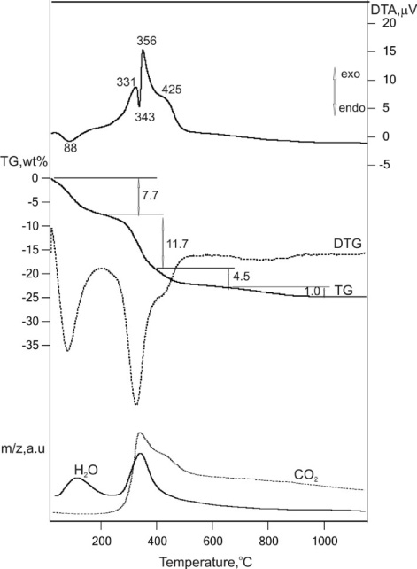
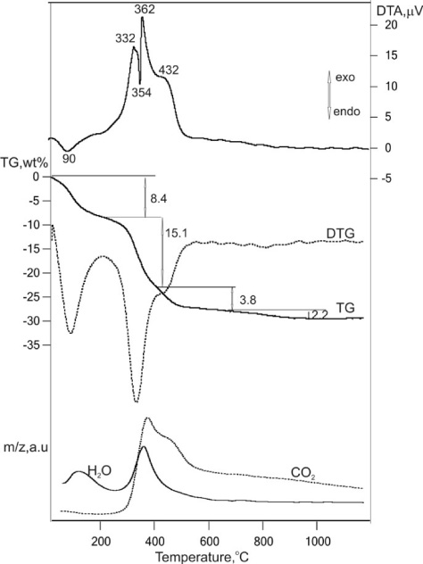
Graphic representation of the results for group G1 (young crown dentin). The observed endo- and exothermic effects with their respective temperatures are shown on the DTA curve. The TG curve represents the weight loss percentage at different temperature stages. DTG, H2O and CO2 curves are also shown.
Graphic representation of the results for group G2 (old crown dentin).
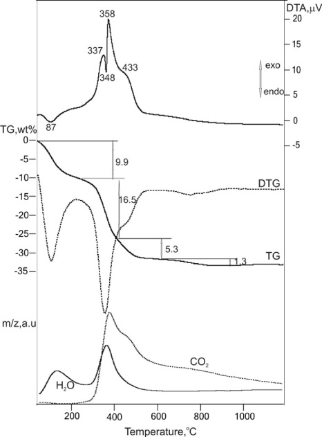
Graphic representation of the results for group G3 (crown dentin from an endodontically treated tooth).
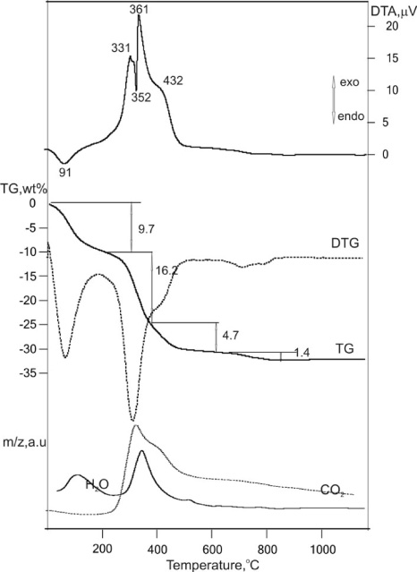
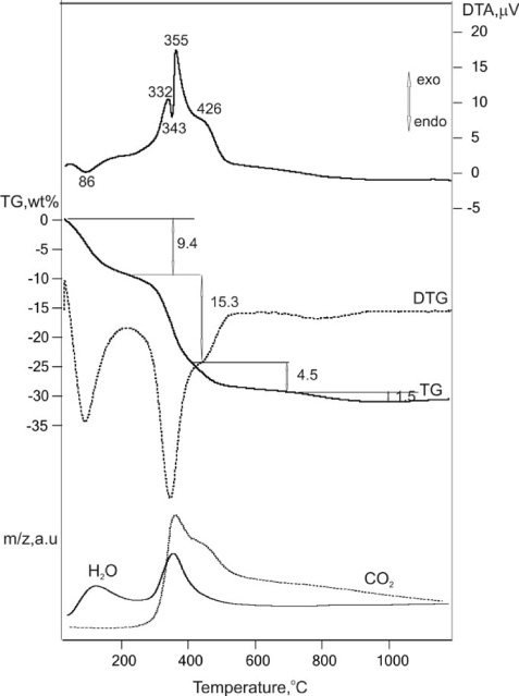
Graphic representation of the results for group G4 (young root dentin).
Graphic representation of the results for group G5 (old root dentin).
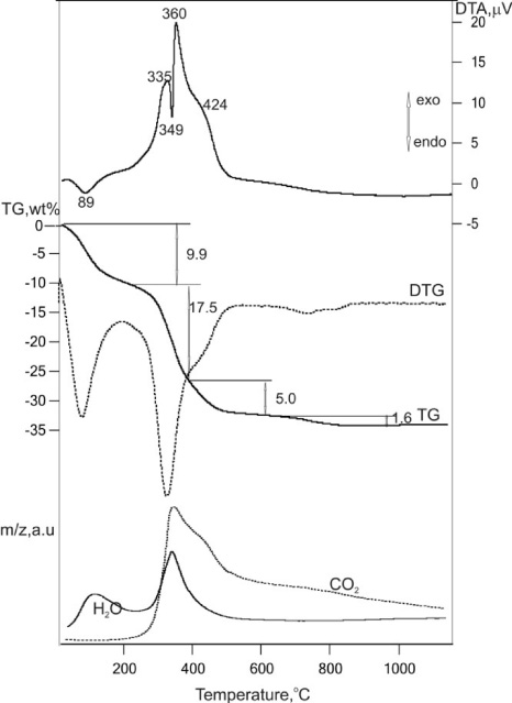
Graphic representation of the results for group G6 (root dentin from an endodontically treated tooth).
The following curves can be observed on the graphics: TG – mass loss curve (wt%); DTG – a derivative of the TG curve. When the curve crosses the inflexion points, it changes to a concave or convex shape, which is a sign of phase change; DTA – indicates the type of reaction - endo- or exothermic; H2O – indicates the released water content; CO2 – indicates the emitted carbon dioxide content.
The TG curve provides quantitative information about the mass loss percentage, whereas the H2O and CO2 curves provide only qualitative information. There were no distinct differences between the graphical representations of the groups. A comparison of the DTA curves showed that endo- and exothermic reactions happened in the same temperature intervals in all of the samples, with slight variations.
There were differences in the mass loss percentage, as presented in Table2.
TG and DTG data in temperature interval 25–1000 °C and thermal decomposition processes of investigated samples
The specimens used in other investigations were ground at low temperatures in order to make them more fragile and easy to manipulate [12]. However, this might damage the interrelationship between the water, the collagen and the mineral phase of dentin. The results of our investigation correspond to the DTA curves of dentin available in the literature [13]. They prove that the main contents of dentin, including water, can be preserved even after grinding of the material.
In the TG analysis of young dentin (G1), the first mass loss (−7.66%) occurred between 25–218 °C (Fig. 1). The reaction is exothermic. The curve for water loss confirms that the water content has evaporated at this interval. The second mass loss occurred between 218 °C and 415 °C. The reaction is characterised by two exothermic effects and an endothermic one between them. Since the CO2 curve showed the most substantial loss in this area of the curve, it can be suggested that the endothermic reaction is a result of the sample’s dehydration. A significant amount of CO2 loss was recorded, coinciding with an exothermic reaction, which corresponds to collagen decomposition. The splitting of the exothermic effect, visible between 340–360 °C, could be attributed to a possible overlap with the endothermic reaction.
The third mass loss (−4.51%) was between 415–807 °C. The reaction is slightly exothermic. The decomposition of the organic matter continued. The fourth mass loss (−0.93%) was in the interval 807–1180 °C. Above 800 °C decarbonization occurs, and the reaction is exothermic.
The changes, observed with the increase of the temperature, were between 0 °C and 1200 °C. Since pure hydroxyapatite does not undergo any changes in this interval, it could be suggested that the emission of CO2 and H2O was not connected to the disintegration of its structure. Therefore, the emitted gasses could be due to degradation of various impurities in the structure of dentin.
The dynamic of the observed thermokinetic effects is confirmed in all of the other groups. The TG data is presented in Table2. All groups exhibit the greatest mass loss between 200–420 °C, with two exothermic and one endothermic effect and the emission of CO2 and H2O. These effects occur at almost the same temperatures (Figs 1–6). The differences are only in the mass loss percentages. The least substantial mass loss was recorded in the “young crown dentin” group (G1), and the most substantial - in the “root dentin” groups, including old and devitalised ones (Table2).
There are two possible explanations:
1. Changes in the inorganic component: presence of inorganic impurities, which are less thermally stable than pure hydroxyapatite;
2. Changes in the organic component: the presence of altered collagen matrix that disintegrates more easily at high temperatures.
According to the mechanisms of crystal growth, low-molecule calcium-phosphate substances can be formed under specific conditions (temperature, moisture, pressure). In agreement with the laws of dentinogenesis, dentin dehydration (physiologic and pathologic) is a strong factor in the formation of apatite and non-apatite impurities - octa calcium phosphate (OCP), tricalcium phosphate (TCP), dicalcium phosphate dihydrate (DCPD).
The hydroxyapatite molecule has a hexagonal structure with a chemical formula Ca10(PO4)6(OH)2. It is the smallest structural entity needed for crystal formation. Each hydroxyapatite crystal has three zones - outer part, inner part and a hydrate layer. Each zone can participate in ion exchange. Calcium can be replaced with magnesium or sodium, phosphate - with carbonate groups, hydroxyl group - with carbonate, fluoride and chlorine [14].
It is suggested that a disruption in the hydrate layer can increase the ion exchange and accelerate the formation of OCP and DCPD in old, as well as in devitalized teeth [15]. Low-molecule calcium-phosphate substances have been observed in this layer. Their composition is similar to that of DCPD [16]. A study performed with Fourier transform spectroscopy showed a spectre, similar to that of OCP [17]. The blood supply of devitalized teeth is cut off, leading to dentin dehydration and temperature change. This can also occur due to dentinal tubules occlusion with age. Dentin dehydration disrupts the hydrate layer and leads to the crystallisation of new phases - OCP and DCPD. In endodontically treated teeth, this process occurs faster than in ageing teeth. Despite this, Raman spectroscopy results confirm larger amounts of non-apatite impurities in old dentin [15]. These impurities are less thermally stable than pure hydroxyapatite. In TCP samples, mass loss begins between 300 and 700 °C with a change in hydrogen phosphate ions, before the apatite decomposition begins [18]. The crystal structure of DCPD loses its stability at 127 °C, starting with the loss of the two water molecules in its composition [19]. Pure OCP disintegrates at temperatures between 0 and 300 °C [20]. It could be suggested that the observed mass loss in the groups of old crown (G2) and devitalized crown (G3) dentin is due to the presence of less thermally stable impurities.
Factors like endodontic treatment and ageing change the temperature, the moisture content and the pressure, which increases the formation of non-apatite impurities (OCP, TCP, DCPD). The dentin loses its elasticity. A reorganisation of its architectonics occurs, characterised by tubule occlusion. Part of the hydroxyapatite is replaced by low-molecule calcium phosphate substances with lesser mechanical strength, which could impact its fracture resistance.
Another possible reason for the greater mass loss in the samples of root dentin - old (G5) and devitalized (G6), could be a change in collagen structure. In contrast to enamel, the organic component of dentin mainly consists of collagen type I. Collagen in the peritubular dentin is scarce, as 90% is located in the intertubular dentin [21]. The formation of apatite crystals in the intertubular dentin is catalysed by specific atomic groups of the collagen fibrils. Thus, crystal formation and deposition are observed in 3 zones - directly on the fibrils, in empty spaces on their surface and in pores inside them. The mineral component of bones is largely located inside the collagen fibrils, with a small amount situated in the spaces between them [14].
Available studies in the literature show that, generally, the mineral content of dentin increases with age [22,23]. This is largely due to the formation of transparent dentin inside the dentinal tubules (with a decrease in tubule density and tubule diameter). However, during the physiological formation and deposition of transparent dentin inside the dentinal tubules, decomposition of peritubular and intertubular crystals occur, as they act as one of the possible mineral sources. The size of intertubular crystals decrease, and intratubular deposits increase [24]. An indication of this is the lack of peritubular dentin in the areas, where transparent dentin is formed [25]. Therefore, it can be suggested that during the deposition of transparent dentin, the number of hydroxyapatite crystals inside the collagen network decreases. Thus, the mineral content inside the tubules in part increases at the expense of the mineral content in the intertubular dentin and the collagen network. This could leave the collagen matrix with less mineral content and, subsequently, decrease thermal stability. There are studies available in the literature, concerning the effect of mineralisation on the thermal stability of dentin. Armstrong et al. establish that the temperature needed for denaturation of mineralized dentin is between 160–186 °C. In contrast, the temperature needed for disintegration of demineralized dentin is in the interval 65–176 °C [26]. The presence of apatite crystals stabilises the collagen matrix [27]. It can be suggested that the loss of apatite crystals in the intratubular dentin due to the formation of transparent dentin inside the tubules could affect the thermal stability of collagen. Overall, the mineral content increases, but at the expense of changes in the organic matrix, which leaves it less thermally stable.
The smallest amount of transparent dentin should be found in young dentin. This would make its collagen matrix the most stable, ensuring minimal mass loss during differential thermal analysis. This corresponds to the findings in our study (group G1). The comparison between the devitalized dentin samples - crown and root dentin showed greater mass loss in the root dentin (G6) (Table2). It can be assumed that ageing and endodontic treatment can damage the collagen-hydroxyapatite interrelation and speed up collagen degradation. The changes affect root dentin and progress coronally. This is confirmed by the literature data since transparent dentin formation begins at the apex and gradually reaches the crown [28,29]. Moreover, a study into the age-related covalent shifts in dentin proteins showed significant changes in amino acid composition, cross-linking and racemization of dentin phosphoproteins, resulting in almost complete dephosphorylation over the functional life of the tooth [30]. This suggest that, as with all tissues, the qualities of dentin decrease with age and that this change is evident in the modifications that occur in the protein matrix. Whether those changes occur in a similar matter in endodontically treated teeth and to what extent they can affect the thermal stability of dentin in general requires further investigation.
Conclusion
Differential thermal analysis and thermogravimetric analysis are suitable methods for tracing and confirming the changes that occur in hard dental tissues. When heated up to 1200 °C, dental samples exhibit mass loss, combined with endo- and exothermal effects. These effects are due to the disintegration of Ca-PO4 and Ca-CO3 substances, different from pure hydroxyapatite. The observed thermal changes are easily traceable, which shows that a number of impurities are significant. This could be related to endodontic treatment, as well as the natural process of transparent dentin formation with age. Thus the thermal stability of dentin could be affected due to changes in the hydrate layer of hydroxyapatite during its dehydration, its decrease in the collagen network and the secondary precipitation into transparent dentin. This leads to water loss and organic component disintegration. The differential thermal analysis results reveal the need for a more thorough investigation into the differences between vital and non-vital teeth.
Footnotes
Ethics approval
Research was approved by the Ethics Committee of Medical Iniversity - Plovdiv.
Conflict of interest
All authors state no potential conflict of interest.
Funding
This research was funded as a part of a PhD program by Medical University - Plovdiv.