
Abstract
BACKGROUND:
Various biomaterials/technologies have been tested for treatment of intervertebral disc (IVD) degeneration (IDD). Only few non-surgical options exist.
OBJECTIVE:
Assessment of efficacy and safety of the hyaluronic acid derivative hydrogel HYADD®4-G in IDD using a well-established rabbit annular puncture model.
METHODS:
Rabbits were punctured at two IVDs to induce IDD. Thirty days after, IVDs were injected with HYADD®4-G or saline. IVD hydration, height, appearance and tissue organization were assessed by radiographs, MRI and histopathology. Safety of HYADD®4-G injection was evaluated in non-punctured IVDs.
RESULTS:
HYADD®4-G injection restored disc height to over 75% of the pre-punctured disc, saline injections led to 50% of initial disc height. Compared to saline, HYADD®4-G treatment resulted in improved water retention as revealed by MRI quantification. 83.3% of HYADD®4-G injected discs had normal appearance and reached grade I of the Pfirrmann scale. Regarding tissue organization and cellularity, HYADD®4-G treatment resulted in significantly lower IDD scores than saline (p < 0.01). HYADD®4-G injected into healthy IVDs did not induce inflammation or foreign body reactions.
CONCLUSIONS:
Intra-discal HYADD®4-G injection is safe and has therapeutic benefits: IDD could be limited through restoration of disc height and hydration and maintenance of normal IVD tissue organization.
Keywords
Introduction
Low back pain is a major musculoskeletal problem in orthopaedics with an overall prevalence of 31% [1]. Causes of back pain often involve diseases of the intervertebral disc (IVD) including IVD degeneration (IDD), clinically characterized by reduction of disc height and osteophyte formation [2,3].
IVD tissues cannot regenerate and different therapies for IVD regeneration or IDD process retardation are in development since conventional surgical interventions like spine fusion or disc arthroplasty remain unsatisfying due to mobility constraints and complications [4,5]. There are different therapeutic strategies focusing on extracellular matrix (ECM) modulation [6,7] such as protein injections [8,9], gene therapy [10,11], and stem cell therapy [12–15] to block or delay IDD up to intermediate stages. Best results were obtained with stem cells delivered in a gel-like matrix (for example collagen or hyaluronic acid (HA) based hydrogels) mimicking the mechanical and hydration properties of the IVD ECM [16–19]. Even cell-free HA hydrogels have shown a therapeutic effect in IDD [20–22].
HA is a highly viscous polysaccharide with 250 to 50,000 repetitions of its disaccharide core. One gram of HA can bind up to 6 L of water which makes it one of the most hydrophilic molecules. It is abundantly present in the extracellular matrix of soft connective tissue and is a major component of synovial fluid and cartilage. HA is also naturally present in the matrix of IVDs where it forms viscous multi-molecular aggregates containing proteoglycans [23–25]. HA derived hydrogels injected ex vivo into pig IVDs have shown potential for disc repair [20]. In cultured bovine IVDs, an HA-derived hydrogel attenuated inflammation and modulated the disc ECM towards anabolic behaviour [26]. In a study on ex vivo rat tail IVDs an injected HA hydrogel restored normal nucleus pulposus (NP) morphology [27]. In addition, intra-discal injection of HA reduced disc degeneration in experimental non-human primate and porcine models [28,29].
The HA derivative HYADD®4-G (Fidia Farmaceutici, Abano Terme, Italy) is a partial hexadecylamine of 500 to 730 kDa HA. In this HA derivative, an aliphatic amine (hexadecylamine) is bound to HA at the carboxylic group of the glucuronic acid (2% substitution). HYADD®4-G rheological and viscoelastic properties are superior to those of human synovial fluid [30]. HYADD®4-G has shown beneficial effects on synovial changes in osteoarthritis and was clinically tested as intra-articular visco-supplementation for shoulder osteoarthritis (OA). It turned out to be a safe treatment for relieving pain and restoring shoulder functionality [31]. Similar results were obtained in clinical trials on patients with knee OA [32,33].
The present study aimed to investigate for the first time the safety and therapeutic potential of intra-discal HYADD®4-G injections in reducing IDD using a well-established rabbit annular puncture model.
Materials and methods
Seeing that mechanical properties of HA-based hydrogels are similar to those of the NP matrix and that HA is a natural component of EMC, we hypothesized that HYADD®4-G intra-discal injections were biocompatible and suitable for IDD treatment. Saline injections were not believed to have therapeutic effects.
To assess safety and efficacy of HYADD®4-G in IDD, 11 adult female HYLA white rabbits (3.0–4.5 kg) were used. 8 animals (group 1) were punctured at two IVDs (L2/L3, L4/L5). 30 days later (D0), these rabbits underwent intra-discal injections of HYADD®4-G at L4/L5 and saline solution at L2/L3.
To assess the safety of HYADD®4-G, 3 non-punctured rabbits (group 2) underwent HYADD®4-G injections into L2/L3, L3/L4 and L4/L5 IVDs. An experimental time course is depicted in Fig. 1.

Experimental time course indicating surgical interventions and data collection over a range of 90 days (D-30 to D60).
Rabbits were anesthetized with medetomidine (100 μg/kg, Dormitor 1 mg/mL, Orion Pharma) and ketamine (50 mg/kg, Narkamon 50 mg/mL, Bioveta), maintained/adjusted through 2% propofol (i.v, 4–12 mg/kg, MCT/ACT Fresenius Kabi). Butorphanol (i.m. 100 μg/kg, Butomidor, Werft, Germany) served as analgesic. After determining the correct level, a 18-gauge needle was inserted 3–4 cm ventral from the midline into the disc space under fluoroscopic control, held for 10 seconds and rotated 180° before removal.
Each rabbit had two discs punctured (L2/L3, L4/L5) [13]. Non-punctured L5/L6 disc served as control.
Intra-discal injection
Intra-discal injections were performed 30 days later. Rabbits were prepared and anesthetized as before, except that the spine was approached from the contra lateral side.
15–20 microliters of HYADD®4-G (Sodium hyaluronate hexadecylamide 8 mg/mL, FIDIA FARMACEUTICI, Italy) or saline solution (0.9%, Sigma-Aldrich, St. Louis, USA) were injected into discs with a 21-G needle, monitored by fluoroscopy.
X-rays (C-arm, Ziehm 8000) of anesthetized rabbits were taken according to Fig. 1. IVD height was expressed as disc height index (DHI) as previously described [34]. Changes in DHI were expressed as percentage DHI (%DHI) of the pre-operative height (%DHI = post-operative DHI/pre-operative DHI × 100).
MRI acquisition and analysis
MRI scans were performed in vivo in sedated animals (i.m. medetomidine 100 μg/kg and ketamine 50 mg/kg) according to Fig. 1.
MRI device was a 3.0-T Siemens Prisma scanner (Siemens Medical Solution, Germany) with 15-channel knee coil for signal amplification. T2-weighted and T2-weighted-STIR (short inversion time inversion recovery) sections (sagittal plane) were obtained using following settings: Fast-spin-echo sequence with repetition time (TR): 3500 msec, echo time (TE): 89 msec, matrix: 512 × 512, field of view: 160 mm, number of excitations: 2, section thickness: 1.8 mm and fast-spin-echo sequence with TR: 2000 msec, TE: 10 msec, inversion time (TI): 230 msec, matrix: 488 × 488, field of view: 160 mm, number of excitations: 2, section thickness: 1.8 mm, respectively.
T2 relaxation time for NPs of injected IVDs was calculated using the following settings: Multi-spin-echo sequence with TR: 3000 msec and TE: 13–104 msec (8 echoes), matrix: 256 × 256 field of view: 160 mm, number of excitations: 1, section thickness: 3 mm.
Black and white MRI images of T2-weighted-STIR sections were converted into RGB/colour images by OsiriX Medical Image software (OsiriX Foudation, Switzerland).
Additionally, MRI T2-weighted images were evaluated for IDD status using the Pfirrmann grading system (Table 1) [35].
MRI grading table of IVD degenerative changes according to Pfirrmann
MRI grading table of IVD degenerative changes according to Pfirrmann
Euthanasia was performed with subcutaneous medetomidine (100 μg/kg) and ketamine (50 mg/kg) followed by a lethal dose of intravenous T61. Experimental IVDs were explanted and fixed.
Histological and histopathological analysis
Fixed IVDs were decalcified, paraffin-embedded and sectioned (5 μm) at 6 different levels. Histological sections were stained with hematoxylin and eosin (H&E), toluidine blue and Masson-Trichrome (MT) for analysis and grading as described previously [34,36]. A scale based on 4 categories of degenerative changes with scores ranging from “normal disc” to “severely degenerated disc”, spanning 12 points (3 points in each category), was used to assess IVD state (Table2) [34].
Histological grading table of IVD degenerative changes [34]
Histological grading table of IVD degenerative changes [34]
Histological grading table of IVD degenerative changes based on the 4 categories. Maximum score is 12 points.
To assess the safety of HYADD®4-G injections, histological sections of IVDs L2/L3, L3/L4 and L5/L6 from the 3 animals of group 2 were examined for inflammatory reactions as previously described [37].
Statistical analyses were performed with GraphPad Prism (GraphPad Software, Inc.), significance threshold was set to P value < 0.05.
Changes in DHI, MRI and Pfirrmann grading were assessed by two-way analysis of variance (ANOVA) and Tukey’s post hoc analysis. Histological grading was analysed by one-way ANOVA plus Tukey’s test for the treatment effect.
Results
One animal died during surgery due to a cardiac arrest and another animal was euthanized before HYADD®4-G or saline injection because of self-mutilation. Four animals presented a reversible paresis for 2 days after surgery, which is common after such spinal interventions. No other clinical signs could be observed. Animal body weights were stable throughout the study.
Radiographic assessment
The percentage DHI (%DHI) is defined as the disc height index at a specific time point compared to the initial, pre-punctured disc height index. Data of non-punctured, non-treated L5/L6 IVDs was used as control.
Thirty days after puncture (D0) both, saline-treated and HYADD®4-G-treated IVDs, showed a significant decrease in DHI from 25% to 30% compared to control, indicating that the puncture indeed led to IDD (Fig. 2). There were no significant differences in %DHI between saline- and HYADD®4-G-treated IVDs at D0.
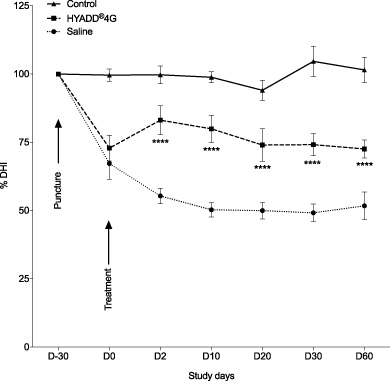
Changes in intervertebral disc height index after needle puncture and treatment (arrows). Thirty days after puncture, saline or HYADD®4-G was injected into discs. The percentage of disc height (with untreated discs as 100% control) was measured at each time point to quantify changes. Values represent the mean %DHI ± standard error of the mean. A significant difference was reached at each time point for saline vs HYADD®4-G (∗∗∗∗ P < 0.0001).
The treatment significantly affected the disc height. Two days after injections, the HYADD®4-G injected discs recovered 10% in height whereas the saline-treated IVDs showed further narrowing of 12% (%DHI at D2: HYADD®4-G: 83.1% ± 5.2%, saline: 55.4% ± 2.8%, p < 0.0001). From D10 to D60, HYADD®4-G treatment restored disc height to a level slightly over 75%, whereas saline treatment led to a stable disc height of 50% (D10, D20, D30, D60: p < 0.0001).
Disc desiccation is a hallmark of IDD. To assess hydration of HYADD®4-G or saline treated IVDs, magnetic resonance imaging (MRI) was performed 2, 30 and 60 days after injections. The T2-weighted images showed that punctured and saline-treated L2/L3 discs rapidly lost water content, whereas HYADD®4-G injected L4/L5 displayed improved water retention (Fig. 3). Quantification revealed a significant enhancement of hydration (T2 values) at each time point for HYADD®4-G versus saline (HYADD®4-G vs. saline. D2: p < 0.0001; D30: p < 0.05; D60: p < 0.001) (Fig. 4).
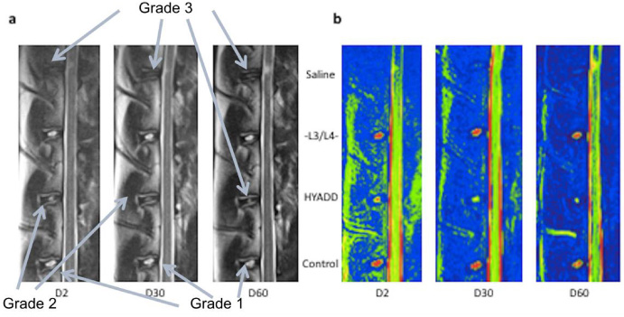
Sagittal T2-weighted MRI of a representative spine (
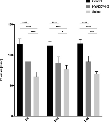
Quantification of MRI T2 values performed at D2, D30 and D60 after treatment. The T2 values of the saline group were the lowest at each time point compared to control or HYADD®4-G treatment and significance was reached at each time point (saline vs control D2, D30, D60 : ∗∗∗∗ p < 0.0001; saline vs HYADD®4-G D2 : ∗∗∗∗ p < 0.0001, D30 ∗ p < 0.05, D60 ∗∗∗ p < 0.001). Values are shown as mean ± standard error of the mean.
Pfirrmann grading on MRI images showed significant differences between HYADD®4-G- and saline-treated IVDs at each time point (Fig. 5). The saline injected L2/L3 IVDs showed grade I (n = 1), grade II (n = 2) and grade III (n = 2) of degeneration at day 2. In these discs, degeneration proceeded and reached grade II (n = 1), grade III (n = 2) and grade IV (n = 2) by the end of the study (D60). In contrast, HYADD®4-G treated L4/L5 IVDs showed only mild degeneration. At the three tested time points, similar to the non-treated L5/L6 control discs, 83.3% of the HYADD®4-G injected discs (n = 5) had a normal appearance and reached grade I of the Pfirrmann scale (Fig. 5 and Table 3). Saline-treated IVDs showed higher grades of degeneration than control IVDs at all time points (Fig. 5; p < 0.0001).
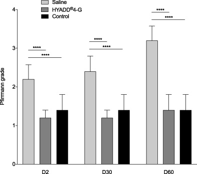
Changes in Pfirrmann grade after treatment. The grades of degeneration in the saline group were significantly higher than those of the HYADD®-4G treated group or control group at each time point (∗∗∗∗ p < 0.0001). Values are shown as mean ± standard error of the mean.
Pfirrmann grading results
Pfirrmann grading results at day 2, 30 and 60 after treatment for L2/L3 (saline), L4/L5 (HYADD®4-G) and L5/L6 (control) IVDs. For each grade, the number of classified discs of the corresponding group is given, followed by the overall percentage in brackets.
To assess IDD with regard to tissue and cell morphology, IVDs were graded histologically. HYADD®4-G-treated, degenerated discs had significantly lower IDD scores than saline-treated IVDs (p < 0.01) (Fig. 6).

Histological grading scores. The grading scores and thus the severity of IDD were significantly higher for the saline group than for the HYADD®-4G treated group (∗∗∗ p < 0.001, ∗∗ p < 0.01). Values are shown as mean ± standard error of the mean.
Saline-treated L2/L3 discs showed marked signs of degeneration. MT staining allowed visualization of the NP-annulus fibrosus (AF) boundary, which was better preserved in HYADD®4-G-treated (Figs 7e and 8b) than saline-treated IVDs (Figs 7b and 8a).
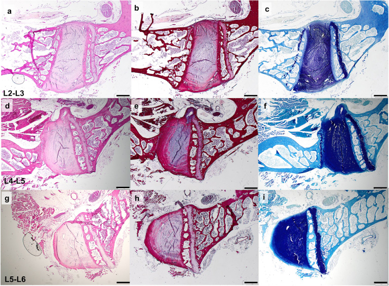
Representative histological H&E (A, D, G), MT (B, E, H) and toluidine blue (C, F, I) staining of non-injected control (G, H, I), saline injected (A, B, C) or HYADD®4-G treated (D, E, F) IVDs of the same animal. Scale bars: 1 mm.
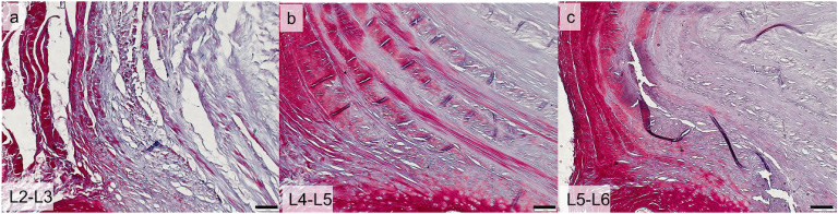
Representative MT stainings showing the NP-AF border in saline treated L2/L3 (A), HYADD®4-G treated L4/L5 (B) and control L5/L6 IVDs. HYADD®4-G injected discs showed a better NP-AF delimitation than saline injected IVDs. Scale bars: 100 μm.
Toluidine blue stains proteoglycans. Saline-treated L2/L3 NPs were less stained (Fig. 7c) (seen as lighter blue), compared to HYADD®4-G-injected or control NPs (Fig. 7f and i).
In order to assess the safety of HYADD®4-G treatment, sections of non-punctured but HYADD®4-G-injected IVDs of group 2 were analyzed for inflammatory signs. None of the IVDs showed any signs of inflammation or foreign body reaction (Fig. 9).

Representative histological H&E (A, D, G), MT (B, E, H) and toluidine blue (C, F, I) staining of non-punctured HYADD®4-G-injected IVDs of the same animal. No signs of inflammation were seen. On the L3/L4 IVD (D, E and F) an osteophyte is visible. Scale bars: 1 mm.
This study aimed to evaluate both the safety and the efficacy of HYADD®4-G injections into degenerated IVDs in the well-established rabbit annular needle-puncture model. A single intra-discal HYADD®4-G injection was safe and effective in restoring DHI, while preserving IVD morphology.
The nature of human disc degeneration is complex and no animal model perfectly reflects it [38–40]. Several animal IDD-models of induced or spontaneous disc degeneration have been used, each presenting advantages and limitations [38–40]. Using rodents remains a technical challenge due to their small size. Further, NP progenitor cells persist in the notochord of adult rodents and pigs but disappear in human [40,42]. The rabbit annular puncture model is a compromise regarding anatomy, composition and costs. This well-established model creates mild and progressive IDD [12,34,41,42]. The time point of 90 days after IDD induction for assessing disc degeneration in rabbits is comparable to the time lines of other studies [42–45].
The vast majority of animal models of intervertebral disc diseases are in quadrupeds, the only bipedal models available usually present ethical dilemmas that preclude their usage in most Institutions. Given that the mechanical loading to which human intervertebral lumbar discs are exposed is significantly influenced by the upright posture, it may be common sense to believe that quadruped animal models would be poorly representative of the real situation in humans. Posture discrepancies notwithstanding, it is an admitted fact that muscle contraction and ligament tension are significant contributors to the load to which intervertebral discs are exposed specially in quadruped animals [46]. In fact, due to the increased complexity in stabilizing a horizontally aligned spine vs. a vertically almost spontaneously and perfectly balanced spine, it has been speculated that the load exerted by these structures may be even greater than that in humans resulting from the bipedal stance [47].
This holds true regardless of the overall size of the quadruped animal considered for a model since the ratio between animal weight and disc surface is kept constant across the range of species available as study models [39]. These results fully requalify quadruped models as suitable choices for extrapolating observations to the bipedal species.
Since HA is predominant in the NP matrix, it seemed appropriate to evaluate HA derivatives for IDD repair. HA is responsible for IVD hydration, viscoelasticity and other mechanical properties [29,48]. Moreover, HA has signaling character and substantially interacts with cellular components and molecules, including the epigenetic machinery to regulate gene expression [16,22,49–54]. HA has shown beneficial effects in several experimental models by slowing down IDD. Pfeiffer et al. [29] demonstrated that HA intra-discal injections reduced IDD progression in a non-human primate nucleotomy model. Similar results were seen in a porcine model of IDD [28]. Nakashima et al. [55] demonstrated that cross-linked HA and cross-linked chondroitin sulfate hydrogels regenerated IVDs in a rabbit model, evidenced by MRI and histology. In addition, two HA derivative products analogous to HYADD®4-G (HYADD3® and HYAFF120®) showed biological and rheological behaviour similar to native NP tissue and prevented IVD changes following nucleotomy in a porcine model [20,56]. The authors also showed that HYADD3® was a suitable support for bone marrow stem cells and HYAFF120® could naturally be infiltrated by functional disc chondrocytes. The advantage in using HYADD®4-G compared to HYAFF120® is the very low-frequenced chemical modification of HA, that allows retaining more of its native biological properties. Moreover, in comparison to HYADD3® [56], this study has demonstrated the efficacy of HYADD®4-G in IDD treatment without an association to bone marrow stem cells.
In this study, annular puncture successfully induced IDD within 30 days, as previously reported [42]. Degeneration was characterized by disc narrowing, NP dehydration and increased Pfirrmann score. Disc degeneration significantly affected IVD morphology and cellularity. Two days after saline injection into degenerated IVDs, a further disc narrowing was observed. Probably, the injection caused a second puncture and thus provoked further degeneration. In contrast, HYADD®4-G prevented further narrowing and increased DHI. Indeed, HYADD®4-G-treated discs showed an increase in DHI that can be explained by an immediate disc sustaining effect of the gelatinous material that is less prone to pouring out than saline solution.
MRI evaluation included a qualitative analysis through Pfirrmann scoring and a T2 relaxation time quantification. Pfirrmann MR grading system is widely used to classify disc degeneration, evaluating disc morphology and signal intensity on MR T2-weighted images. The signal intensity on T2-weighted images can reflect biochemical as well as structural changes during IDD. Both MRI analyses revealed that HYADD®4-G allowed a significant restoration of disc hydration and structure. Pfirrmann grading showed that HYADD®4-G but not saline preserved discs from further degeneration.
Histopathology demonstrated that IVD tissue was better organized after HYADD®4-G treatment compared to saline. Further, toluidine blue staining confirmed re-hydration in HYADD®4-G- but not saline-injected discs. This was probably due to an increased proteoglycan content, eventually triggered by HYADD®4-G administration since HA has shown a dose dependent positive effect on glycosaminoglycan and aggrecan production [57].
As seen through the evaluation of inflammation or foreign body reaction in 9 IVDs from 3 non-punctured animals, a single intra-discal HYADD®4-G injection should be safe.
In the present study, adjacent vertebra of one HYADD®4-G treated and one saline treated IVD in two different animals developed osteophytes (data not shown). This is likely due to osseous injury during puncture and/or injection and is not believed to be a side-effect of the injected material. Different groups have reported systematic osteophyte formation near punctured rabbit IVDs [41,42,58]. In this study, osteophyte formation was rather weak. Interestingly, this would confirm a tendency of reduced osteophyte formation in a HYADD®4-G treated mouse OA model [59]. However, we cannot rule out that saline and/or HYADD®4-G had some influence on osteophyte formation.
Possible mechanisms responsible for the beneficial effects of HYADD®4-G are related to biochemical and physical properties of HA. In IDD glycosaminoglycan and water contents of IVDs decrease, resulting in fibrotic changes. Possibly, an increased loading stress in the disc, obtained by HYADD®4-G injection [27], prevented further degeneration and could have triggered tissue repair programs. It is known from previous studies that HA stimulates ECM biosynthesis, cell proliferation, cell migration and phenotype maintenance of chondrocytes and NP cells [60,61]. The beneficial effects of HA administration in restoring proteoglycan aggregation has also been demonstrated in non-human primate lumbar spines [29] where this accumulation was crucial for water retention in the NP [62].
Long-term studies in larger animals with biomechanical tests are recommended, particularly to determine the effect duration after a single intra-discal injection. The demonstrated efficacy of HYADD®4-G in this study is interesting, considering that HYADD®4-G is already safely used intra articularly in patients with degenerative joint disease.
This pilot study in a rabbit IDD-model showed that HYADD®4-G has high therapeutic potential. IDD was attenuated through restoration of DHI, hydration and normal tissue organization, however the use of a quadruped species puts some limitations on the transferability of the results to the human situation.
Footnotes
Acknowledgements
We thank Prof. Lawrence Bonassar for his help.