
Abstract
BACKGROUND:
A burn wound is one of the most frequent and devastating injuries for patients which requires extensive care. Early treatment of burn wounds improves healing significantly.
OBJECTIVE:
This study was designed to investigate the efficacy of amnion and collagen-based hydrogels on cutaneous burn wound healing in rats with covering membrane.
METHODS:
We prepared a novel cell free hydrogel comprising human amnion, rabbit collagen, carboxymethyl cellulose sodium salt, citric acid, methyl paraben, propyl paraben, glycerin and triethanol amine. The wound covering membrane was developed from rabbit collagen and prawn shell chitosan. Beside swelling ratio, water absorption, equilibrium water content, gel fraction and spreadability analysis, in vitro cytotoxicity and biocompatibility tests were performed for the formulated hydrogels. Following the skin irritation study, second-degree burns were created on the dorsal region of the rats and the gels were applied with/without covering membrane to study the wound contraction and re-epithelialization period.
RESULTS:
The formulated hydrogels were observed non-cytotoxic and compatible with human blood cells. No erythema and edema were found in skin irritation assay confirming the safety and applicability. Hydrogel consisting in a combination of amnion and collagen demonstrated significantly rapid wound healing, driven by complete re-epithelialization (16.75 ± 0.96 days) and closure by wound contraction (72 ± 3.27%, P < 0.0000009) when wound dressing membrane was used, whereas this gel alone healed about 62.5 ± 4.43% (P < 0.00001) and required 18.75 ± 0.50 days to complete re-epithelialization. Additionally, the gel with covering membrane treated group had maximum average body weight, food and water intake.
CONCLUSION:
The amnion and collagen-based blended gel offers alternative possibilities to treat skin wounds when covered with film, which could overcome the limitations associated with modern therapeutic products such as high costs, long manufacturing times, complexities, storing, and presence of living biomaterials.
Introduction
Burn injury is considered one of the most common and devastating medical burdens worldwide since the ancient period of human history. Globally, 265,000 individuals die every year because of burns [1]. The vast majority of the burn incidents occur in low- and middle-income countries and almost half happens in the South-East Asia region. For instance, according to WHO, nearly 173,000 children are moderately or severely burned every year in Bangladesh [2]. The standard burn wound treatment, currently clinically practised, is the engrafting of an autologous split thickness skin graft and amniotic mebrane allograft which results in a clearly advantageous outcome [3–5]. However, the limited availability of healthy donor skin, amniotic graft, high cost, complex manufacturing, and complex storage are the difficulties which hinder the incorporation of these grafts into routine clinical use. In addition, the requirement of tissue banking facilities, patients’ need to come to the clinic, and lack of expertise of the medical personnel in graft transplantation are the limitations. Furthermore, human amniotic membrane dressings perform better if replaced every other day [6]. Despite recent tremendous advances in wound care products such as stem cells, Integra, AlloDerm and Biobrane, recombinant growth factors and cellular tissue-engineered skin substitutes-based wound treatments [7–9], these commercial advanced treatments are associated with high costs for patients in low-income countries. Traditional therapies based on discarded biomaterials such as amniotic membrane extracts, collagen, chitosan, blended hydrogel, are considered to be an interesting and promising alternative [10–16].
Amnion is widely used as a burn wound healing material and known as an ideal biological burn wound dressing material [17]. It acts as a scaffold for proliferation and differentiation of native skin cells due to the presence of fibronectin, elastin, nidogen, collagen types I, III, IV, V and VI and hyaluronic acid which finally lead to the promotion of neo-epithelialization [18]. The structural protein-collagen is involved in all three phases of the wound-healing cascade [19]. Collagen stimulates cellular migration and contributes to form new healthy granulation tissue very quickly over the burn, helping to heal rapidly. Collagen-based biomaterials have been reported to stimulate and recruit specific cells, such as macrophages and fibroblasts, along the healing cascade to enhance and influence wound healing [19]. A cationic biopolymer chitosan was reported to disrupt the outer membrane of bacteria and hamper the growth of infectious substances [20] which is a beneficial biological property for wound healing by protecting the wound from microbial infection. These chitosan and collagen are eco-friendly biomaterials as they do not produce any harmful residues. These biomaterials can provide moisture or absorption, depending on the mode of preparation and delivery system [13]. In developing countries, people would like to treat burn injuries at home unless the complications increase to a certain level. However, patients respond best when rapid treatments are available for fast closureing and protecting wounds. There is a need of a low cost and effective fast aid product to manage burn injuries [4] that is easy to produce and store, is physiologically effective and its application does not require medication. The limitations related to modern therapeutics, increasing numbers of burn injuries, and the advantages of naturally available biomaterials rationalize to formulate the hydrogels consisting of amnion and collagen. Here, we have demonstrated the usefulness of amnion and collagen blended hydrogel which can significantly accelerate wound closure through contraction and re-epithelialization in an animal model for skin burns in the presence of biomembrane.
Materials and methods
Preparation of amnion powder
Amniotic sacs were collected from a sero-negative donor (HIV, Syphilis, Hepatitis B and C viruses) after caesarean deliveries with the written consent of the donor. After collection, the amnion extracts were prepared as described previously [12]. In short, the amnion membranes were carefully separated from chorionic by forceps and the separated amnion membranes were washed with PBS containing streptomycin and penicillin. After washing, the membranes were freeze dried at −55 °C overnight. The freeze dried membranes were then minced into small pieces and sealed into plastic bags for radiation sterilization at 10kGy using cobalt 60𝛾 source. The small pieces of membrane were then aseptically processed into powder form which was later used for gel preparation.
Isolation of collagen from rabbit skin
Rabbit skin collagen was isolated according to the method as described before [21]. Pretreated rabbit skins were cut into small pieces (approx. 1 cm2 each) and suspended in 0.5 M acetic acid solution at a solid-to-solvent ratio of 1:10 (w/v). After shaking at 200 rpm for 24 hrs at RT, the sample was filtered and the liquid soluble portion was called acetic acid soluble collagen. The non-dissolved residue was used for pepsin soluble collagen extraction as follows: the residues were re-suspended in a new solution of 0.5 M acetic acid at a concentration of 1:10 (w/v) and pepsin at a concentration of 2 mg/ml. All samples were filtered and stored at 4 °C at the end of every single extraction. Both collagens were precipitated with a solution of 2.6 M NaCl. After being centrifuged at 10,000 rpm for 30 min, the samples were re-suspended in five volumes of 0.5 M acetic acid and were dialysed for 48 hrs in DW and dialysis membrane tubing with 6–8 kDa Mw cut-off. Finally, the resulted dialyzed collagen was freeze dried at −55 °C for further use.
Extraction of chitosan from shrimp shells
Chitosan was extracted from shrimp shells according to the method established by Khan et al., 2005 [22]. In short, waste prawn shells were washed with hot DW and were dried in an oven at 105 °C for 72 hrs. Dried shells were ground using a blender and deproteinized with 3% NaOH and demineralized with 3% HCl. The deproteinized and demineralized chitin was then neutralized and dried in oven at 105 °C for 24 hrs. Chitosan was obtained by deacetylation of chitin with 50% NaOH where the ratio of chitin: NaOH
Formulation of hydrogel
Amnion and collagen powder was dissolved in 1% citric acid. When these amnion and collagen powders were completely homogenized, other ingredients (i.e. antibacterial ingredients such as methyl paraben and propyl paraben, and moisturizing agent glycerin) were added. Subsequently, triethanolamine was added dropwise to neutralize pH 7. In one glass tube, all the components were added under continuous stirring except carboxymethyl cellulose sodium (CMC-Na). In another tube, different concentrations of gelling agent- CMC-Na were prepared at 60 °C which formed a transparent solution. Finally, both solutions were mixed gently at 40 °C under aseptic condition. The formulated hydrogels (HG) were then placed to cool down to 30 °C. The ingredients and concentrations are listed in Table 1.
The composition of the nine types of formulated gels (HG1-HG9)
The composition of the nine types of formulated gels (HG1-HG9)
Collagen-chitosan blended membranes were prepared using the solvent evaporation method as described by Indrani et al. 2017 [13]. Chitosan was dissolved in 100 mL of 1% acetic acid by stirring at RT for 6h and neutralized with 1 M NaOH. Separately, collagen was dissolved in 0.5 M acetic acid by stirring for 12h at RT. These two polymeric solutions were then mixed in a ratio as listed in Table 2 and stirred for 24–48 h at RT. From the resultant polymer blend 5 mL of each blend was poured into a petri dish and freeze dried. The freeze dried membrane was washed several times with DW, and freeze dried again at least for 12 h. Finally, the dried membranes were subjected to gamma-ray irradiation.
The concentration of collagen and chitosan for developing the blended biomembranes
The concentration of collagen and chitosan for developing the blended biomembranes
Measurements of the swelling capacity of the hydrogels were performed as described by Abdallah, 2019 [23]. The swelling ratio was calculated according to the following equation: Swelling ratio = [Ws − Wd]/Wd, where Ws is the weight of sweollen gel and Wd is the weight of the dry gel.
The percentage of the water absorption was estimated according to the methods of Bhuiyan et al., 2015 [24]. Water absorption % was calculated according to the following equation: Water absorption [%] = [Wt − Wi] × 100, where Wt is the weight of swollen gel sample at time ‘t’ and Wi is the initial weight of dry gel samples.
Determination of gel fraction and equilibrium water content
The percentage of the gel fraction was determined according to the method as described previously [25]. The gel fractions of the samples were calculated gravimetrically according to the following equation: Gel fraction (%) = [Wd/Wi] × 100, where Wd is the weight of dried gel after extraction and Wi is the initial weight of dried gel.
Equilibrium water content (EWC) was estimated as described by Kim et al., 2003 [26]. EWC was calculated using the following formula: EWC [%] = [(Weq − Wi)/Weq] × 100. Where, Weq is the weight of swollen gel at equilibrium and Wi is the initial weight of dry gel.
Spreadability assesment
Spreadability assesment was performed according to method of Abdul et al., 2013 [27]. In short, 1 gm of gel was placed within a circle of 2.3 cm diameter pre-marked on a glass plate of 20 × 20 cm, over which a second glass plate was placed. A weight of 500 gm in the form of water-filled beaker was allowed to rest on the upper glass plate for 5 min. The increase in the diameter due to gel spreading was noted.
In vitro cytotoxicity and red blood cell compatibility test
In vitro cytotoxicity test was performed on brine shrimp as described by Bundela and Bajpai, 2008 [28]. The final gel samples were dissolved in artificial seawater at different concentrations and the active nauplii were inoculated. After overnight incubation, the nauplii were counted. Here, 0.5 mg/ml of Vincristine PCH (Pharmachemie BV, the Netherlands) was used as positive control and sea water was served as negative control.
Heparinized human blood was used to test the biocompatibility of the selected gel samples as described by Bhowmik et al., 2017 [29]. Tested samples were prepared using blood and selected gel at 3:1 ratio. Blood sample of the same donor was also diluted at the same ratio with distilled water and normal saline which served as positive and negative control, respectively. After mixing, the mixtures were kept in an incubator for 2 h at 37 °C. The samples were then spread on glass slides and observed under an inverted microscope (20× magnification).
Skin irritation studies
To assess in vivo irritability and applicability of the gels, the dorsal skin-hairs of the selected female Wistar rats were shaved on the date of experiment [30]. In total eight animals were examined and were randomly assigned to four groups (i. amnion gel, ii. gel consisting of amnion and collagen, iii. without dressing membrane, and iv. collagen gel). The animals were treated with 1 ml gel daily up to four days and finally the treated skin was visually examined for erythema and edema.
Artificial burn induction, wound contraction and re-epithelialization
In total, 24 healthy female Wistar rats of 100–150 g body weight were randomly assigned into six experimental groups: i. 1% silver sulfadiazine (Burnsil, Beximco Pharmaceuticals Ltd, Bangladesh) as positive control, ii. without gel served as negative control, iii. treated with collagen gel, iv. treated with amnion gel, v. treated with amnion and collagen blended gel with and vi. without covering membrane. Each rat was anesthetized with Ketamine HCl solution 100 mg/kg (Gonoshasthaya Pharmaceuticals Ltd, Bangladesh) body weight by intraperitonial injection. Subsequently, the hair at dorsal region was trimmed and properly shaved with a sharp blade and cleansed with an alcohol swab.
Burns were created using a piece of aluminum (2.5 cm diameter) heated to 100 °C for 5 min. The hot aluminum was gently applied for 15 seconds on the shaved area [31]. The animals were treated with 1 ml of gels on a daily basis, topically, for a period of 16 days. Gross changes in the wounds were evaluated from day 0 to 16 post burn. Images were taken regularly using a Canon IXUS 130 camera, and the morphological evaluation such as appearance of the wound was recorded. Re-epithelization was monitored by recording the number of days required for crust to fall away, leaving no raw wound behind [12]. To monitor wound contraction, progressive changes in the wound area were measured. The wound margin was traced at 4 days intervals by millimeter measuring scale and measurements were continued up to 16 days. After every four days, the healed area was calculated by subtracting initial wound area to the unhealed area [11]. Using the formula below, the percentage of wound contraction was calculated on the respective day. The percentage of wound contraction was calculated taking the initial area of wound (625 mm2) as 100%.
Statistical analysis
All statistical analyses were calculated by one way independent test using SPSS version 22.0 (SPSS Inc., Chicago, IL, USA) on a Microsoft Excel 2010 platform. All quantitative data presented in this study include mean (±) standard deviations (SD). P < 0.05 was considered statistically significant.
Results
General observations of the formulated hydrogels and biomembrane
Full-term human placentas were collected during cesarean sections. The erythrocytes were depleted and the amnion was separated from chorion membrane for further processing. Afterwards, amniotic membranes were lyophilized, freeze dried, blended and finally gamma irradiated to obtain powder and make amnion solution (Fig. 1A). Collagen was isolated from rabbit skin and freeze dried to extract powder for further use (Fig. 1B). With the addition of several ingradients including distinct concentrations of amnion solutions, collagen solutions, and CMC-Na solutions, in total nine types of hydrogels were prepared (the composition of hydrogels is shown in Table 1). The gels were presereved in frozen conditions (Fig. 1C). All of the formulated hydrogels were found to be semi-solid and homogeneous in nature with good consistency (Fig. 1D). The pH of all nine gel formulations was measured and ranged from 6.56 to 7.3. The color of gels containing only amnion extracts were off-white while the gels containing both amnion and collagen were light white. In case of gel containing collagen alone, the color was found to be deep white (Fig. 1E). However, all gels formulations were semi-transparent, viscous and flexible visually (Fig. 1F). The chitosan-collagen blended biomembrane were prepared from the rabbit skin derived collagen and prawn shell derived chitosan (Fig. 1G–J). We observed that a mixture of collagen concentration (10 mg/ml) and chitosan concentration (10 mg/ml) formed a transparent biomembrane (Fig. 1K).
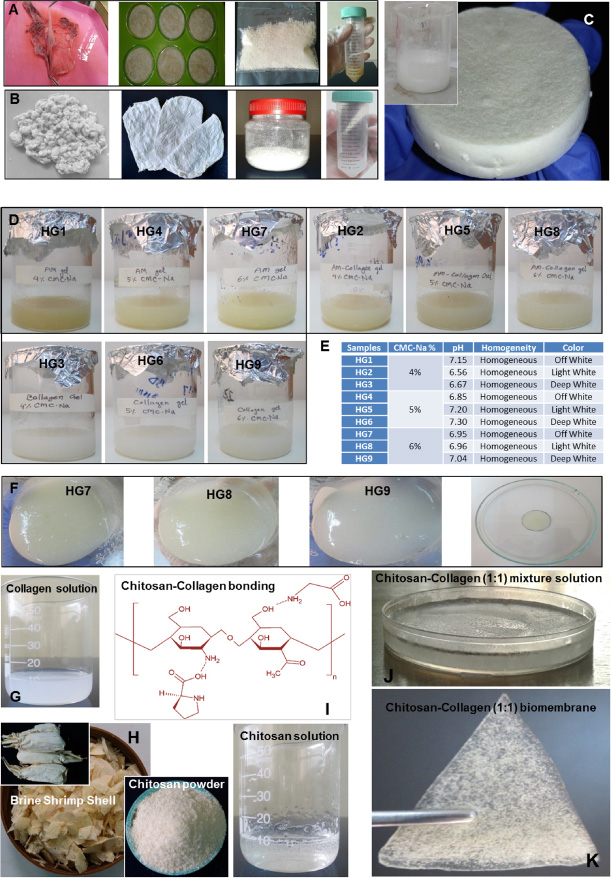
Preparation amnion, collagen and chitosan; and the physical appearance of the formulated hydrogels and biomembrane. (A) Seperation of amnion from chorion. The dried lyophilized amniotic membrane was 10 kGy gamma irradiated and amnion solution. (B) Rabbit skin derived freeze dried powder was used to formulate collagen solution. (C) Representative picture of frozen hydrogel. (D) Physical appearance of all nine types of the gels. (E) pH, color and homogenecities of the formulated gels. (F) Six percent CMC-Na hydrogels appeared as semi-solid like flexible lubricants, viscus, and semitransparent. Gel sample placed within circle of petridish. (G) The preparation of chitosan and collagen blended bio-polymer, bonding between chitosan and collagen, membrane solution before freeze drying and the representative image of prepared biomembrane sheet.
One of the crucial ingredients of the formulation of hydrogel is the gelling agent. Different percentages of gel formulations were tried in order to select the best gelling agent. Gels containing 4.0% of Na-CMC formed a very thin gel. With 5.0% gelling agent somewhat better gel was obtained but gel containing 6.0% of Na-CMC formed uniform and smooth gel. Thus, 6.0% of Na-CMC was selected as the optimized concentration of gelling agent. However, we have prepared nine types (HG1–HG9) of hydrogel gels contained 1–2% of amnion powder, collagen powder and amnion-collagen together with 4–6% of Na-CMC (Table 1). The swelling ratio of the gels ranged from ∼1.26 to ∼2.59 (Fig. 2A). The water absorption and equilibrium water content of these formulated gels were observed to increase from ∼19.23% to ∼25.85% and ∼55.7% to ∼72.1%, respectively with the incraesing percentage of Na-CMC (Fig. 2B, 2C). The gel fraction of hydrogel was denoted to vary from ∼70.83% to ∼87.30% (Fig. 2D). The value of gel spreadability of the formulated gels was measured which ranged from ∼3.9 to ∼6.3 cm (Fig. 2E). Spreadability of 6% gels was found lower because high concentration gels tend to be thicker and spread less. Although three distinct concentrations of CMC-Na were added to form the hydrogels, but 6% of CMC-Na gels possed better physical properties in terms of swelling ratio, water absorption, equilibrium water content, gel fraction and spreadability. The chitosan-collagen blended biomembrane (Table 2) were evaluated based on physical properties such as swelling ratio, water absorption (%) and equilibrium water content (%). We observed that the membrane (BM1) composed of the equal concentrations of chitosan (10 mg/ml) and collagen (10 mg/ml) possed highest water absorption, equilibrium water content and swelling ratio whereas BM8 (Collagen 16 mg/ml: Chotosan 4 mg/ml) were lowest (Fig. 2F, 2G, 2H).

Physical properties of the hydrogels and biomembranes in terms of swelling ratio, water absorption, equilibrium water content, gel fraction and spreadability. Distinct hydrogel samples at different CMC-Na concentrations (A) swelling ratio, (B) water absorption (%), (C) equilibrium water content (%), (D) gel fraction (%), and (E) spreadability in cm. Fabricated biomembrane samples (F) water absorption (%), (G) equilibrium water content (%) and (H) swelling ratio at various chitosan: collagen ratio.
Upon incubation of human blood with gel samples in vitro, less than 2% hemolysis was observed and the red blood cells remained intact in 3:1 ratio which indicated biocompatibility of the gel samples. On the other hand, cell damage observed when distilled water (positive control) incubated with the same ratio (Fig. 3A). The brine shrimp lethality assay was performed to observe the in vitro cytotoxic effect of the final formulated gel samples. We did not observ any death of the nauplii, not even at the higher doses of gel samples apart from amnion-collagen blended hydrogels and amnion-collagen blended gel in presence of biomembrane. Similar results were found in the case of saline water incubation (negative control). However, in these cases, no death of the nauplii was noticed when the doses limited to 0.25 and 0.75 mg/ml. At a concentration of 1 mg/ml about 15% deaths of the nauplii were observed (Fig. 3B). To analyze the applicability of the prepared gel, skin irritation assay were performed in vivo using a rat model. After topical application of all nine types of hydrogels for a period of 48 hrs, it was observed that the gel did not induce any edema or erythema (Fig. 3C). To test the biotic and abiotic stress tolerability, at least for few days, we expose the experimental gel and membrane samples to normal environmental conditions in a room. We did not observed any shrinkage, deterioration or any macroscopically visible microbial growth on it (Fig. 3D). This result indicated the safety of gels to be applied topically. We also observed that the hair formation was normal compared to non-treated rats.
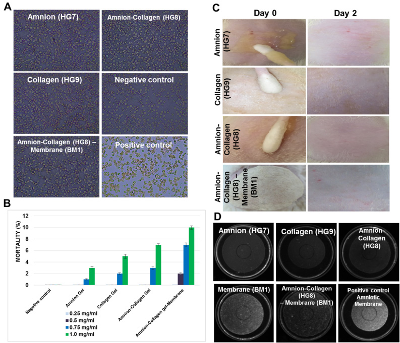
Biocompatibity, cytoxicity and irritability analyis. (A) Human blood biocompatibility were shown by RBC morphology of heparinized blood incubated with distinct hydrogels with 6% CMC-Na and biomembrane. Microscopic view of blood cells at 20× magnification. (B) Brine shrimp lethality bioassay shows the mortality percentages of brine shrimp. (C) Topical application of the gels for in vivo skin irritation assay on shaved rats. (D) To observed the samples self-deterioration when expose to environmental stress for 4 days. Amniotic membrane graft served as positive control.
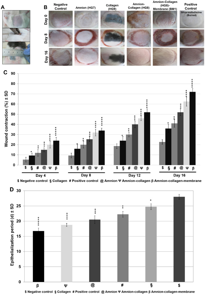
Initially on day 0, using a 2.5 cm diameter piece of aluminum, a second-degree burn was created in all experimental anesthetized rats (Fig. 4A) which produced a painful injury, with no bubbles, white color and subsequently hyperemia occurred into the damaged tissue area. After 30 minutes of burn injury, respective gel was applied in each group of rats except negative control. On day 4, negative control and collagen group showed wet crust where amnion, amnion-collagen blended with/without biomembrane and positive control group showed slight dry and thick dry crust, respectively. On day 8, negative control and collagen group were observed with less detachment of crust edge rather than all treated rat groups. On day 12, scar tissue and contraction became clearly visible in each group. On day 16, crusts were all disappeared and re-epithelialization was completed in amnion-collagen blended with/without biomembrane group showed better healing rate with no scar tissue while other groups showed moderate healing with scar tissue and a minor wound (Fig. 4B).
For caption see next page.
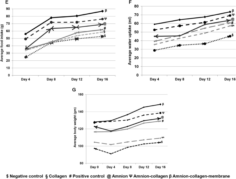
Macroscopic evaluation of wound closure and quantitative measurement of wound contraction, re-epithelialization and behaviour. (A) Burn induction process and initial wound area measurement. (B) Macroscopic wound healing representative images of Wistar rats on day 0, 8, and 16 which were treated with amnion, collagen, amnion-collagen blened gel with or without covering films, and controls. (C) Clinical evaluation of wound healing regarding wound contraction in rats at day 4, 8, 12, and 16. (D) Complete re-epithelialization period required for different groups of treated animals. ∗indicates the level of significance against negative control group. The changes in behaviours factors such as average (E) food intake, (F) water uptake and (G) body weight over the courses of wound healing treatment.
The rate of wound contractions varied from group to group. From the begining of topical gel treatment, for instance on day 4, amnion-collagen blended treated group was noticed with maximum healing rate in presence of biomembrane, which was four folds to control group and statistically most significant (P < 0.0002). However, amnion-collagen in absence of biomembrane and amnion gel treated groups were also statistically significant (amnion-collagen P < 0.0004, amnion P < 0.0006). After 8 days of post burning, amnion-collagen blended gel when covered with biomembrane contracted about 34 ± 2.31% (P < 0.000006). Negative control group had least wound contraction rate of 9.33 ± 2.31% while amnion and amnion-collagen without biomembrane treated groups were 25.33 ± 2.31% (P < 0.0007) and 32 ± 3.27% (P < 0.00007), respectively. Similar pattern of wound contraction were visualized also on day 12 and 16. On 12th day of treatment, amnion-collagen blended gel with biomembrane group the contraction were reached to 52 ± 3.27% (P < 0.000003). Healing rate of amnion-collagen and amnion treated groups were 46 ± 2.31% (P < 0.000007) and 40 ± 3.27% (P < 0.00007), respectively. On day 16, amnion-collagen blended gel including biomembrane treated group showed superiority 72 ± 3.27% (P < 0.0000009) while negative control group healed only 22.67 ± 2.31%. Amnion-collagen, amnion, positive control and collagen treated groups were healed 62.5 ± 4.43% (P < 0.00001), 52 ± 3.27% (P < 0.00004), 41 ± 3.83% (P < 0.0004) and 36 ± 4% (P < 0.001), respectively (Fig. 4C).
In cases of average re-epithelialization period in all experimented groups, biomembrane covered amnion-collagen blended gel treated group required only about 16 days while amnion collagen, amnion, positive control, collagen and negative control groups needed 19, 20, 22, 24 and 28 days, respectively. The amnion-collagen in presence of biomembrane (P < 0.000002) and amnion-collagen without membrane (P < 0.000007) treated groups were statistically more significant than amnion treated group (P < 0.0001) and positive control group (P < 0.0002) (Fig. 4D). During wound healing process the behaviours factors such as body weight, water and food intake have been influced. We observed that body weight gaining was highest when applying the amnion-collagen blended gel with dressing membrane. For the cases of food and water intake, we also noticed better food and water intake in amnion-collagen blended gel when covered by membrane (Fig. 4E–G).
Traditional therapies based on discarded biomaterials are interesting alternatives for dressing burn and skin wounds, which improves healing, is readily available, easily applicable, economical, prevents infection and desiccation, and facilitates healing [32]. Previously, we studied burn wound healing gel products consisting of a combination of amnion and Aloe vera extract [12]. The application of collagen over the injured burn improved the healthy granulation tissue which enabled rapid healing [33,34]. Here, we described that a gel consisting of a combination of amnion and collagen extract has the potential as a burn wound healing product in hydrogel format which is easy to produce and apply. Again, chitosan has been reported as an antimicrobial biomaterial which could prevent penetration of bacteria into the wound [35]. The blend of chitosan and collagen could form a membrane which prevents bacterial infection and maintains the moister around the wound as well [13,36]. So, we believe that covering the wounded area with the chitosan-collagen biomembrane onto the amnion-collagen hydrogel may speed up the healing process by serving as a synergistic function and protecting the wound area from the external environment.
One of the crucial ingredients of the formulation of hydrogel is the gelling agent. Thus 6% Na-CMC was selected as the optimized concentration of gelling agent. Another study also showed that gel containing 6.0% of Na-CMC formulated uniform gel that was effective for wound healing [12]. Considering all the formulated gel types, the gels consisting of amnion and collagen in CMC-Na (6%) has been observed with promising physical properties in most instances. These three formulations were found to be homogeneous, viscous and flexible in nature with good consistency. Additionally, lower values of spreadability indicating that they spread less and possed the tendency to stick to the wound area over a longer period of time which is eventually expected for topical application [37,38]. The spreadibility could easily be increased by a small amount of shear [39]. Furthermore, we experienced that it was hard to maintain the gel texture above 6% of CMC-Na. the outer surface of our body acts as a defense barrier where pH level increase from epidermis to dermis requires to formulate a gel with a pH range of 6.5--7.3 which is desirable for the skin [38]. The pH of the finally selected formulations (n = 3) was found to be between 6.95 and 7.04 which implies that gels were suitable for skin wound healing therapy and the formulation can thus be used without the risk of irritation to the skin.
Regarding the fabricated biomembrane, the equal concentrations of chitosan (10 mg/ml) and collagen (10 mg/ml) could formed a dressing film which possess better biophysical properties as a protective barrier [13]. The biocompatibility, safety and applicability of the blended gel and biomembrane were confirmed by in vitro cytotoxicity, human red blood cell compatibility, and skin irraitation assay [12]. In vivo no nauplii death was observed in presence of gel samples at lower concentrations. However, with increasing concentrations the lethality of the gels attributed due to formation of viscous layer on the gills of nauplii and the reduction of dissolved oxygen in water as the high amount of the gel samples led to high viscosity [40]. In vitro RBC did not undergo lysis or coagulation in contact with gels indicating its biocompatibility and ansence of osmotic shock [12,40]. After applying the amnion-collagen gels with and without covering membrane on a shaved rat, no edema or erythemas were found [12].
Wound contraction and re-epithelialization are an integral part of the healing process, which closes wounds and keeps it safe from the external environment and shrinkage of the wounded area by repairing the tissues [41]. However, we observed that the rate of wound contraction and re-epithelialization varied from group to group but maintained a similar pattern. The wound contraction on day 16 for the amnion-collagen group with biomembrane showed a statistically more significant (72 ± 3.27% with P < 0.0000009) contarction rate in comparison to the other groups. Furthermore, the amnion-collagen treated group showed faster re-epithelialization (16.75 ± 0.96 days) in comparison to other rat groups when the wound was covered by membrane. Our results indicate that the combination of amnion and collagen imparted exclusive wound healing properties of both biomaterials [10,11,42], and the addition of membrane top served as an excellent wound covering material which protected the wound area from external stress and maintained moisture as well when the membrane was composed of chitosan and collagen [43]. We did not notice any negative effects on rats’ behaviour during the treatment period. The amnion-collagen blended gels with or without dressing membrane rats group were observed with significant average body weight, food intake and water uptake. The protective barrier of biomembrane [13] in combination with hydrogels may help the rats to be healthier and maintain their body weight and eating behaviour. We did not add any growth factors or cells to the gels and membranes. The addition of skin progenitor cells could significantly increase the healing rate. For optimal wound healing, a combination of skin stem cells and biomaterials therefore need to be tested.
Conclusion
All biomaterials used in this study were obtained from naturally available bio-waste. The procedures used for the preparation of hydrogels and biomembrane are biologically safe and economically desirable. For instance, we have extracted amnion from C-section derived placenta, collagen from slaughtered rabbit skin, and chitosan from prawn shell. We developed a biocompatible wound healing gel with a combination of amnion and collagen which significantly enhances the efficacy of burn wound healing in the presence of chitosan-collagen wound dressing biomembrane. However, further investigation is required to study the underlying molecular mechanisms of wound healing, in particular which factors are responsible for the healing.
Footnotes
Acknowledgements
The authors acknowledge the technical support and animal facilities from the Institute of Tissue Banking and Biomaterial Research, Atomic Energy Research Establishment, Bangladesh. SM Asaduzzaman acknowledges the support from the Government of Bangladesh, and the International Atomic Energy Agency, Austria. In addition, Md Shaifur Rahman acknowledges support from the German Academic Exchange Service (DAAD-91607303).
Conflict of interest
None to report.
Contribution
MMR and MSR conceived the idea and designed the experiments. MMR and MAU performed the experiment. MMR, MSR and MAU analysed the data. MSR and MMR wrote the manuscript. AS and MLH provided and nurished the animals. MSA, MZH and SMA supervised the works and finally edited the manuscrpt. All authors approved the final version.