
Abstract
BACKGROUND:
Human induced pluripotent stem cell (hiPSC)-derived hepatocytes are an attractive alternative cell source to primary human hepatocytes for tissue regeneration.
OBJECTIVES:
This study presents an application of lactose-silk fibroin conjugates (Lac-CY-SF) bearing 𝛽-galactose residues as a substrate for culture of hiPSC-derived hepatocytes. A comparison of hiPSC-derived hepatocytes cultured on three different substrates; Lac-CY-SF conjugates, Matrigel and type I collagen was performed.
METHODS:
Cell morphology, viability, maturation and albumin secretory function were assessed by phase-contrast microscopy, tetrazolium-based colorimetric assay, immunofluorescence staining and enzyme-linked immunosorbent assay.
RESULTS:
Morphological characteristics of the cells cultured on the conjugates resembled those on Matrigel throughout the 6-day culture period. The number of viable cells cultured on the conjugates was comparable to that on Matrigel at day 2 and 6. The protein expression of mature hepatocyte markers, asialoglycoprotein receptor 1 and albumin, by the cells cultured on the conjugates resembled that by the cells cultured on collagen at day 2 and 6. Albumin secretory function per cell cultured on the conjugates was higher than that on collagen and comparable to that on Matrigel.
CONCLUSIONS:
These limited results suggest that Lac-CY-SF conjugates may be as useful as Matrigel and collagen for cultivation of hiPSC-derived hepatocytes.
Introduction
Hepatocytes are the chief functional cells of the liver and perform a number of metabolic, endocrine and secretory functions. Culture of primary human hepatocytes provide useful in vitro models of drug metabolism, protein synthesis and other liver-specific functions. However, primary human hepatocytes in culture have some disadvantages, such as their scare availability, inter-individual variability and rapid de-differentiation [1–3]. Hepatocytes derived from human induced pluripotent stem cells (hiPSC) have recently been developed to overcome these shortcomings [4–6]. hiPSC-derived hepatocytes exhibiting many of the key functional features of primary human hepatocytes are proposed as an attractive alternative to primary human hepatocytes [4–7]. Due to the recent commercialization of hiPSC and hiPSC-derived cells, hiPSC-derived hepatocytes have become widely used for investigation in regenerative medicine, tissue engineering and drug toxicity screening [7].
Since hepatocytes are anchorage-dependent cells, they need to attach to a suitable substrate for growth and survival [3,8]. Various natural extracellular matrices (ECM) such as type I collagen, fibronectin, laminin and Matrigel have been used as a substrate for hepatocyte culture [3,8]. Type I collagen is most commonly used for hepatocyte culture [3]. Matrigel is a high-salt urea extract of Engelbreth-Holm-Swarm mouse sarcoma consisting primarily of laminin and type IV collagen with lesser amounts of fibronectin, entactin and heparan sulfate proteoglycan [3,9,10]. Matrigel has been shown to be one of the most effective culture substrates for the maintenance of highly differentiated hepatocytes [3,10]. Moreover, Matrigel has been used for induction of hepatocytes from hiPSC [5,6]. On the other hand, it is known that hepatocytes bind specifically to 𝛽-galactose residues by the asialoglycoprotein receptor (ASGPR) on their surfaces [8,11,12]. Many studies have shown that galactosylated substrates derived from synthetic polymers and natural polymers such as galactose-carrying polystyrene [8,13] and gelatin [8,14] can promote hepatocyte attachment and functional maintenance. Therefore, galactosylated substrates have been proposed as an alternative to natural ECM in hepatocyte culture. A recent study has found that galactose-carrying polystyrene is useful for differentiation of mouse embryonic stem cells toward hepatocytes and maturation of mouse embryonic stem cell-derived hepatocytes [15].
Silk fibroin (SF) is a natural fibrous protein created by the silkworm. SF fibers have been used as a textile material for thousands of years and as a surgical suture thread for centuries. An aqueous SF solution prepared by dissolving SF fibers in highly concentrated salt solution can be reformed into films, fibers, gels and sponges [16,17]. The reformed SF has been recently exploited as a substrate for cell culture and tissue engineering [16,17]. In our previous studies, SF was galactosylated via covalent conjugation of SF and lactose bearing the 𝛽-galactose residue using cyanuric chloride (CY) as a coupling reagent [18–20]. We showed that rat primary hepatocytes attached well and were maintained on the lactose-SF conjugates (Lac-CY-SF) [18,19]. In the present study, we investigated the ability of Lac-CY-SF to maintain differentiated hiPSC-derived hepatocytes. hiPSC-derived hepatocytes cultured on Lac-CY-SF were characterized by assessment of cell morphology, viability, maturation and functional performance, and compared to the cells cultured on natural ECM, Matrigel and type I collagen.
Materials and methods
Preparation of substrate-coated plates
An aqueous solution of SF was prepared by dissolving degummed silk cocoon fiber in 9M LiBr aqueous solution [21] and used as a starting material for Lac-CY-SF glycoconjugates. Based on our procedure, chemical modification of tyrosine and lysine residues in SF with lactose was carried out using CY as a coupling reagent to obtain Lac-CY-SF (Fig. 1) [18–20]. A 20% weight increase from SF starting material was observed for Lac-CY-SF [18,20]. Lac-CY-SF conjugate-coated plates were prepared as described in our previous paper [18,19]. An aqueous solution of 0.1% (w/v) Lac-CY-SF was sterilized by filtration using Millex-HV syringe-driven filter unit (pore size 0.45 μm, Merck Millipore, Darmstadt, Germany). An aliquot (0.40 mL) of the filtrate was placed in each well (15.5-mm-diameter dish) of polystyrene flat-bottomed 24-well plates for tissue culture (Iwaki Science Products Dept., AGC Techno Glass, Tokyo Japan) at room temperature for 1 h to permit Lac-CY-SF proteins to be absorbed on the surface of the well. After the removal of the Lac-CY-SF solution, 0.70 mL of Dulbecco’s phosphate buffered saline (without calcium chloride and magnesium chloride) (D-PBS(-)) was added to each well. Lac-CY-SF conjugate-coated plates were stored in D-PBS(-) until use.
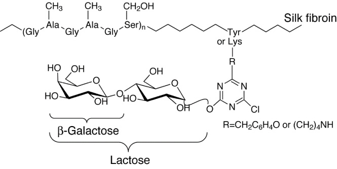
Chemical structure of lactose-silk fibroin conjugates (Lac-CY-SF) prepared using cyanuric chloride (CY) as a coupling reagent. Figure modified from reference [18].
As a positive control, Matrigel-coated plates were prepared by coating each well of polystyrene flat-bottomed 24-well plates for tissue culture (Iwaki) with 0.50 mL of 0.03% (w/v) solution of Matrigel (Becton Dickinson, Franklin Lakes, NJ, USA). Matrigel was allowed to absorb for 3 h, and the excess solution was removed before plating the cells. Commercially available collagen-coated plates (Iwaki), which were prepared by coating polystyrene flat-bottomed 24-well plates for tissue culture (Iwaki) with type I collagen, were used as another control.
hiPSC-derived hepatocytes used in this research were the commercially available product (ReproHepato TM ; REPROCELL Inc., Yokohama, Japan). In-house hiPSC were generated from human adult somatic cells [22], that were obtained from a commercial source, and differentiated into hepatocytes, ReproHepato TM using a proprietary protocol [6]. All cells were differentiated from a single-donor hiPSC clone and gave consistent and reproducible results across multiple assays. The cells were pre-cultured on Matrigel-coated 12-well culture plates with Maintenance Medium (REPROCELL) containing 10% fetal bovine serum (FBS) and 1% Maintenance Medium Supplement (REPROCELL) according to the manufacturer’s recommendation and trypsinized. The cells were seeded onto each substrate-coated well of a 24-well plate at a density of 3.12 ×10⁵ cells/well with 0.50 mL of Maintenance Medium containing 10% FBS and 1% Maintenance Medium Supplement, and cultured in a humidified incubator at 37 °C and 5% CO2 for a prescribed time. The medium was replaced with 0.50 mL of fresh medium every other day. Cell morphologies after 2 h, 2 days and 6 days of culture were observed by phase-contrast microscope (IX70; Olympus, Tokyo, Japan).
Cell viability assessment
Viable cell number on a substrate-coated well was relatively assessed using a cell counting kit-8 (CCK-8; Dojindo Laboratories, Kumamoto, Japan) based on the conversion of a water-soluble tetrazolium salt, 2-(2-methoxy-4-nitrophenyl)-3-(4-nitrophenyl)-5-(2,4-disulfophenyl)-2H-tetrazolium, monosodium salt (WST-8), to a water-soluble formazan dye upon reduction by dehydrogenases [23]. After 2 or 6 days of culture, the medium was replaced by 1 mL of fresh medium. Then, 0.10 mL of CCK-8 solution was added to each well of the plate and incubated for 2 h in a CO2 incubator. The absorbance of the CCK-8 assay solution was measured at 450 nm using a microplate reader (EnSpire 2300 Multilabel Plate Reader; PerkinElmer, Inc., Waltham, MA, USA) and background subtraction was performed.
Immunofluorescence staining
Cells were fixed in cold methanol for 10 min and washed twice with D-PBS(-). After being blocked with D-PBS(-) containing 2% bovine serum albumin (BSA) for 1 h, the cells were incubated with primary antibodies (diluted 1:100 in D-PBS(-) containing 0.1% BSA) at 4 °C overnight. The primary antibodies were asialoglycoprotein receptor 1 (ASGPR1) antibody (8D7) (Santa Cruz Biotechnology, Inc., Dallas, TX, USA) and anti-human albumin cross-absorbed antibody (Bethyl Laboratories, Inc., Montgomery, TX, USA). The cells were washed twice with D-PBS(-) and incubated with secondary antibodies (diluted 1:100 in D-PBS(-) containing 0.1% BSA) for 1 h at room temperature. The secondary antibodies were Alexa Fluor 488-conjugated goat anti-mouse IgG (Invitrogen, Carlsbad, CA, USA) and Alexa Fluor 594-conjugated donkey anti-goat IgG (Invitrogen). Cell nuclei were counterstained with 4 ′ ,6-diamidino-2-phenylindole dihydrochloride (DAPI) (Life Technologies, Carlsbad, CA, USA) in D-PBS(-) containing 0.1% BSA for 3 min at room temperature. The marked cells kept in D-PBS(-) were examined using fluorescence inverted microscope (Eclipse Ti; Nikon, Tokyo, Japan).
Albumin secretion analysis
On day 6, hiPSC-derived hepatocytes were refreshed with new culture medium and further incubated for 24 h. An aliquot (0.10 mL) of the medium was withdrawn from each well for analysis. The amount of albumin secreted into the culture medium for 24 h by the cells present on a substrate-coated well was measured by enzyme-linked immunosorbent assay (ELISA) using a human albumin ELISA quantitation kit (Bethyl) according to the manufacturer’s protocol.
Statistical analysis
Data were expressed as mean ± standard deviation (SD) from more than three samples. Statistical analyses were performed using an unpaired Student’s t test with KaleidaGraph software (HULINKS Inc., Tokyo, Japan) to compare different material samples. Differences with p < 0.05 were considered significant.
Results
Cell morphology
The morphologies of hiPSC-derived hepatocytes cultured on the Lac-CY-SF conjugate-coated wells for 2 h, 2 days and 6 days were compared to those on the wells coated with Matrigel or type I collagen (Fig. 2). The well-known shape change, from a round to a flattened morphology, slowly proceeded on Lac-CY-SF and Matrigel compared to collagen. After 2 h of culture, most of the cells on Lac-CY-SF and Matrigel showed a round morphology, whereas the cells on collagen showed round and flattened morphologies. At day 2, a number of cells retained a round shape on Lac-CY-SF and Matrigel. In contrast, the cells on collagen were flattened and formed a confluent monolayer. From day 2 through day 6, most of the cells on Lac-CY-SF and Matrigel became flattened; however round cells were still observed at day 6. Monolayers of viable cells continued to remain on collagen at day 6.
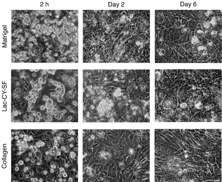
Phase-contrast micrographs of hiPSC-derived hepatocytes on wells coated with Matrigel, Lac-CY-SF or collagen after the cultivation for 2 h, 2 days and 6 days. Scale bar indicates 50 μm.
Cell viability of hiPSC-derived hepatocytes on substrate-coated wells at day 2 and 6 was quantitatively assessed using WST-8 assay. Figure 3 shows the absorbance of formazan dye produced by the cells on the well, which was proportional to the number of viable cells. At day 2 of culture, there were no significant differences in the viable cell numbers on the wells coated with Lac-CY-SF, Matrigel or collagen. Since the absorbance of WST-8 assay by the cells on each substrate-coated well at day 6 was comparable to or higher than that at day 2, it was confirmed that the cells remained viable on three kinds of substrate-coated well at day 6. At day 6, the viable cell number on Lac-CY-SF was almost the same as that on Matrigel, but slightly lower than that on collagen. No significant difference in viable cell numbers between Matrigel and collagen was observed at day 6.

WST-8 colorimetric assay for assessing viable cell numbers of hiPSC-derived hepatocytes present on wells coated with Matrigel, Lac-CY-SF or collagen at 2 days and 6 days. ∗ p < 0.05.
The protein expression of important mature hepatocyte markers, ASGPR1 and albumin, in the cytoplasm was assessed by immunofluorescence staining (Fig. 4). ASGPR1-positive cells were detected on three kinds of substrates at day 2. Regardless of the kind of substrate, ASGPR1 expression increased with many positive cells at day 6. Although a remarkable difference in ASGPR1 expression was not observed for the cells cultured on three kinds of substrates at day 2, ASGPR1 expression by the cells cultured on Matrigel was higher than that by the cells cultured on Lac-CY-SF and collagen at day 6. On the other hand, many cells on three kinds of substrates were albumin-positive at day 2. In line with the results of ASGPR1 expression, most of the cells were albumin-positive and an increase in albumin expression was observed on all substrates at day 6. The cells cultured on Matrigel showed somewhat higher expression of albumin compared to the cells cultured on Lac-CY-SF and collagen at day 6.
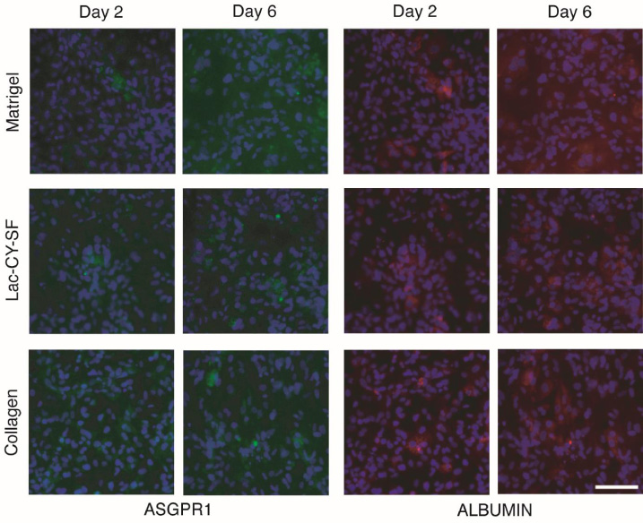
Fluorescent images of hiPSC-derived hepatocytes immunostained for ASGPR1 and albumin after 2 days and 6 days of culture on wells coated with Matrigel, Lac-CY-SF or collagen. Nuclei were counterstained with DAPI (blue). Scale bar indicates 100 μm.
Albumin secretion was investigated as a hepatocyte differentiated function. Figure 5(A) shows the amount of albumin secreted into the medium for 24 h from day 6 to day 7 by hiPSC-derived hepatocytes cultured on a substrate-coated well. The amounts of albumin secreted by the cells cultured on Lac-CY-SF and collagen were the same, but slightly lower than that by the cells cultured on Matrigel. In order to evaluate the amount of albumin produced by a single cell, namely, albumin secretory function per cell, albumin data were normalized to WST-8 assay data at day 6, proportional to the number of cells present on a substrate-coated well. Data normalization showed that albumin secretory function per cell on Lac-CY-SF was comparable to that on Matrigel and higher than that on collagen (Fig. 5(B)).
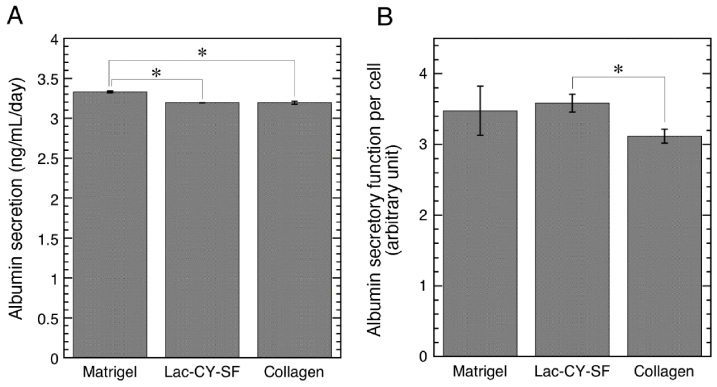
Albumin secretion by hiPSC-derived hepatocytes cultured on wells coated with Matrigel, Lac-CY-SF or collagen at day 7 of culture. (A) Amounts of albumin secreted into the medium per day by the cells present on a substrate-coated well. ∗ p < 0.05. (B) Evaluation of albumin secretory function per cell on a substrate-coated well. Albumin values were normalized to WST-8 assay data proportional to the number of cells present on a substrate-coated well. ∗ p < 0.05.
Our previous studies showed that the incorporation of 𝛽-galactose residues into SF matrix improved attachment and maintenance of rat primary hepatocytes and human hepatocellular carcinoma-derived cells whereas SF matrix inhibited their attachment [18–20]. In accordance with these results, light microscopic images of cultured hiPSC-derived hepatocytes demonstrated that the cells attached to Lac-CY-SF conjugate-coated wells at 2 h, and the cells on Lac-CY-SF were retained until day 6 as well as on the wells coated with Matrigel or collagen (Fig. 2). Following to microscopic findings, WST-8 assay results clarified that the viable cell number of hiPSC-derived hepatocytes cultured on Lac-CY-SF was comparable to that on Matrigel at day 6 (Fig. 3). These results suggest that Lac-CY-SF conjugates have the same level of ability to maintain hiPSC-derived hepatocytes as Matrigel.
For monitoring the maturation of cultured hiPSC-derived hepatocytes, the protein expression of ASGPR1 and albumin was analyzed. Regardless of the kind of substrate, the expression of both ASGPR1 and albumin increased from day 2 to day 6 (Fig. 4). This means that the culture period affects maturation of hiPSC-derived hepatocytes. Six-day cultivation was more desirable for maturation of the cells used in this research compared to two-day cultivation. On the other hand, immunofluorescence images at day 6 showed that ASGPR1 expression by the cells cultured on Matrigel was higher than that by the cells cultured on Lac-CY-SF and collagen (Fig. 4). These results indicate that the maturation of hiPSC-derived hepatocytes was time-dependently induced on Lac-CY-SF as well as collagen although the maturation was most efficiently promoted on Matrigel. The findings concerning the maturation of hiPSC-derived hepatocytes on Lac-CY-SF are consistent with previous reports that galactosylated substrates induced maturation of mouse and human embryonic stem cell-derived hepatocytes [15,24].
hiPSC-derived hepatocytes cultured for 6 days synthesized and secreted albumin in the same way as matured hepatocytes perform metabolic functions. The cells cultured on Lac-CY-SF showed a higher level of albumin secretory function compared to the cells on collagen (Fig. 5(B)). Although no statistically significant difference in albumin secretory function was observed for the cells cultured on Matrigel and collagen, the cells on Matrigel tended to exhibit a higher level of albumin function compared to the cells on collagen. On the other hand, the cells cultured on collagen possessed more flat morphology than those on Lac-CY-SF and Matrigel (Fig. 2). Many studies reported that spherical or cuboidal hepatocytes have a higher performance of liver functions compared to flat hepatocytes [10,25]. Therefore, our limited studies seem to indicate that Lac-CY-SF substrate can induce less flat morphology accompanying higher functional performance compared to collagen. Moreover, the clinical use of the cells cultured in contact with animal-derived material is not preferable from a regulatory point of view due to the risk of contamination by prions, viruses or other zoonoses. Animal-derived Matrigel and collagen are not ideal substrates for cell culture in regenerative medicine. For that reason, the results of albumin secretory function support the hypothesis that insect product Lac-CY-SF could be used as an alternative substrate to animal-derived ECM for the culture of hiPSC-derived hepatocytes in regenerative medicine. Further functional studies are required to verify this interpretation of the results.
In addition to the findings presented here, Tasnim et al. reported that a three-dimensional culture using galactosylated cellulosic sponges enhanced the functional performance of hiPSC-derived hepatocytes [26]. Thus, a three-dimensional culture system using previously developed Lac-CY-F sponges [20] instead of a two-dimensional Lac-CY-SF substrate may further increase the function of hiPSC-derived hepatocytes.
Conclusions
In summary, our findings showed that hiPSC-derived hepatocytes cultured on Lac-CY-SF bore a resemblance to hiPSC-derived hepatocytes cultured on Matrigel in cell morphology and viability as well as functional performance of albumin secretion, whereas those on Lac-CY-SF bore a resemblance to hiPSC-derived hepatocytes cultured on collagen in cell maturation. Moldable Lac-CY-SF could be used as an alternative substrate to animal-derived ECM for the culture of hiPSC-derived hepatocytes in liver regenerative medicine and tissue engineering.