
Abstract
BACKGROUND:
Previously we found that a group of phosphorylated proteins (SIBLINGs) in bone binds with the Ti-device, and increases the early bone formation around the Ti–implants remarkably. From these results, we explained the biochemical mechanism of a strong bond between living bone and Ti, which was discovered by Brånemark and colleagues. For the clinical application of our findings, we need a large amount of these proteins or their substitutes.
OBJECTIVE:
We aimed to create a new molecule that equips with essential functions of SIBLINGs, Ti-binding, and bone enhancement around the Ti implant.
METHODS:
We chemically phosphorylated chitin and obtained a soluble form of phosphorylated chitin (P-chitin). In this solution, we immersed the Ti-devices of web-form (TW) which we previously developed and obtained the P-chitin coated TWs. Then we tested the P-chitin coated TWs for their calcification ability in vitro, and bone enhancing ability in vivo, by implanting them into rat calvaria. We compared the P-chitin coated TW and the non-coated TW in regard to their calcification and bone enhancing abilities.
RESULTS:
Ti-devices coated with phosphorylated-chitin induced a ten times higher calcification in vitro at 20 days, and four times more elevated amount of bone formation in vivo at two weeks than the uncoated Ti-device.
CONCLUSIONS:
Phosphorylated chitin could be a partial substitute of bone SIBLING proteins and are clinically applicable to accelerate bone formation around the Ti implants, thereby achieving the strong bond between living bone and Ti.
Introduction
A remarkable discovery was made in the 1950s by Brånemark et al. [1,2], who found that solid-state Titanium (Ti) and the living bone tissues are firmly bound together. They found this phenomenon when they observed the bloodstream in a rabbit bone marrow using a light microscope, which was equipped with a Ti objective lens holder. This finding attracted the attention of orthopedic and dental researchers with expectations of creating new artificial bone and teeth. More than 50 years later, medical applications of Ti for reconstruction in artificial joints and roots of teeth developed universally. However, to date, the mechanism of how bone formation is initiated and how the interface between bone and Ti are maintained is not understood.
A strong bond between the living bone and Ti, which was discovered by Brånemark and colleagues, is highly valuable for regenerative medicine for bone and tooth reconstruction. However, there has been one crucial shortcoming: it takes many months to acquire a practical bond strength. This fact is reasonable since the bond is not based on the simple chemical reaction but on the biological process of bone formation around the surface of Ti. So far, numerous researchers tried to accelerate the fixation of the Ti implant into the bone, which includes roughening the Ti surface [3–5], 3D geometrical structures [6], chemical modification of the Ti surface [3,6], and coating the surface with various substances such as hydroxyapatite [7].
In 2012, we found that a group of phosphorylated and RGD-contained proteins, i.e. the SIBLING family, in bone tissue possessed binding ability with Ti [8]. Moreover, when the bone SIBLING proteins were coated on the Ti-device and implanted into animal bone, the amount of early bone formation was promoted 100 times higher than the non-coated Ti-device [9]. These results explained the fundamental mechanism of preferential bone formation on the surface of Ti, and we call the SIBLING proteins in the bone “implant proteins”. However, natural SIBLING proteins are not purified easily from animal bone. Also, their biosynthesis by genetic technology is highly expensive. Therefore, if we create a substitute molecule for SIBLING, it will grant favors to reconstruction therapy of bone and tooth in which Ti is used.
Chitin is the most abundant biopolymer second to cellulose [10], the structure of which was elucidated by chemically and X-ray diffraction data [11,12]. We know that chitin is highly insoluble in the common solvents. However, we found that chitin became soluble and bounded with Ti when we chemically phosphorylated it [13,14]. This finding was a highly significant breakthrough since the soluble form of chitin can be manipulated by many biochemical processes. Therefore, in this study, we tried to examine the functions of soluble phosphorylated chitin (P-chitin) by combining it with the Ti-device. We followed the previously reported protocol [9,16] and soaked the Ti-device of web form (TW) in the solution of P-chitin to obtain the P-chitin coated TW, and checked the chitin coated TW for two functions. First, we checked whether the P-chitin coated TW promotes in vitro calcification. Second, we studied in vivo whether P-chitin coated TW promotes bone formation when we implanted them into the animal bone and compared them with the result of non-coated TW.
Materials and methods
As mentioned above, we decided to create a new synthetic material that fulfills two essential characters of SIBLING proteins, multiple phosphate groups, and an RGD sequence, an example of which is shown in Fig. 1 [17].

Partial amino acid sequences 201-317 of a typical SIBLING family protein, human bone sialoprotein. Total amino acids of human bone sialoprotein is 317 residues, out of which 74 residues are serine (s) and threonine (t). They are potentially phosphorylated and bind with Ti, and one RGD sequence, which binds with osteoblasts. From 5 to 15 residues of serine per molecule are reported to be phosphorylated. Amino acids susceptible to phosphorylation are presented in bold, and the cell adhesive sequence rgd is surrounded by a rectangle, each corresponding to the two important functions of SIBLING proteins, Ti binding, and cell-binding abilities. Cited from [1].
Chitin is a product from Fuji-Film Wako, Tokyo. We chemically phosphorylated the methods previously [13–15]. In short, we phosphorylated chitin with phosphorous pentoxide by using dimethyl sulfoxide (DMSO) as a solvent, and methane sulfonic acid as a catalyst. Two grams of chitin powders were suspended in 40 ml of DMSO, which contained 4.5 g of P205 and 1.5 ml of methane sulfonic acid beforehand. The molar ratio of P205 against acetyl glucosamine residue was about 3:1. The mixture was continuously stirred at 70 ∼ 80 °C for 6 h. The reaction was stopped by the addition of acetone to obtain a final concentration of 60% in volume. The reaction product was collected by filtration and rinsed by acetone several times and air-dried. We dissolved the dried product in 50 ml of distilled water followed by dialysis against distilled water in a refrigerator at 10 °C for 48 h and lyophilized. We charged the phosphorylated chitin on a Ti-beads chromatography [8,9,16], and the fraction retained in the column was used for the experiments of the calcification in vitro and the enhancement of bone formation in vivo.
Ti-devices used in this study had a 3D unwoven texture in a web form (TW) and are composed of thin Ti fibers of 80 μm, initially developed in this laboratory [18,19] and available in various sizes (Zellez®, Hi-Lex Corporation-Funakoshi, Japan). This material has already been verified as one of the ideal 3D Ti scaffolds for bone ingrowth and formation [18,19]. The TW of the disc-forms of 13 × 1.5 mm was used for the calcification experiment, and those of 5 × 1.5 mm for the bone formation experiments, both with 87% porosity. In combination, the TW and the purified P-chitin, the 20 pieces of the TW, were immersed in 2 ml of the 0.2% solution of the purified P-chitins and incubated at 15 °C for 15 min. After immersion, P-chitin coated TW were air-dried in a clean bench and stored at 5 °C.
In vitro calcification of the P-chitin coated TW
Calcifiability of P-chitin coated TW was tested by the incubation method in the calcifying solution with high Ca and phosphate ions concentration to obtain hydroxyapatite, which has been reported previously [20,21]. In short, this method includes the incubation in the calcification solution, which provides for high Ca ions and phosphate ions than the physiological fluid. This solution has been verified to induce hydroxyapatite on the solid substrates rapidly under the described conditions.
Pieces of P-chitin coated TW (5 × 1.5 mm) were immersed in the calcifying solution, whose contents were 15 mM CaCl2, 9 mM K2PO4, 0.7 M NaCl and 0.02 M NaHCO3, pH of which was adjusted to 6.01 by the passage of gaseous carbon dioxide. The bottles that contained the samples were tightly sealed and incubated for 48 h at 37 °C. After the first immersion, the coated samples were rinsed with distilled water, lyophilized, and weighed. These samples were then immersed again in a new calcifying solution, as described above. We continued the immersion for 20 days with a change of the solution every other day.
Implantation into rat calvaria
Over 20 eight-week-old male rats (Wistar, 200–230 g) were anesthetized with Xylazine (90 mg/kg) and Ketamine (10 mg/kg) per body weight. The P-chitin coated TWs were implanted into the holes with a diameter of 5.2 mm created by dental trepan bur. After implantation, periosteum and skin were carefully placed back and sutured. The Committee for Animals approved the protocol of the animal experiments (“Experiments in Kanagawa Dental College”). The experiment was carried out under the guidelines proposed by the Institutional Animal Care and Use Committee. We implanted one piece of either the P-chitin coated TW or TW alone into each rat's calvaria. After two and four weeks we retrieved the samples for examination, as described previously [9,16].
Histological observation
We implanted one TW per rat into their calvaria, using more than 20 TWs in total. There were four groups of the retrieved TW samples: The two-week retrieved ones were divided into the P-chitin coated (group 1) and the non-coated TW (group 2). Likewise, the four-week retrieved samples were divided into the coated (group 3) and the non-coated TW (group 4). For each group, we prepared five histological sections from five rat calvaria, totaling more than 20 sections. For the evaluation of bone formation in the histological sections, we used Image-J and measured the arias of total TW and the area occupied by the new bone in each section. We found that the ratio of bone-occupied area to the total TW area (bone occupancy) in each section was the extent of bone growth.
We fixed the excised pieces of cranial bone which contained the implant in 10% neutral formaldehyde and embedded it in a resin (Technovit 8100 Heraeus Kulzer, Germany) to prepare undecalcified sections, according to the manufacturer’s protocol. Three-μm-thick of the vertical section from the central parts of the TW disc was obtained from each specimen using an Exakt grinding machine, assisted with a mechanical digital micrometer (Exakt, Norderstedt, Germany). We stained the section with Villanueva Osteochrome bone stain (Polysciences, Inc., Arrington, PA, USA).
Statistical analysis
We compared the ratio of bone-occupied area to the total TW area (bone occupancy) in the P-chitin coated and non-coated TW group using Student’s t-test. A p-value of <0.05 was considered to be statistically significant.
Results
Enhanced calcification on the chitin coated Ti-device
Figure 2 shows the scanning electron micrographs with different magnifications of the intact TW (A, C, E, and G) and the P-chitin coated and calcified TW after incubation in the calcifying solution for 20 days (B, D, F, and H). At lower magnification, crystals of precipitated materials seem to be flat or irregular half-bulbar shapes on the surface of TW (Fig. 2D). However, in the higher magnifications (Fig. 2F and H), these structures are found to be composed of assembled leaves or plate shapes, growing from the surface of TW.

Scanning electron micrographs of the untreated original TW (2 × 1.5 mm) (A, C, E, and G) and the same TW which was coated with P-chitin and incubated in the calcifying solution for 20 days (B, D, F, and H).
Figure 3 summarizes the increase in weights of the precipitation in the P-chitin coated TW, compared with non-coated TW. We can observe a remarkable growth in the masses in the coated TW, which was 6.7, 4.8, and 4.9 times higher than those of the uncoated TW at 6, 12, and 20 days of incubation, respectively.
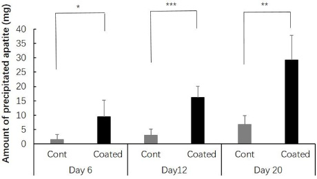
Ability to enhance calcification on the P-chitin coated TW. The P-chitin coated TW (19 × 1.5 mm) were incubated in the calcification solution for 6, 12 and 20 days, and an increase of the weights (mg) were expressed as the ratios to the average weight of the uncoated TW without incubation, which are 6.7, 4.8 and 4.9, respectively. Student’ P values were 0.0359, * (<0.05), 0.00587, ** (<0.01) and 0.00267, *** (<0.005) between the average values of uncoated control and coated groups, at 6, 12 and 20 days, respectively.
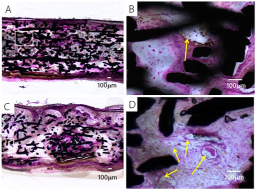
Figures 4A–D show the histological observation of the P-chitin coated TW (group 1) and un-coated TW (group 2) as control, which we implanted into rat calvaria and retrieved after two weeks. The P-chitin coated TW (Fig. 4A and B) induced more active new bone formation within the framework of TW, compared with TW alone, which scarcely produced new bone formation (Fig. 4C and D). Results demonstrated that the bone formation (stained in brown) and capillaries development (indicated by yellow arrows) were more active than those of the non-coated TW at two weeks after implantation. There was some heterogeneity in the bone formation within the TW. The Ti fibers are observed as the black colored materials in Fig. 4. The bone formation was significantly active around the high density areas of Ti fibers. In the higher magnification (Fig. 4D), many capillaries were growing (indicated by yellow arrows).

Histological observation of P-coated (A and B) and non-coated (C and D) Ti-devices, TW implanted into rat calvaria for two weeks. B is the enlarged picture of the area defined by a rectangle in A, and D is the same in C. Yellow arrows indicate capillaries. Black bodies are Ti-wires (80 μm) that compose TW.
These enhancements of bone formation by the P-chitin coated TW were again clear four weeks after implantation. Figures 5A–D show the histological observation of the P-chitin coated TW (group 3) and the non-coated TW (group 4) as control implanted into rat calvaria and retrieved four weeks after implantation. As described in the method section, we calculated the ratios of bone-occupied area to the total area of TW and compared them among the four groups.
Histological observation of the P-chitin coated (A and B) and non-coated (C and D) TW implanted into rat calvaria for four weeks. B is the enlarged picture of the area defined by a rectangle in A, and D is the same in C. Yellow arrows and black bodies are the same as Fig. 3.
Since we found the lowest bone occupancy in group 2 (the non-coated TW at two weeks), we divided the bone occupancy of the other three groups by that of group 2. The proportion of bone growth in all groups is listed in Fig. 6. At two weeks, bone occupancies of the chitin coated TW (group 1) was 4.0 times higher than those of the un-costed TW (group 2) at two weeks. At four weeks, the bone occupancy ratio of the coated TW (group 3) was still 1.6 times higher than that of control (group 4). These results are the first evidence that proves P-chitin on the surface of Ti enhanced bone formation.
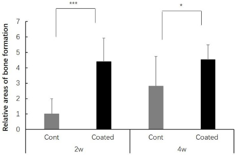
Enhancement of bone formation in the P-chitin coated TW compared with those of the non-coated TW when they were implanted into rat bone for two and four weeks. In each tissue section of the retrieved TW, the ratio of area occupied bone to the total area of TW were analyzed and compared. We compared the bone occupancy ratio in each section against the bone occupancy ratio of the uncoated TW at two weeks, which was the lowest among the analyzed. At two weeks, bone occupancies of the chitin coated TW was 4.0 times higher at two weeks, and 1.6 times higher at four weeks, than those of the uncoated TW (n = 5).
As shown in Fig. 2, the enhanced effect on calcification of the P-chitin coated TW compared with non-coated TW was remarkable. The calcifying solution used in this study proved that the formation of hydroxyapatite is highly effectively on the surface of solid substances such as collagen or Ti [20,21]. In this solution, the phosphate groups in the P-chitin coated TW obtained an ideal situation for calcium-phosphate aggregation and effectively induced calcification on the surface of Ti. We think this is one of the reasons why the coated TW obtained a higher calcification than the non-coated TW.
Previously, only a few researchers analyzed the P-chitins for their ability to induce calcification in vitro [22,24]. Yokogawa et al. phosphorylated the commercial products of fiber-typed chitin and soaked it in saturated Ca(OH)2 for one week, and then soaked it in 1,5 × SBF (1.5 times simulated body solution) [22]. They stated that the phosphate groups are most likely bound to the C6 area of N-acetyl glucosamine (2-acetamido-2-D-glucose) residues of this P-chitin, and concluded that P-chitin is a reasonable material for calcification, coinciding with our present results. The chemical structure of phosphorylated chitin can be found in Fig. 7. We assumed that the introduction of numerous phosphate groups into the C6 areas of this polymer might cleave the inter-chain hydrogen and hydrophobic bonds, thereby inducing the local and total conformational changes. These changes might offer a feasible circumstance for calcification. On the P-chitin coated TW, P-chitin was bound and immobilized on the surface of the Ti-device, which might further strengthen the working environments for calcification. These possible mechanisms are worth further investigation.

Putative structure of phosphorylated chitin in this study. Two identical N-acetyl glucosamine residues linked by a 𝛽 1-4 glucoside bond, and two phosphate groups bound to C6 are shown.
In terms of the mechanism of the bone enhancing effect of P-chitin coated TW, we noticed that in these experimental periods of two and four weeks, there was no sign of cartilage formation. Instead, more active capillaries appeared in the P-chitin coated TW than in non-coated TW. Also, there was some tendency that in the areas where Ti wires were dense, bone and capillaries formation were more active. These observations suggest the favorable actions of P-chitin for bone formation on the TW or the active molecules released from the surface of TW. Since the active development of capillaries is an essential pre-requisite for bone formation, there is a possibility that P-chitin itself may have some functions to induce vascularization, together with the bone producing activity. Wang et al. [23] reported an increased growth and differentiation of osteoblasts and fibroblasts by the addition of soluble P-chitin in the culture media, suggesting the same idea. Also, Yokogawa et al. [24] reported the positive effects of P-chitin on cultured cells. They cultured L-929 cells and the MC3T3-E1 cells on the phosphorylated and the Ca(OH)2-treated commercial chitin fibers and self-made chitin sheets. They stated that these phosphorylated coated and Ca(OH)2-treated materials induced higher cell-adhesive strength and osteoblastic differentiation than the control polystyrene culture disks without these materials.
We hypothesized that the introduction of a large number of phosphate groups into C6 areas of chitin molecules will split the inter-chain hydrogen or hydrophilic bonds, thereby inducing a local and whole crystal conformational change of chitin as already described (Fig. 7). One of the results of these changes was the increased solubility after phosphorylation, which we previously observed. Again, the immobilization of P-chitin on the TW in our system might specifically enhance the biologically feasible environments for growth and differentiation of osteoblasts to create bone. These hypotheses are soon confirmed in in vitro cell culture experiments in our laboratory.
There is a more definite explanation for the increased bone formation in the P-chitin coated TW. Phosphate groups in P-chitin promoted local calcification on the surface of TW in vivo, as shown in vitro in this study (Fig. 2). This local formation of apatite invites osteoblasts to adhere more easily and start bone formation more rapidly than the naked surface of TW. This explanation was partially verified in this study.
Our results show that the phosphorylated-materials coated on Ti surface remarkably accelerate the increase of calcification in vivo. Moreover, it was demonstrated that the binding between Ti and new formed bone will strengthen. Also, we found that the P-chitin coated Ti-device more effectively induced bone formation than the non-coated control. Both results indicate that the Ti-device, when it is coated with P-chitin, will become a promising tool for regenerative medicine of bone and teeth.
Our study is the first to report that P-chitin coating is highly useful to enhance early bone formation. Since this method is composed of coating on the surface of the Ti-implant device with rather pure substances of P-chitin, its clinical applications seem to be highly valuable. The fact that P-chitin coating enhanced earlier bone formation is especially suitable for resolving the problem of bone loss after implantation. Although the detailed mechanism for enhancement of bone formation is still to be conclusive, we can conclude that P-chitin is a highly active biomaterial for calcification and bone formation when combined with the Ti-device.
Footnotes
Acknowledgements
The authors appreciate the valuable advice given by Dr. Arthur Veis, Professor Emeritus, North Western University, USA. This study was supported in part by a Grant-in-Aid for Challenging Exploratory Research (23659907) and a Grant-in-Aid for Research (C) (15K05554) from the Ministry of Education, Culture, Sports, and Technology in Japan.