
Abstract
BACKGROUND:
The design and fabrication of hemocompatible and low-toxicity formulations remains a challenging task. Hydrogels are of considerable importance for biomedical applications since they are highly compatible with living tissue, both in vivo and in vitro.
OBJECTIVE:
The present study aimed to develop and evaluate the characterizations and in vitro hemocompatibility of a hydrogel using polyvinyl alcohol and gelatin with different concentrations.
METHODS:
The gelling process was realized by cross-linking the polyvinyl alcohol and gelatin. The morphological and structural examinations of the synthetic hydrogels were done by scanning electron microscopy (SEM) and X-ray powder diffraction (XRD). The swelling behavior of the prepared hydrogels in water was evaluated. Prothrombin time, activated partial thromboplastin time, and thrombin time were measured, and a hemolysis test was done to evaluate the hemocompatibility of prepared hydrogels.
RESULTS:
The increase of the gelatin concentration in polyvinyl gelatin hydrogel increases the porosity and enhances the absorptivity of the prepared hydrogel. The measured hematological parameters indicated enhancement of hemocompatibility as the gelatin concentration was increased in the prepared hydrogel.
CONCLUSIONS:
The results obtained from this study confirm that gelatin was able to improve the properties of the polyvinyl alcohol–gelatin hydrogel and enhance the hemocompatibility. Thus, the prepared hydrogel could be used in a variety of biomedical applications.
Introduction
Hydrogels are used for various biomedical applications. These polymeric hydrogels do not dissolve in water at physiological temperatures. pH. applications of hydrogels in the biomedical field include contact lenses, artificial corneas, wound dressing, coating for sutures, catheters, and electrode sensors. The manipulation of the physical properties of hydrogels leads to a wide range of applications. Since hydrogels absorb water, they are usually biocompatible and are non-irritating to the soft tissues when they come in contact with them [1,2].
Polyvinyl alcohol (PVA) mixtures have long been used with other natural polymers because of their ability to form films. The performance properties of PVA are influenced by the molecular weight and the degree of hydrolysis. The molecular mass of PVA is ∼160 kDa. PVA has a planar zigzag structure like polyethylene [3]. All PVA classes are easily dissolved in water and are dependent on factors such as molecular weight, particle size distribution, and particle crystallinity. As a hydrophilic polymer, PVA exhibits perfect water retention properties. Optimum solubility takes place at 87% to 89% hydrolysis [4–6].
Gelatin is obtained by the thermal denaturation of collagen from animal skin, bones, and, rarely, fish scales. It includes at most the residues of three amino acids glycine (arranged every third residue), proline, and 4-hydroxyproline in its structure. Gelatin contains higher levels of pyrrolidines, which results in the formation of stronger gels [7,8]. The existence of triple helixes leads to gelatin film strength. The greater the triple-helix content, the higher the strength of the film, and the lower the swelling property in water [9,10].
Hemocompatibility is one of the primary standards, which limits the clinical applicability of blood-contacting biomaterials [11]. These materials take place in an adjacent link with blood, which is a complex ‘organ’ comprising of 55% plasma, 44% erythrocytes, and 1% leukocytes and platelets. Thus, reverse interactions between recently developed materials and blood should be extensively analyzed to stop activation and destruction of blood components. The initially adsorbed protein layer on the biomaterial surface mainly triggers the adverse reactions, such as the activation of coagulation via intrinsic pathway, the activation of leukocytes, which produce inflammation, and the adhesion and activation of platelets [12,13].
This study aims to prepare polyvinyl alcohol–gelatin (PVAG) hydrogel with different gelatin concentrations in order to improve the biocompatibility properties of the hydrogel. The characterizations and hemocompatibility of the prepared PVAG are examined.
Materials and methods
Polyvinyl alcohol–gelatin hydrogel preparation
Polyvinyl alcohol (PVA) with a molecular weight of 85000 Da was supplied by the laboratory Reagent (Techno Pharmchem Haryana, India). The gelatin used in this study was obtained from Kose Chemical Co., Ltd, Japan. One gram of PVA was added to 100 ml of triply distilled water at 80 °C and was continuously stirred to obtain a clear transparent solution. An aqueous gelatin solution was obtained by adding the proper weight of gelatin in 50 ml of triply distilled water at 80 °C. The mass fraction of gelatin (wt.%) was calculated according to the following equation:
SEM provides high-resolution imaging of surface morphology. One cm2 of each PVAG was coated with gold. The coating was required to obtain a clear image/micrograph of the insulating material. The coating layer was very thin (200 Å) so that it did not hinder the identification of specific minerals. Then all films were observed under the SEM at an amplification of 5000X.
XRD characterization
In order to determine the crystallinity of all the PVAGs, XRD (STOE & Cie GmbH, Darmstadt, Germany, 𝜃-𝜃) was performed using Cu radiation generated at 40 kV and 40 mA. The range of the diffraction angle was 10 to 70° 2𝜃. The 0.02° step size of 2𝜃 was maintained at a scan speed of 2 s/step. PVA alone was used as a supplementary control.
Swelling behavior
The PVAG films were cut, and the initial weight was determined (w
i
). The samples were then immersed in distilled water (swelling medium) at room temperature. The samples were picked up from distilled water at regular intervals of four hours and then dried under vacuum. The weight after swelling was determined (w
f
). The swelling rate (SR) was calculated as a percentage as follows:
Changes in blood clotting factors were measured before and after the endothelialization of PVAG with a monolayer of endothelial cells, followed by exposure of blood containing acid citrate dextrose as an anticoagulant. Coverage of PVAG with endothelial cells was done by culturing human umbilical vein endothelial cells (HUVEC) (HUVEC, Single Donor, in EGMTM Lonza, USA) on PVAG for four days. PVAG pieces were exposed to blood for one hour. Blood was collected after exposure, and plasma was separated by centrifugation at 3500 rpm. Prothrombin time (PT), activated partial thromboplastin time (APTT) and thrombin times (TT) were measured by the semi-automated analyzer (Huma Clot Duo Plus, HUMAN Gesellschaft für Biochemica und Diagnostica mbH, Germany).
Hemolysis test
Blood samples were collected on sodium citrate and then diluted with PBS. A piece of 1 cm2 from PVAG was incubated with diluted blood at 37 °C for 60 min. PVAG was removed from the blood. Blood was then centrifuged, and the supernatant was transferred to the cuvette, and the optical density (OD
test
) of the tested supernatant was measured at 545 nm with the spectrophotometer. 0.2 ml of diluted blood was mixed with 10 ml of 0.1% sodium carbonate solution for 60 min at 37 °C, and optical density was measured at 545 nm and used as a positive control (OD
+ve). For negative control, 0.2 mL of diluted blood was mixed with 10 ml of normal saline solution and incubated for 60 min at 37 °C.
Figure 1(a–d) shows the scanning electron microscope micrographs of pure PVA and the blend with 5, 15, and 25 wt.% of gelatin. It can be seen that with an increased concentration of gelatin, the average size of pores in the PVA hydrogel became increasingly large and a relatively loose network structure was formed. Gelatin was chosen for this study due to its RGD motifs, which act as cell attractants and enhance cellular adhesion to the matrix. Therefore, RGD motifs have been found to be ideal for wound healing and various tissue engineering applications [14,15]. In the PVA-gelatin polymer mixture, the crosslinking formation between the aldehyde groups that present in glutaraldehyde and the free amino groups of gelatin can be obtained during the blending process. Such crosslink is obtained as a result of the strong reactions between the active functional groups found within the gelatin and PVA structure. These groups are carbonyl (C=O), NH, and hydroxyl (OH) for gelatin and PVA, respectively. As a result of these interactions, a formation of covalent bonds between these groups can be obtained [16]. So, the behavior of PVAG samples depends on the crosslinking formation between the functional groups of the two polymers. An apparent effect on the morphology of the PVA surface was observed with varying gelatin concentrations from 5, 15, and 25 wt.% within the PVA structure. The sponge construction within the sample with least gelatin concentration, 5 wt.%, showed the smallest pores in the surface, which was analyzed using a scanning electron microscope (Fig. 1(b)). However, with the increase of gelatin ratio inside the PVA sample to 15 and 25 wt.%, the pore diameter was found to improve, and pores were more homogeneously distributed (Fig. 1c,d).
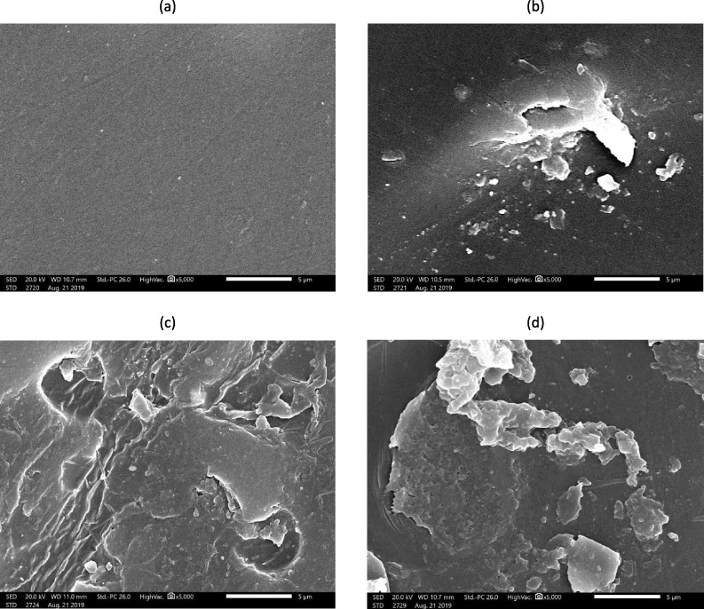
SEM micrographs representing the topographic view of the prepared hydrogels: (a) Pure PVA; (b) PVAG 5%; (c) PVAG 15%; (d) PVAG 25%.
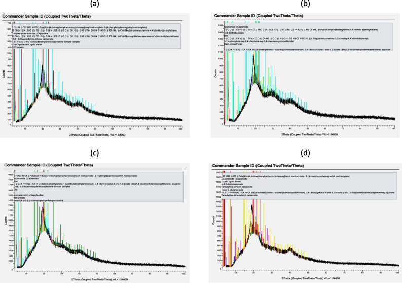
X-ray diffractograms of the prepared hydrogels: (a) Pure PVA; (b) PVAG 5%; (c) PVAG 15%; (d) PVAG 25%.
The crystallinity of pure PVA that blends with 5, 15, and 25 wt.% of gelatin was characterized by XRD. The XRD curves are shown in Fig. 2(a–d). The XRD pattern of pristine PVA (Fig. 2(a)) exhibits sharp diffraction peaks at 2𝜃 = 20, 30, and 44, respectively. These results provide evidence that PVA has a semi-crystalline nature that both has a crystalline and amorphous structure [17]. The crystalline nature of PVA is assigned to the presence of strong inter- and intra-molecular hydrogen bonding between the different molecules of PVA chains [18]. The XRD patterns of PVA that contain 5, 15, and 25 wt.% of gelatin are depicted in Fig. 1(b–d), respectively. It is clear that PVAG blends show a broad peak at 2𝜃 = 20, which is typical for semi-crystalline polymers such as PVA [18]. In addition, the two peaks obtained at 2𝜃 = 30 and 40 disappeared entirely. This demonstrates that the molecules of gelatin were effective in destroying the crystal structure of PVA. Gelatin molecules can replace PVA inter- and intra-molecular hydrogen bonds, and form stable hydrogen bonds with PVA. This indicates a significant change in the degree of crystallinity, combined with an increase in the amorphous regions of the PVAG blend composite [19]. This confirms the excellent compatibility and miscibility of the two polymer blend components due to the crosslinking formation within the investigated sample as a result of a strong interaction between the hydroxyl group of PVA and NH and C=O, as mentioned before [18].
Figure 3 displays the swelling behavior of PVAG compared to pure PVA. The swelling rates of PVAG with deferent concentrations of gelatin were higher than pure PVA. Also, the highest swelling rate was obtained for PVAG: 25%. This indicates that the increase of the gelatin concentration improves the ability of PVAG to absorb water. The swelling rate was over 100% for the PVAG 5%, 5%, and 25% after six hours. The swelling rate is used for biomedical materials, especially the ones used in wound healing, to decide the time of substitution of a wound dressing [20,21]. The highly absorbent hydrogels are usually biocompatible with the soft tissues when in contact with them [2]. Our findings indicate that as the gelatin concentration in PVAG increases, the water absorption increases, which may increase the biocompatibility of PVAG.
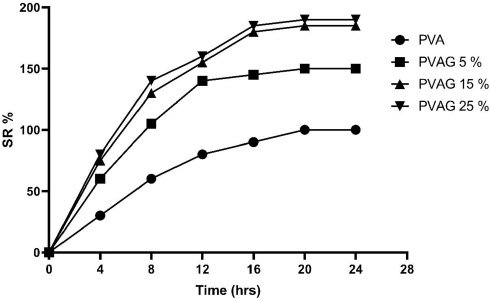
Swelling behavior of the prepared polyvinyl alcohol–gelatin hydrogel.
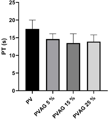
Comparison of prothrombin time of blood due to incubation with a polyvinyl alcohol–gelatin hydrogel.
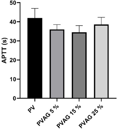
Comparison of activated partial thromboplastin time of blood due to incubation with a polyvinyl alcohol–gelatin hydrogel.
One of the critical assessments of the biomaterials is their interaction with blood or hemocompatibility [22]. There are three hemocompatibility test models: the static blood incubation model, the agitated blood incubation model, and the shear flow model [11]. In the second and third models, biomaterials are tested under the flow of blood under controlled conditions [23,24]. In the first model, materials are incubated with blood under the static condition for a certain time. This model was chosen in the present study [25]. Figures 4–6 represent the coagulation effect of pure PVA and PVAG. PT, APTT, and TT of PVAG with different concentrations were lower than pure PVA. The findings of this study showed that the addition of gelatin might reduce the coagulation effect of pure PVA. It is known that the interaction between blood collagen gel and dextran sulfate leads to the transformation of Factor XII to the active enzyme Factor XIIa. The mentioned mechanism is the first step in the coagulation cascade [26]. The interaction of the examined biomaterial with blood may cause a shortening of the coagulation time due to the activation of the intrinsic pathways of coagulation [23]. The coagulation results of PVAG in the present study showed a reduction in clotting time when compared to pure PVA. The monitored clotting times of blood were in the normal range for PVAG with different concentrations of gelatin.
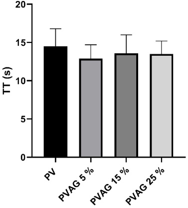
Comparison of thromboplastin time of blood due to incubation with a polyvinyl alcohol–gelatin hydrogel.
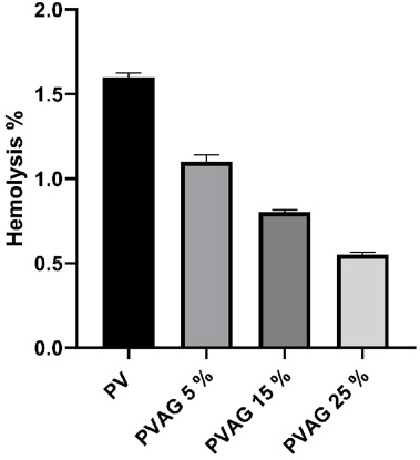
Hemolysis assay of blood after incubation with a polyvinyl alcohol–gelatin hydrogel.
In the present study, the in vitro hemolysis test was performed to examine the effect of PVAG on the major component of blood cells; red blood cells. As shown in Fig. 7, the hemolysis percentages were 1.6 ± 0.02%, 1.1± 0.04%, 0.8 ± 0.02%, and 0.6 ± 0.016% for pure PVA, PVAG 5%, PVAG 15%, PVAG 25% respectively. According to the hemolysis percentage, biomaterials could be classified in three different categories: hemolytic resulting in over 5% hemolysis, slightly hemolytic resulting in between 5% and 2% hemolysis, and nonhemolytic resulting in below 2% hemolysis [27]. Our findings indicate that PVAG is a nonhemolytic material and also show that a clear decrease in the hemolysis percentage is associated with the increase of the gelatin concentration in PVAG.
The use of gelatin as a channeling agent allows the preparation of polyvinyl alcohol–gelatin hydrogel with desirable absorbent properties. The results obtained from this study confirm that the gelatin was able to improve the properties of the polyvinyl alcohol–gelatin hydrogel. The hemocompatibility of PVAG samples increases as the percentage of gelatin increases inside the PVAG structure. All measured hematological parameters were found to be in the normal ranges and lower than that of pure PVA. These findings indicate that increasing the gelatin concentration enhances the biocompatibility of PVAG with human blood.
Footnotes
Conflict of interest
None to report.