
Abstract
BACKGROUND:
Bone volume augmentation is a routine technique used in oral implantology and periodontology. Advances in the surgical techniques and the biomaterials field have allowed a greater accessibility to these treatments. Nevertheless, dehiscence and fenestrations incidence during dental implant procedures are still common in patients with bone loss.
OBJECTIVE:
The main objective is to evaluate in a pilot experimental study the biological response to mesoporous silica (MS) hybrid scaffolds and its regenerative capacity in different formulations.
METHODS:
Two defects per rabbit tibia were performed (one for control and other for test) and the biomaterials tested in this study have been used to fill the bone defects, prepared in two different formulations (3D hybrid scaffolds or powdered material, in 100% pure MS form, or 50% MS with 50% hydroxyapatite (HA). Euthanasia was performed 4 months after surgery for bone histopathological study and radiographic images were acquired by computerized microtomography.
RESULTS:
Results showed that radiographically and histopathologically pure MS formulations lead to a lower biological response, e.g when formulated with HA, the osteogenic response in terms of osteoconduction was greater.
CONCLUSIONS:
We observed tolerance and lack of toxicity of the MS and HA, without registering any type of local or systemic allergic reaction.
Introduction
Dental implants and prosthesis are widely common treatments which have been increasingly used in recent years. Advances in the surgical techniques and the biomaterials field in traumatology and dentistry have allowed a greater accessibility to these treatments, increasing functional and aesthetics demands from patients. Nevertheless, dehiscence and fenestrations incidence during dental implant procedures and articular prosthesis implementation are still common in patients with bone loss, regardless their origin [1–3]. In these situations, a bone volume augmentation is a routine technique used in implantology and periodontology for functional and aesthetic aspects. However, the filling material may initiate the host tissue response by increasing osteoclastic activity and macrophages inflammatory response [4]. Therefore, in certain specific cases, inert biomaterials can cause significant delay in healing through host tissue interaction. In this regard, the effect at a cellular and molecular level in the tissue depends on the morphology, composition, chemistry, porosity and particle size of the material [5].
Bone graft and/or membrane can promote selective cellular growth to restore the defect. However, the normal healing process can be impaired or accelerated depending on the type of filling material [6]. The combination of organic and inorganic phases produces hybrid porous scaffolds that combine mechanic strength, porosity, flexibility, cohesion, and tailored degradation, key properties to promote hierarchical tissue growth and recovery of biological functionality in tissue regeneration applications [7,8] .
At the forefront of bioactive and resorbable formulations, chitosan shows paramount mucoadhesive and hemostatic abilities, that combined with K-carrageenan, produce elastic scaffolds with highly interconnected porous architecture that facilitate the formation of biosimilar grafts [9,10]. Moreover, the incorporation like hydroxyapatite (HA) and mesoporous silica (MS) nanostructures, have demonstrated to enrich the scaffold with all inherited benefits of the biocompatible inorganic phases and a rough surface prone to cell adhesion [11]. Specifically, HA provides the hybrid scaffold with mineral material as source for the bone tissue regeneration and mechanic strength, while mesoporous silica nanostructures with 2D hexagonal mesoporous structure, large surface area, and hydrothermal stability endow the scaffold with a large set of theragnostic activities, (paramount capacity for allocating therapeutic moieties for prolonged release applications, versatile surface for grafting MRI/PET contrast agents, etc.) [11].
Our main objective in this work is to evaluate in a pilot experimental study, by scanning electron microscopy (SEM), the enriched formulation of chitosan & k-carrageenan 3D scaffolds with mesoporous silica (MS) nanostructures with the incorporation of hydroxyapatite (HA) nanocrystals and its regenerative capacity in different formulations.
Materials and methods
Animals
Two albino New Zealand male rabbits were used (class: mammal, order: lagomorph, genus: leporidae and oryctolagus, and species: cuniculus). These animals are characterized by having a cylindrical body and a slightly nervous temperament. They were supplied by the Laboratory Animal Service (SAI, no REGAES: 300305440012) of the University of Murcia. These standardized animals had a determined genetic-sanitary composition and, were fed in controlled environment. The animals were treated following the European Union Guide (86/609/CEE, regarding the protection of animals used for experimental and other scientific purposes), UE law (BOE 223/1988 and 265/1990, to manipulate experimental animals), and national law RD 1201/2005 of October 10, on the protection of animals used for experimentation and other scientific purposes, and Law 32/2007 of November 7 for the care of animals in their exploitation, transport, experimentation and slaughter.
The animals presented an initial weight comprised between 3.5 and 4 kg, 28–32 weeks old, skeletally mature e.g. having a good capacity to withstand surgical trauma and between 15–18 weeks of age (ages close to physeal closure when reach the adequate bone volume for the study).
Formulations
The biomaterials tested in this study have been used to fill in bone defects at different concentrations (see experimental design, Fig. 1), and were prepared in two different formulations as 3D scaffolds or as a powdered material. Based on a previously used route to produce chitosan & k-carrageenan 3D scaffolds dropped with mesoporous silica nanostructures [11], in this work we have enriched the formulation with the incorporation of hydroxyapatite nanocrystals.

Experimental design and surgical protocol. (A) Soft tissue separation and tibia exposition. (B) Performing bone defects with trephine at low speed. (C) Biomaterial application. (D) Wound suturing.
3-Aminopropyl(diethoxy)methylsilane (≥97%), tetraethyl orthosilicate (98%), iron(III) chloride hex hydrate (97%), hydrochloric acid (37%), phosphoric acid (85%), cyclohexane (99.8%), Igepal CO-520 [Polyoxyethylene (5) nonylphenylether, branched], glycerol solution (86–89%), Pluronic® (Sigma-Aldrich, St. Louise, MS, USA) P123[triblock-copolymer-PEO20:PPO70:PEO20,Poly(ethyleneglycol)-block poly(propyleneglycol)-block-poly(ethyleneglycol)], average M n ≈ 5800), isooctane (C8H18 ≥ 99%), 2-propanol (≥99.5%, IPA), Tween® 20 (viscous liquid, polyethylene glycol sorbitan monolaurate), paraformaldehyde (PFA, reagent grade, crystalline).
Chemicals for the hybrid scaffold preparation
The natural polysaccharides 𝜅-Carrageenan (Gelcarin GP 812NF) and chitosan (High molecular weight, Deacetylated chitin) were supplied by IMCD Sinochem (Hong Kong) and Quingdao Co. LTD (Qingdao City, Shandong Province, China), respectively; Hydroxyapatite was provided by Fluka; Simvastatin was obtained from Fagron Iberica; 1,4-Butanediol diglycidyl ether crosslinker (BDDE, >95%) was obtained from Aldrich. Milli-Q (Merck-Millipore®) deionized water was used in all the experiments. Other chemicals of analytical grade employed in the synthesis of the magnetic mesoporous scaffolds were used without further purification and were supplied by Aldrich.
Synthesis of mesoporous silica nanostructures
Prior to the development of the scaffolds, mesoporous silica nanostructures were prepared following a soft template method [12]. To this end, Triblock copolymer, Pluronic P123 was used as template in the presence of glycerol solution, while tetraethyl orthosilicate (TEOS) was added as the silicate precursor at acidic conditions. In a typical synthesis, 7.8 g of both P123 and glycerol were dissolved in 300 g of an aqueous acidic solution 2 M of HCl/H3PO4 (2:1). The mixture was stirred at 35 °C until surfactant was completely dissolved, and 16.4 mL TEOS (80.61 mmol) was added under vigorous magnetic stirring. The stirring was stopped after 10 min and the reaction continued in static conditions for 24 h, followed by an aging at 100 °C for 24 h. The final product was filtrated, washed with water, and dried at 60 °C. The resulting material was extracted with a mix of isooctane/ethanol and water/acetone to remove surfactant.
Synthesis of hydroxyapatite-enriched magnetic scaffolds
Following the procedure recently reported [11], 5 mL of deionized water at 70 °C were used to dissolve 100 mg of 𝜅-carrageenan under vigorous magnetic stirring, and poured into a second dispersion made of chitosan, mesoporous silica nanostructures and hydroxyapatite nanocrystals (300 mg, 170 mg and 170 mg, respectively) dispersed in a slightly acetic acid solution. The new mixture was homogenized by blending with an Ultraturrax T25 at 24,000 rpm and crosslinked afterwards by adding 0.4 mL of BDDE (crosslinking agent). The homogeneous sample was slowly poured in a syringe and freezed at −20 °C for 24 h. Once frozen, the scaffold with cylindrical shape was introduced in a potassium chloride solution (1% w/v) at 60 °C for 24 h to finish the crosslinking reaction. Lastly, the stable 3D hybrid scaffold was washed with deionized water and freeze-dried. Afterwards, some of the 3D hybrid scaffolds were milled to produce powdered formulations.
Characterization techniques
The structural and morphological characterization of the hybrid scaffolds were studied with the help of Transmission electron microscopy (TEM) images, obtained using a JEOL JEM-1011 microscope (100 kV) and scanning electron microscopy (SEM) using a Zeiss FE-SEM ULTRA Plus microscope employing secondary electrons and operating at 5 kV.
Experimental procedure
This procedure was approved by Ethic Committee of University of Murcia. The two rabbits’ paws were used, in each of them a control and a test were performed (sponge or powder mesopore 100%, or 50% with 50% hydroxyapatite (HA) (Fig. 1). The surgery protocol was performed at the Laboratory Animal Service of the University of Murcia (Fig. 1).
Briefly, antibiotic prophylaxis was administered with amoxicillin (Clamoxyl LA, Laboratorios Pfizer) unique dose of 0.1 ml/k intramuscularly (i.m.) 30 minutes before procedure. Premedication with sedative effect, atropine sulfate (0.3 mg/kg, i.m.), was administered after 10–15 minutes, aiming at reducing excessive upper respiratory secretion, intestinal motility, as well as bradycardia produced by certain anesthetic agents. Chlorpromazine hydrochloride (10 mg/kg, i.m.) to reduce anesthetic agents. After 10–15 minutes a sedative effect and muscle relaxation was reached, reducing animal anxiety.
Anesthetic induction
Ketamine hydrochloride (Merial) (50 mg/kg, i.m.) was used as a fast acting hypnotic and powerful pain reliever, with little action as a muscle relaxant, which was potentiated with chlorpromazine. It does not cause respiratory depression, nor does it have cumulative effects, achieving sufficient analgesic action for surgery at 15–20 minutes. The maintenance regimen was on demand by administering 20 mg/kg, i.m. at the slightest sign of agitation of the animal. 1% atropine: although it is used as a local anesthetic in dentistry, in this study it has been used as a local anesthetic in both the tibia and the jaws, due to its anesthetic effect and its great degree of vasoconstriction. The depth of anesthesia was evaluated in response to ear-pinch and palpebral and corneal reflexes.
Shaving and antisepsis
After premedication of the animal, we shaved the area with an electric razor, both hind legs were shaved in an area between the middle 1/3 of the thigh and the middle 1/3 of the leg. All shaved hairs were cleaned immediately to avoid contamination. Subsequently, for asepsis of the surgical field, 10% iodinated polyvidone (Betadine®) was applied to the area.
Surgery
A skin incision of about 30 mm was made on the ventral side of the joint, using a scalpel (blade number 22). The proximal and distal bone reliefs of the tibial tuberosity were taken into account as anatomical references. The incision was performed down to the bone, separating the periosteum with a Freer’s periosteum, exposing the bone to be treated. This approach is non-traumatic, avoiding profuse bleeding and significant decreases in blood volume. We marked the area where the trephine beds would be practiced with a gratin tip, making sure that both circular lines were separated by a space of at least 10 mm, to avoid contamination of the product under study. With 4 mm diameter trephine drills mounted on a micromotor at low revolutions and irrigation with physiological serum, we perforated the cortex in all its thickness to the bone marrow, making two defects per tibia, subsequently washing the perforations with Ringer Lactate to remove all the bone particles resulting from bone milling. Immediately, we deposited the materials under study in the corresponding defects according to the experimental design, or they were left empty in the case of the control defects. Next, the wound was closed carefully. We started with the periosteum and the deep muscular plane with loose spots of Coated Vicryl® 4/0, in this way, we contributed to the stability of the biomaterial in the receptor bed avoiding its extrusion, as well as the formation of a significant hematoma. Next, we sutured the cutaneous plane thanks to a continuous suture and simple stitches with Vicryl rapide® 3/0, Finally, the surgical wound was washed with saline solution, the povidone-iodine antiseptic was reapplied and a Nobecutan® type plastic spray dressing was applied.
Post-operative care
The animals were deposited in the cages to avoid untimely manipulations as well as uncontrolled reactions by the animal in the first hours after the intervention. We ensured that the recovery room was noise-free, with an adequate temperature and dim light. Assessments for postoperative pain were carried out by: assessment of motor activity; changes in appearance, such as curling, bristling hairs, and discharge from the eyes or nose; changes in temperament, increased aggression, reluctance to interact; changes in the sounds made, gnashing or gnashing of teeth, increase or decrease in the sounds made; changes in the amount of food or water consumed, weight loss, decreased urination, and stool production; and physiological changes to heartbeat, respiratory rate, blood pressure, oxygen saturation, and skin color. The surgical site was evaluated to detect erythema, edema, discharge, etc.
Collection of samples
Euthanasia was performed 4 months after surgery by administering an intravenous overdose of Thiopental Sodium, 2% solution, 1 g in 50 mL of distilled water (Pentothal) causing cardiorespiratory arrest. Once the death of the animal was verified, it was surgically addressed and, after detaching the soft tissues, a horizontal osteotomy was performed, perpendicular to the tibial axis, with an oscillating saw, extracting the portions that covered the holes with surrounding remaining bone tissue.
Image acquisition by computerized microtomography and analysis of samples
Samples were processed individually using a SPECT/CT Albira II ARS (Bruker Corporation, Billerica, MA, USA) multimodal scanner. Acquisition parameters were: 45 Kv, 0.2 mA, voxel 0.05 mm. Acquisition cuts were axial 0.05 mm thick and 800 to 1000 images were obtained from each piece through a 2400 × 2400 pixel flat panel digital detector and a 70 × 70 mm FOV. The acquired images were reconstructed in the three planes of space using an FBP algorithm (Filtered Back Projection) or underwent advanced reconstruction using the Amide image analysis program (AMIDE, UCLA University, LA, USA), and a 3D reconstruction using the Volview image analysis program (Kitware Inc., Clifton Park, NY, USA). The images were analyzed, first independently and then jointly by two researchers separately, performing a descriptive, not a quantitative analysis.
Pathological method
After μCT analysis, bone samples were preserved 48 hours in neutral formol (4%). Blocks were sectioned with a 2 mm circular saw on each side of the bone defects and decalcified with hydrochloric acid (Osteomoll®, Merck Chemicals, Darmstadt, Germany) for 24 hours to be embedded in paraffin. After roughing the paraffin and the more peripheral sections of the defect, 5 μm cuts were made of the central part, which were stained with Hematoxylin-Eosin and Masson’s trichrome by the usual method used by the Chair of Pathological Anatomy at the University of Murcia.
Results
Physical aspects
The selection of chitosan and 𝜅-carrageenan mixed with inorganic phases, HA and mesoporous silica nanostructures, as components to develop a resistant biocompatible 3D porous microstructure is based on their chemical features that allow them to interact strongly. On one hand, the amine groups of chitosan behave as a reactive cationic polyelectrolyte with high charge density below pH 6.5 [13] easily adhering to the negatively charged 𝜅-carrageenan molecules [14], providing a high degree of mixing of both biopolymers. On the other hand, the additional incorporation of a crosslinker agent (BDDE) to the scaffolds formulation and their submission to a basic-saline environment at 60 °C, completes the covalent linking of the whole structure by ionic gelation, improving their stability and resistance, as was explained by De Boulle et al. [15]. Additionally, another contribution to the porosity of these scaffolds, arises during the freezing stage process in which small ice crystals are created that, on their turn, produce voids during the subsequent sublimation of the primary drying phase. The presence of mesoporous silica matrices inside the biopolymers structure further interferes with the ice crystals formation, contributing to the wide size range of porosity obtained.
The porous nature of the three-dimensional hybrid mesoporous scaffolds is shown in Fig. 2. In Fig. 2A, a frontal view of dried samples of mesoporous silica nanostructures is shown together with a frontal view of the hybrid scaffold picture (Fig. 2B) evidencing the that the cylindrical shape is preserved after the demoulding process and that the arrangement of pores is completely random and the wide range of porosity sizes observable at macroscopic length. In Fig. 2C, we can observe a hybrid scaffold immersed in biological degree water to assess their integrity under physiological moisty conditions and despite being immersed for more than one month, the aqueous medium remains transparent and no leaching of magnetite NPs can be observed.
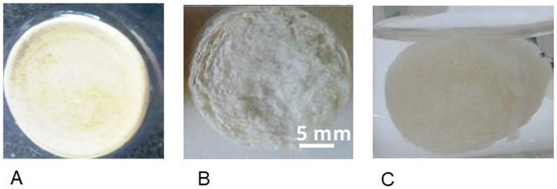
Photographs of hybrid mesoporous scaffolds. (A) sample of dried mesoporous silica nanostructures (B) frontal view of the hybrid scaffold picture evidencing the wide range of porosity sizes observable at macroscopic length, (C) hybrid scaffold immersed in water under to assess their integrity under physiological moisty conditions.
The microstructure and morphology of the scaffolds were analysed by TEM and SEM microscopy (Fig. 3). Figures 3A and 3B show a TEM micrograph of a dried preliminary mixture of mesoporous silica nanostructures and hydroxyapatite nanocrystals, performed to assess their nanometric features. On Fig. 3A, a detail of the ordered channels structure of mesoporous silica matrices, can be observed while on Fig. 3B, HA nanocrystals distributed on the left side of micrograph can be observed showing their characteristic spike morphology, with a moderate size dispersion and an average length above 100 nm.

TEM and SEM micrograph of (A) mesoporous silica nanostructures with their ordered channels structure and (B) together with hydroxyapatite nanocrystals with spike morphology on the left side of the image. (C) SEM micrographs at low magnification showing the availability of a wide hierarchy of pores, and (D) with high magnification displaying the roughness of the organic matrix owing to the inclusion of inorganic nanostructures.
Figure 3C shows highly porous hierarchical structures with a remarkable surface roughness and intricate pore walls. This special surface topography is given by the uniform distribution of inorganic nanostructures throughout the bioactive matrix surface Fig. 3D, to which additionally, mesoporous silica contributes with pores at the nanometric scale and creating a significantly interconnected structure. These particles are partially embedded by the polysaccharides film, which keeps them well-integrated to the chitosan/carrageenan matrix. These images also evidenced the existence of a large interparticle porosity associated with pores of small sizes. Pores <3 μm can improve cell–surface interaction and anchorage-dependent cell–cell communication [16], as reported that could have critical implications in bone regeneration, enhancing cellular function [17] and increasing the initial attachment and seeding efficiency [18].
In clinical terms, we observed tolerance and lack of toxicity of the mesoporous silica and HA, without registering any type of local or systemic allergic reaction.
The following aspects could be observed in the radiological study: Figs 4A and B show a tomography performed 4 months after surgery, where the presence of defects produced in the tibia are clearly observed. The presence of a radiopaque structure in the proximal defect area stands out, compatible with the biomaterial used. Figure 4C shows a 4D reconstruction of the tibia showing an irregular bone surface, highlighting the presence of periosteal perilesional sclerosis at the proximal end of the tibia. In Fig. 4 (lower part) different sections of the tibiae of the two rabbits and scaffold materials are seen, highlighting in its proximal portion a bone defect compatible with the experimentally produced defect, in which an isolated radiodense structure is seen, compatible with regenerative bone sequestration. Radiologically, it appears that the powder forms showed more advanced states of healing and that the 3D hybrid scaffold generates a complete reconstruction of the external tibial cortex.

(A) Tibia tomography at 4 months axial section. (B) Tibia tomography at 4 months sagittal section. (C) 3D Tibia reconstruction. Rabbit 1 left paw. Lower part: microCT images of biomaterials and rabbits. (D) Sponge mesopore 100%. (E) Powder mesopore 100% powder. (F) Sponge mesopore and HA 50%. (G) Powder mesopore and HA 50%.
In the histopathological autopsy study, microphotograph of the right tibia of rabbit 1 (3D hybrid scaffold 100% mesoporous silica, Figs 5A and B upper side) showed a peripheral area with mature lamellar bone, surrounding a central area with biomaterial remains, neoformation of a fine trabeculate inside it and osteoid formation with little structure, delimiting the area almost completely. In the microphotograph of the left tibia of rabbit 1 (Powdered hybrid scaffold 100% mesoporous silica, Figs 5C and D upper side), we could observe an area of medullary necrosis with destructuring of the normal trabeculate, the presence of sequestrations and the formation of granulation tissue with the presence of an inflammatory infiltrate lymphoplasmocyte with some eosinophils.
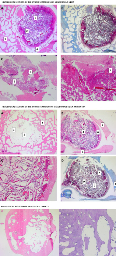
Upper part: Histological sections of the hybrid scaffold 100% mesoporous silica. (A) Sponge, HE, 10×. (B) Sponge, TM, 10×. (C) Powder, HE, 10×. (D) Powder, TM, detail to 20×. (1) Peripheral area with mature lamellar bone (2) Central zone with biomaterial remains. (3) Neoformation of a fine trabeculate inside the biomaterial. (4) Osteoid formation with little structure, almost completely delimiting the intervened area. (5) Unstructured normal trabeculate. (6) Bone sequestration. (7) Granulation tissue with lymphoplasmocyte infiltrate. Central Part: Histological sections of the hybrid scaffold 50% mesoporous silica and HA 50%: A) Sponge, HE, 10×. B) Powder, HE, 4×. C) Powder, HE, 20×. (D) Powder, TM, 4×. (1) Few remains of biomaterial. (2) Ripe bone trabeculae. (3) Incipient trabeculae next to the biomaterial. (4) Trabeculate with greater organization, distribution and connection between trabeculae occupying the entire medullary area of the defect. (5) Mature cortex. (6) Thick ostoid layer. (7) Maturity of the internal trabeculae, similar to that of the peripheral osteoid. (8) Cortical mature bone (9) Internal focus of ossification. Lower part: Histological sections of the controls. HE: Hematoxylin eosin; TM: Masson's trichrome.
The defect in the right leg of rabbit 2 (3D hybrid scaffold 50% mesoporous silica and HA 50%, Fig. 5 central side) showed few remains of the biomaterial and a new formation of more mature bone trabeculae that alternate with incipient ones that are arranged next to the biomaterial (Fig. 5A central side). The left tibia of rabbit 2 (Powdered hybrid scaffold 50% mesoporous silica and HA 50%) showed remains of the biomaterial surrounded by a fine immature trabeculate in which a greater organization, distribution and connection between trabeculae could already be seen occupying the entire medullary area of the defect (Fig. 5B central side). This was surrounded by a mature cortex next to which there was a thick layer of osteoid (Fig. 5C central side). With Masson’s trichrome we could see the degree of maturity of the internal trabeculae, similar to that of the peripheral osteoid. In blue, the mature cortical bone and some internal focus of calcification are observed (Fig. 5D central side).
The photomicrographs of the control defects (Fig. 5, lower side), indicated, in general, an almost complete closure of the cortical part, with the presence of a compact bone also inside the medullary canal and a less numerous but slightly more internal trabeculate mature than in defects with 100% mesoporous silica.
Mesoporous sillica nanoparticles are a new material generation for tissue engineering due to biocompatibility, osseointegration, high stability under pH and temperature variations. Besides, the material shows chemical versatility for surface functionalization, interesting structural and textural properties, and high drug loading capacity [19]. Although there is a profound knowledge about their physical properties, few clinical trials have been developed.
In this pilot study, we tested the tolerance and biological response of mesoporous silica in a rabbit animal model, formulated with or without HA and in two forms of presentation, powder and sponge. In view of the results, it appears that both radiographically and histopathologically, pure mesoporous silica formulations lead to a lower biological response. That is, when formulated with HA, the osteogenic response in terms of osteoconduction is greater. This seems to respond to the role of mesoporous silica as a support material that allows the integration of other drugs and/or materials, apart from regenerative material itself [19–22].
In vivo studies results indicated that mesoporous bioactive hybrid materials produce minimal toxicity to the body. Moreover, biodegradability makes them more suitable for application in bone tissue engineering [23] even in 3D printed scaffolds on controlled architecture which offer a great compression resistance, in vivo osteogenesis and adecuate degradability [24].
An importante limitation in the development of scaffolds to bone regeneration has been the limited vascularization to the tissue regenerating. For this reason, molecules as Recombinant Human Bone Morphogenic-2 (rhBMP-2) improved vascularization and osteogenesis in a rabbit femur defect repair model [25].
Taking all these results into account the key factor seems to be linked to the composition of the different mesopores formulations, like tricalcium phosphate [6], antibiotics [22], bioglasses [20], or HA [26].
The main limitation of this study is obviously the small sample size, so it is necessary to repeat the experiment with a greater number of animals to develop a statistic study. In this sense, although the leporine model is the most used animal in musculoskeletal research [27], is a bulky animal, difficult to handle, relatively expensive and due to its size it cannot be introduced into the CT before slaughter, where the legs separated from the rest of the animal’s body are already studied. For this reason, for future studies we propose the rat femur model, where bone defects of 2–3 mm could be performed, which could also be assessed by microCT throughout the study, allowing us to have more data. The results could vary in different study times, so it is advisable to design an experiment with sacrifices at different times, in case it is carried out in rabbits, or periodic evaluation by microCT if it is carried out in rats (which do fit live on the device).
Conclusions
This pilot study reveals that MS hybrid scaffolds used in defects of the tibia, produced a significant periosteal reaction with a bone involvement compared to the control defects. It seems that the state of bone regeneration at 4 months is more advanced and with less compromised, when 50% MS mixed with 50% HA hybrid scaffolds are used. Scanning electron microscopy have shown in HA nanocrystal a characteristic peak morphology on micrographs with size dispersion and MS has shown to contribute creating an interconnected structure, highlighting the existence of a high interparticular porosity. More studies with different formulations and healing times are necessary in order to advance towards a more efficient bone regeneration material.
Conflict of interest
None to report.
Funding
This research did not receive any specific grant from funding agencies in the public, commercial, or not-for-profit sectors.