



Abstract
BACKGROUND:
Transforming growth factor-β1 (TGF-β1) plays an important role in chondrocyte growth and the synthesis of extracellular matrix (ECM). Due to the rapid metabolism, controlled release systems for TGF-β1 have attracted increasing interest recently.
OBJECTIVE:
In this study, a silk fibroin (SF)/chitosan (CS) scaffold incorporated with TGF-β1-loaded microspheres (MSs) was created for cartilage reparation.
METHOD:
The optimal proportion of the SF/CS composite scaffold was determined by evaluating their micromorphology and the proliferation rate of fibroblasts on the surface. Then, SF/CS/TGF-β1-loaded MS scaffolds were prepared by the adsorption method. TGF-β1 release capacity, degradation patterns, cytocompatibility and in vivo implantation were evaluted.
RESULTS:
The SF/CS/TGF-β1-loaded MS scaffold showed good TGF-β1 release over more than 16 days, which could sequentially stimulate chondrocyte synthetic activity. In vitro cell proliferation experiments showed the SF/CS/TGF-β1-loaded MS scaffold could promote chondrocytes adhesion, growth, proliferation and maintained the cellular morphology. An in vivo study demonstrated that a low inflammatory response was observed in rats and that the materials exhibited good biocompatibility.
CONCLUSION:
the results indicated that our SF/CS/TGF-β1-loaded MS scaffold constitute a promising therapeutic option for cartilage reparation.
Introduction
Cartilage damage can be caused by a variety of conditions, such as trauma [1], arthritis [2], and sports-related injuries [3]. However, Cartilage injuries are difficult to treat because of the limited capacity of cartilage to heal. Over the past few decades, the concept of tissue engineering has provided a promising approach for cartilage tissue reconstruction and regeneration. By applying this approach, an engineered cartilage tissue, which was based on cells, biomaterial scaffolds and growth factors, could integrate into native tissues and restore functions lost due to trauma, disease or aging [4–6]. The ideal cartilage scaffold should simulate the extracellular matrix (ECM) of cartilage in terms of structure, composition and function. It should have good tissue compatibility, maintain a dynamic balance between its degradation rate and tissue regeneration rate, and have a suitable porosity. The final mechanics of the scaffold should be close to cartilage tissue, and can be effectively combined with subchondral bone [7].
Silk fibroin (SF), derived from silk fiber, is a natural biomaterial with good biocompatibility and biodegradability and is able to support appropriate cellular activity without eliciting immune activation in the host [8]. SF was also shown to exhibit good mechanical properties, such as high strength and toughness and ease of processability, which make it similar to natural cartilage tissue; thus, SF has been widely investigated and tested for cartilage tissue engineering applications. However, pure SF is too brittle to be used by itself [9,10]. Chitosan (CS) has been widely used to overcome defects in the mechanical properties of SF. It has been reported that CS could induce the conformational transition of SF from a random coil to a β-sheet structure [11]. This transition makes SF attractive as a biomaterial because SF with a β-sheet structure is resistant to water and has good mechanical properties [9,12]. In addition, CS presents some characteristics similar to those of several glycosaminoglycans (GAGs) and hyaluronic acid, which are commonly found in articular cartilage [13–15]. However, the high swelling property of CS limits its application in cell culture and implantations [16]. The blending of SF with CS overcomes the defects of pure SF and CS and shows remarkable biocompatibility, appropriate hydrophilicity, and suitable mechanical properties [17]. The preparation and characterization of SF/CS scaffolds, such as SF/CS thin films [16] and SF/CS sponges [18], has been studied previously.
In this study, we report the development of SF/CS scaffolds incorporated with TGF-β1-loaded CS microspheres (MSs) for cartilage reparation by blending SF with CS to produce a foam structure. The TGF-β1-loaded SF/CS scaffold takes advantage of TGF-β1’s ability to stimulate chondrocyte synthetic activity and decrease the catabolic activity of IL-1 [19]. The CS MSs also release TGF-β1 of extended periods of time. The results demonstrated that SF/CS scaffolds incorporated with TGF-β1-loaded CS MSs could provide a biocompatible environment to promote chondrocyte growth and attachment, and these scaffolds have potential for various applications in cartilage reparation.
Materials and methods
Materials and reagents
CS was purchased from Golden-Shell Pharmaceutical Co., Ltd., China. Raw silk was purchased from Yi Xian Raw Silk Factory, China. TGF-β1 was purchased from PeproTech CO., Ltd. Fibroblasts (L929 cells) were obtained from Hunan Fenghui Biotechnology Co., Ltd. Chondrocytes were a gift from Peking University Shenzhen Hospital, China. All other reagents and solvents used in our study were of analytical grade.
Preparation of SF/CS scaffolds
SF was extracted from raw silk as described in our previous work [20,21]. The CS solution was prepared by dissolving CS (80% deacetylated, molecular weight: 3 × 10⁵ Da) at a 3 wt% concentration in 0.2 M acetic acid.
To prepare SF/CS composite scaffolds, the 3 wt% SF solution and 3 wt% CS solution were blended at different proportions (SF/CS = 3/1, 1/1, 1/3), and the composite solution was mixed for 15 min. Then, the SF/CS solution was poured into polytetrafluoroethene columniform molds (diameter: 10 mm; height: 25 mm), frozen overnight at −20 °C, and lyophilized for 48 hours. The dry samples were removed from the molds and treated in methanol for 4 hours. The final SF/CS scaffolds were prepared by an additional 48 hours of lyophilization to remove the remaining methanol.
Preparation of SF/CS/TGF-β1-loaded microsphere scaffolds
CS MSs were prepared using an emulsion-ionic crosslinking method. Briefly, 100 mL of liquid paraffin and 2 mL of Span 80 were mixed for 10 min by mechanical stirring at 1000 rpm using a stirrer. Then, a 4 wt% CS solution was added dropwise to the above mixture. After mechanical stirring for another 20 min, 10 mL of sodium tripolyphosphate solution (10% w/v in distilled water) was further added dropwise to stabilize the CS MSs through electrostatic interaction with TPP. The MSs, precipitated in the mixture solvent, were repeatedly washed with excess amounts of isopropyl alcohol and petroleum benzine. A sieve mesh diameter of 20 μm was utilized to filter the MSs. Lyophilization was also used to remove the remaining isopropyl alcohol and petroleum benzine.
TGF-β1-loaded MSs were prepared by absorption of a TGF-β1 solution. The TGF-β1 solution (2 μg in 100 μL of phosphate-buffered saline) was added to 100 mg freeze-dried MSs, on average, and absorbed by water absorption. Then, the MSs were stored at 4 °C for 24 hours and lyophilized.
The SF/CS scaffolds were sliced into round (diameter: 10 mm; thickness: 2 mm) samples. The TGF-β1-loaded MS (MS@TGF-β1) suspension (5 mg/mL) was fabricated by adding MS@TGF-β1 into water and undergoing ultrasonic stirring. Then, 100 μL of the MS@ TGF-β1 suspension was added into every SF/CS scaffold sample and immediately lyophilized, after which each sample was loaded with 0.5 mg of TGF-β1.
Scanning electron microscopy (SEM)
The materials were sprayed gold with a thickness of approximately 5 × 10⁻⁶ cm using a Hitachi IB-2 coating unit under a vacuum of 0.1 Torr. The coated samples were examined using a scanning electron microscope (TESCAN, MIRA3, Czech Republic).
Fourier transform infrared spectroscopy (FT-IR) analysis
FT-IR analysis was conducted by a Thermo Scientific Nicolet-iS50 FT-IR spectrophotometer. Freeze-dried scaffolds were placed onto a “Golden Gate” diamond window and analyzed within the spectral region of 700–1800 cm−1.
TGF-β1 release capacity
To investigate the release kinetics of TGF-β1 from the MSs in vitro, MSs (50 mg) loaded with TGF-β1 and 1 mL of phosphate buffered solution (PBS) solution was added to a 2 mL centrifuge tube. Then, the tube was put into a 37 °C incubator and shaken at a frequency of 80 rpm. After incubation for 1 hour, 3 hours, 6 hours, 12 hours, 1 day, 2 days, 3 days, 5 days, 7 days, 10 days, 13 days and 16 days, the MS suspension was centrifuged at 13500 rpm to collect 500 μL of the supernatant for analysis, followed by the addition of a fresh 500 μL of PBS into the MS suspension. The amounts of TGF-β1 released from the MS were evaluated by using the mouse TGF-β1 ELISA kit (Shanghai Enzyme-linked Biotechnology Co., Ltd., China), and the release kinetics of TGF-β1 from the MSs in vitro were obtained. To determine the total loading amounts of TGF-β1, the loaded MSs were dissolved in acetic acid, and an ELISA kit was used as the tracer.
Degradation patterns
The degradation behaviors of the materials were determined after incubation in PBS (pH 7.4, 37 °C) and with an enzyme (1 U) for several different periods of time. The dry weight (Wo) of the scaffolds was also obtained before they were immersed in solution. On the 7th, 14th, 21st, 28th and 35th days, the scaffolds were removed and rinsed in distilled water; then, they were freeze-dried before being weighed (Wr). There were 4 parallel samples for each group. The residue mass ratio was determined using the following formula:
Cytocompatibility and cell adhesiveness
The cytotoxicity of the SF/CS scaffolds was assessed using a mouse fibroblast (L929) Cell Counting Kit-8 (CCK-8) assay. Prior to the cell test, the scaffolds were sterilized using electron beam sterilization radiation followed by immersion in PBS overnight. The sterilized scaffolds (10 mm in diameter, 2 mm in thickness) were placed into 48-well plates, and 300 μL of cell suspension (with a density of 1.5 × 10⁵ cells/mL) was added to every sample. After 4 h incubation at room temperature (∼25 °C), 500 μL of culture medium (MEM, HyClone, Logan, UT, USA) was added to each well, and the cells were continuously incubated at 37 °C with 5% CO2. The culture medium was changed every 2 days.
The proliferation rate of L-929 cells on scaffolds was tested by a CCK-8 assay (Dojindo Molecular Technologies, Kumamoto, Japan) following the manufacturer’s protocol. For the LIVE/DEAD staining assay, the cells were stained with calcein-AM and propidium iodide (PI). Cell growth was observed using a confocal laser scanning microscope, and live cells (calcein-AM-stained green) were distinguished from dead cells (PI-stained).
Coculture of chondrocytes
Chondrocytes were cultured with SF/CS-MS@TGF-β1 scaffolds to evaluate their potential for cartilage tissue engineering. SF/CS-MSs (without TGF-β1-loaded MSs) were used as the control group. The scaffolds (∼10 mm in diameter, 2 mm in thickness) were placed into 48-well plates, and 200 μL of chondrocyte cell suspension (with a cell density of 105 cells/mL) was added into each of the sample wells. The RPMI 1640 culture medium (11875-093, GIBCO) was changed every 2 days.
The proliferation rate was obtained by a CCK-8 assay following the manufacturer’s protocol.
To observe the viability of chondrocyte cells on scaffolds, they were incubated in 2 μM calcein-AM (to stain live cells) and 2 μM PI (to stain dead cells) in PBS for 15 min at room temperature (∼25 °C), and confocal laser scanning microscopy (CLSM) (Leica TCS SP8, Oberkochen, Germany) was used to observe images of live and dead cells at scheduled time periods (days 1, 3, 7 and 14). The glycosaminoglycan (GAG) contents were measured using the 1,9-dimethylmethylene blue (DMMB) colorimetric assay.
In vivo implantation
All experimental designs for animal studies were reviewed and approved by the Shenzhen Test Centre of Medical Devices. Eight female SD rats weighing approximately 250 g were maintained under general anesthesia by injecting 3% pentobarbital (1 mL/kg). After barbering and disinfection treatment, two scaffolds (2 mm in diameter, 5 mm in thickness) per rat were symmetrically implanted in the dorsal subcutaneous area. At the specified time points of 2, 4, 8 and 12 weeks after implantation, the rats were humanely euthanized, and the implants and surrounding tissues were excised for histological evaluation.
For histological analysis, the collective samples were fixed in 4% formaldehyde in PBS, dehydrated with a gradient series of ethanol, and subsequently immersed in xylene and paraffin. Paraffin sections (4 μm) were prepared using a microtome (Leica RM2235, Germany) for routine hematoxylin–eosin (HE) staining.
Statistical analysis
Experimental data were analyzed using SPSS. The results are presented as the mean ± SD (standard deviation). Mann-Whitney U test was used for statistical comparison of the data between two groups, Bonferroni’s correction test was used for other statistics more than two groups. When the P value was less than 0.05 (P < 0. 05), the data were considered significantly different.
Results and discussion
Characterization of SF/CS scaffolds
Figure 1 shows that the composite scaffold created by the freezing drying method has a porous and interconnective structure, which could be beneficial for cell proliferation and adhesion. The FT-IR spectra of pure SF and SF/CS scaffolds at different SF/CS ratios are shown in Fig. 2a. It is easy to distinguish the characteristic peaks of SF and CS. The absorption bands of pure CS were at 1153 cm−1, 1325 cm−1, and 1595 cm−1, which could be attributed to amide III, NH2 and the asymmetric stretching of the C–O–C bridge of CS, respectively. [16,22] Compared with those of CS, the SF absorption bands were at 1653 cm−1 (amide I), 1539 cm−1 (amide II), and 1,239 cm−1 (amide III), attributed to SF with a random coil conformation. As the SF content of the blended SF/CS scaffolds decreased from 75 wt% to 25 wt%, the amide III band of SF shifted from 1238 cm−1 to 1257 cm−1, indicating the conformational transition of SF from a random coil to a β-sheet structure. Figure 2b shows the viability of L929 cells cultured with SF/CS composite scaffolds with different ratios. With the increase of cell proliferation over time, the addition of chitosan significantly promoted the proliferation rate of fibroblasts on the surface of the scaffold, mainly due to the weaker cell adhesion properties of silk fibroin than chitosan. In the composite scaffold, silk fibroin provides support and elasticity, and chitosan provides biocompatibility, which can give play to their respective advantages. Finally, we chose 1/3 SF/CS scaffold because the cell proliferation rate and fluorescent staining picture of the scaffold were relatively the best among the several groups.

Microscope morphologies of four SF/CS scaffolds with different material ratios. SF/CS=1:0 (a, b); 3:1 (c, d); 1:1 (e, f); 1:3 (g, h).

Characterization of SF/CS scaffolds. (a) FT-IR spectra. (b) Results of the CCK-8 assay for the L929 cells on different scaffolds on days 1, 4 and 7. n = 4. ∗ p < 0. 05.

LIVE/DEAD staining of the L929 cells on different scaffolds on days 1, 4 and 7.
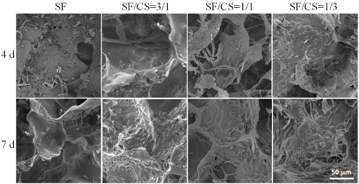
SEM images of L929 cells on scaffolds on days 1, 4 and 7.
The MSs loaded with TGF-β1 were prepared by the emulsion method, which has been used to create many drug-bearing MSs. The MSs were spherical in shape and had an average diameter of approximately 10 μm (Fig. 5b), which made the MSs uniformly distributed in the porous SF/CS composite scaffold (Fig. 5c).
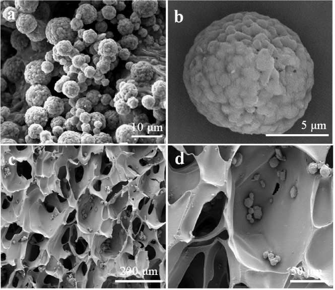
SEM images of TGF-β1-loaded microspheres (a, b) and SF/CS/TGF-β1-loaded microsphere scaffolds.
The release profile for TGF-β1 from the MSs in vitro is shown in Fig. 6a. There was a phenomenon of sudden release at the first stage of delivery for approximately 12 hours, and the cumulative released quantity reached 35.7%. After, the release rate decreased over time. Approximately 85.1% of TGF-β1 was released from the MSs in 16 days. This demonstrated that the MSs had the ability to control TGF-β1 release over a long period of time.
The degradation rate of SF/CS-MS@TGF-β1 scaffolds in vitro is shown in Fig. 6b. The scaffolds lost approximately 80% of their weight for the entire 35-day experimental period when they were immersed in lysozyme solution. However, the scaffolds lost 50% of their weight when the solution was PBS. The results demonstrate that our composite scaffolds allowed for a relatively long release of TGF-β1 in vitro to promote the growth of chondrocytes.

Characterization of SF/CS/TGF-β1-loaded microsphere scaffolds. (a) In vitro release curve of TGF-β1 from the scaffolds; (b) Mass loss ratio of scaffolds with or without an enzyme.
To demonstrate the potential of loaded TGF-β1 scaffolds for cartilage reparation, the viability of chondrocytes cultured with SF/CS-MS@TGF-β1 and SF/CS-MS scaffolds was investigated to demonstrate their biocompatibility in terms of both cell proliferation and morphology. Figures 7a to 7d shows the laser confocal images of cells incubated with the SF/CS-MS@TGF-β1 scaffolds and Figs 7e to 7h show SF/CS-MS scaffolds after incubation for 1, 3, 7 and 14 days, respectively. To reveal their viability, live cells were fluorescent green after being stained with fluorescent calcein-AM, and the dead cells were fluorescent red after being stained with PI. The results clearly demonstrated that an increasing number of live cells attached to the scaffolds with increasing culture time, indicating obvious proliferation. After incubation for 7 days (Figs 7c, 7d, 7g and 7h), live cells almost completely covered the scaffolds without a difference in chondrocyte cell proliferation on SF/CS scaffolds with or without TGF-β1. However, a large difference appeared in the chondrocyte cell morphology after the cells had been incubated for over 14 days. For the SF/CS scaffolds with TGF-β1, the cells became rounded, and the cells incubated with the SF/CS scaffolds without TGF-β1 became spindled (Fig. 7d and 7h inset pictures), which demonstrated that the TGF-β1 SF/CS scaffolds promoted cell proliferation that is similar to that of the cartilage cell state in the body. The rate of chondrocyte cell proliferation on the scaffolds was evaluated by using a CCK-8 assay for up to 14 days, and the results are shown in Fig. 7i. Both scaffolds had good cell proliferation ability after incubation for less than 7 days. When incubated for 14 days, the numbers of cells did not increase notably, and for the different scaffolds, there was no large difference in the cell proliferation results. GAG plays a critical role in regulating the expression of the chondrocytic phenotype and in supporting chondrogenesis both in vitro and in vivo, [15,23,24] so the values of GAG were evaluated by using the DMMB method for up to 21 days, and the results are shown in Fig. 7j. The GAG value of the SF/CS-MS@TGF-β1 scaffolds was larger than that of the SF/CS-MS scaffolds as the incubation time increased, which indicated that the SF/CS-MS@TGF-β1 scaffolds had a better ability to promote cartilage reparation. There was no significant difference in the proliferation experiment and GAG analysis, indicating that the addition of TGF-β1 had little effect on the proliferation of chondrocytes and the secretion of GAG, but the obvious circular chondrocytes observed by laser confocal (compared to the spindle type of the control group) showed that TGF-β1 has a great influence on maintaining the phenotype of chondrocytes. In addition, HE and toluidine blue staining were used to assess the growth status of chondrocytes in the scaffolds (Fig. 8). It can be seen from the figure that chondrocytes not only grew on the surface of the scaffold but also adhered inside the scaffold to a great extent. There is no obvious difference on 14 days, but the number of chondrocytes observed at 21 days in the loaded SF/CS-MS@TGF-β1 group was great larger than that in the TGF-β1-free group, indicating that the addition of MSs and the release of TGF-β1 contributed to the growth of chondrocytes.
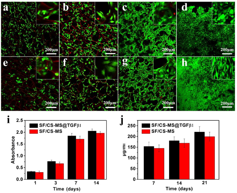
Laser confocal images of chondrocyte cells incubated with the SF/CS-MS@TGF-β1 scaffolds (a, b, c, d) and SF/CS-MS scaffolds (e, f, g, h) for 1 day (a, c), 3 days (b, f), 7 days (c, g) and 14 days (d, h). (i) The rate of chondrocyte cell proliferation on the SF/CS-MS@TGF-β1 and SF/CS-MS scaffolds. (j) The GAG production values of chondrocyte cells on the SF/CS-MS@TGF-β1 and SF/CS-MS scaffolds.

HE and toluidine blue staining of chondrocyte scaffold cocultured for 14 and 21 days.
The results of chondrocyte cells cultured with the SF/CS-MS@TGF-β1 scaffold in vitro demonstrated that these scaffolds have good biocompatibility. To investigate the degradation inflammatory response of these scaffolds in vivo, SF/CS-MS@TGF-β1 scaffolds were implanted subcutaneously in rats. Figure 9 shows the HE staining images of the implanting position after 2, 4, 8 and 12 weeks. It can be seen from the figure that there are many inflammatory cells near the scaffold at the initial stage of implantation (2w), which is a normal immune response after the foreign body is implanted in the body; as time increases, the inflammatory response gradually weakens, new blood vessels and dermal tissue appeared inside of the scaffold indicates that the scaffold can promote the growth of tissues and blood vessels in the body and has good biocompatibility. Scaffold fragments can still be observed after 12 weeks of implantation, indicating that the degradation time of the material is longer than 12 weeks. This longer degradation time is conducive to the healing and regeneration of slow-growing tissues such as cartilage.

HE staining of the SF/CS-MS@TGF-β1 scaffold explants after 2 (a, c), 4 (b, f), 8 (c, g) and 12 (d, h) weeks of implantation. (Black arrow: materials; blue arrow: collagen fibers.)
In this study, SF/CS-MS@TGF-β1 scaffolds were created with an optimal proportion of SF/CS of 1/3; these composite scaffolds showed potential for cartilage reparation. The incorporation of TGF-β1-loaded MS permitted the SF/CS-MS@TGF-β1 scaffolds to release TGF-β1 over a long period of time, enhancing chondrogenesis. Chondrocytes adhered and grew on the surface and inside of the scaffolds, showing that these scaffolds have potential for cartilage tissue engineering. Further animal experiments also demonstrated that the SF/CS-MS@TGF-β1 scaffolds have good biocompatibility in vivo. The study demonstrated here provides a promising therapeutic option for cartilage reparation.
Footnotes
Acknowledgements
This work was supported by the Shenzhen Science and Technology Projects (JCYJ20200-109150605937, 20180309163834680, JSGG20180504170419462, JCYJ20180306170521243) and the National Key Research and Development Program (2018YFE0194300).
Conflict of interest
None to report.