
Abstract
BACKGROUND:
Microbeads for bone repair have been widely studied because they can be conveniently used in clinical applications.
OBJECTIVE:
This study concerns the preparation, physical properties and in vitro characterisation of different types of alginate/calcium phosphate (CaP) ceramic microbeads, which were designed for use as drug delivery systems and bone-regeneration matrices.
METHODS:
Hybrid microbeads were successfully prepared from sodium alginate and various CaP, namely 𝛼-tricalcium phosphate, 𝛽-tricalcium phosphate and hydroxyapatite using the liquid droplet method.
RESULTS:
Porosity, swelling properties and in vitro degradation of the microbeads in the aqueous environment were significantly changed by the added CaP. The compressive strength of the blocks fabricated from the beads was around 120 MPa irrespective of the type of CaP. The initial release rate of the model drug methylene blue was suppressed by the addition of CaP.
CONCLUSION:
The alginate-CaP composite beads hold promising potential as an encapsulation carrier of drugs and component of bone substitutes.
Introduction
Calcium phosphates (CaP) are recognized as a useful biomaterial in hard tissue regeneration [1] because of their osteoconductivity and efficacy in most clinical applications in orthopaedics and dentistry [2]. Hydroxyapatite (HAp; Ca10(PO4)6(OH)2) exhibits excellent biocompatibility because of its similarity in composition to natural bone [3]. Apart from HAp, 𝛼-tricalcium phosphate (𝛼-TCP; 𝛼-Ca3(PO4)2) and 𝛽-tricalcium phosphate (𝛽-TCP; 𝛽-Ca3(PO4)2) have been widely studied as materials for bone repair due to their outstanding biocompatibility, bioactivity and high osteoconductivity. For all forms of bone graft, a spherical shape of the material could be useful for uniform packing in sporadically shaped defects [4].
Recently much attention has been paid to the binding of ceramic bone grafts with bioactive molecules for osteoinductivity and osteogenesis [5], as well as the refining osteogenic properties of bone grafts used for bony defects. The binding of biomolecules to ceramics that allows a controlled release of the laden biomolecules via the binding mode has proven to be difficult. Numerous biomaterials and their derivatives such as alginate [6,7], chitosan [8,9], agarose [10,11], gelatin [12,13], collagen [14,15] and fibroin [16] have been studied for regeneration of osteochondral interfaces or articular cartilage tissues because of their inherent biocompatibility, non-toxicity and biodegradability.
Although natural materials allow favourable biological interactions with host tissues, the low mechanical strength and instability of the materials compared with the native cartilage sometimes complicate their clinical applications [17]. Alginate is considered to be biocompatible, non-toxic, non-immunogenic and biodegradable. Given its natural abundance and low cost, it has been widely used as thickener and emulsifying agent in the food industry and as tissue engineering material. The choice of alginate as a polymeric vehicle can be attributed to its favourable properties and versatility. Sodium alginate and most other alginates from monovalent metals are soluble in water, forming solutions of considerable viscosity. Alginate can be easily modified in any form such as hydrogels, microbeads, microcapsules, sponges, foams and fibres, allowing increased applications in various fields such as tissue engineering and drug delivery. Alginate can be chemically or physically modified for tuning of its biodegradability, mechanical strength, gelation property and cell affinity towards respective applications [18].
Various alginate-CaP hybrids have been developed as synergetic systems for bone regeneration in the form of microspheres [4], cement [19,20], and bulk porous scaffold [21–23]. In the case of microsphere preparation, HAp and calcium titanium phosphate were selected as and inorganic phase and enzyme release was evaluated [4]. However, no comparative studies of alginate-based hybrid beads with various CaP such as HAp, 𝛼-TCP and 𝛽-TCP prepared by the same experimental conditions have been reported on applications in interfacial osteochondral defects or drug delivery vehicles for hard tissue regeneration. In addition, although several hybrid microspheres have been developed from CaP and polylactide, a representative synthetic biodegradable polymer [24,25], effects of CaP have not been sufficiently revealed. If such points are clarified, it is expected that fundamental design for the hybrids with varied biological properties and drug delivery ability can be established.
In our pursuit, we performed a comparative study by combining 𝛼-TCP, 𝛽-TCP or HAp with sodium alginate, a natural polysaccharide that has been shown to be biocompatible. This work describes the preparation and initial characterisation of the alginate microbeads with and without CaP, designed as injectable drug delivery matrices and bone filling materials.
Materials and methods
Materials
Sodium alginate, calcium chloride (CaCl2), and methylene blue were purchased from Wako Pure Chemical Industries, Ltd., Osaka, Japan. 𝛼-Tricalcium phosphate (𝛼-TCP-B) and 𝛽-tricalcium phosphate (𝛽-TCP-100) were purchased from Taihei Chemical Industrial Co., Osaka, Japan and HAp was purchased from Central Glass Co., Ltd., Tokyo Japan. Ultrapure water (prepared using Direct-Q; Nihon Millipore K.K., Tokyo, Japan) was employed for experimental purposes.
Preparation of alginate and alginate hybrid beads
Sodium alginate was dissolved in deionized water to a final concentration of 4% (w/v) with and without equal weight ratio of 𝛼-TCP, 𝛽-TCP or HAp. Pure and hybrid alginate beads were prepared by dripping the 4 mass% sodium alginate solution into CaCl2 under constant stirring at room temperature. The droplets were left for 1 hour in the gelling bath before being removed and rinsed with distilled water.
Material characterisation
The surface structural changes of the samples were characterised using a scanning electron microscope (SEM; Model S-3500N; Hitachi Co., Tokyo, Japan), an energy dispersive X-ray analyser (EDX; Model EX-400; Horiba Co., Kyoto, Japan), X-ray diffractometer (XRD; MXP3V; Mac Science Ltd., Yokohama, Japan) and Fourier-transform infrared spectrometer (FT-IR, FT/IR-6100; JASCO Co., Tokyo, Japan). For FT-IR, the gels were first pulverised and mixed with KBr powder at a mass ratio of 1:100. A thin film was prepared by uniaxially pressing the mixed powder to measurement.
Porosity and density measurements
The density and porosity of the prepared samples were measured by water displacement. A sample with a known weight w
1 was immersed in a beaker holding a known volume v
1 of water. A series of brief evacuation-repressurisation cycle were performed to force the water into the samples. Then the total volume of the water plus the water-impregnated samples (v
2) and the volume of the residual water after the water-impregnated samples were removed (v
3) were recorded. The density of the sample (d) and the porosity of the sample (𝜀) are expressed as follows:
The weights of the dried beads and beads immersed in deionized water for 48 h at room temperature were determined. In brief, the pre-weighed, dried hybrid beads were immersed in 50 ml of ultrapure water at 25 °C for 24 h. At a regular time interval (1 h), the hybrid beads were taken out from ultrapure water, the surface water was blotted off with tissue paper and then reweighed. The experiment was carried out until the equilibrium weights of the films were reached. The swelling ratio was evaluated using:
The alginate/alginate hybrid hydrogels were incubated in 20.0 ml of saline solution at 37 °C. Gel degradation behaviour was monitored by measuring the dry weight loss over time. At least four samples at each time point were used to obtain the weight loss curve. Experiments were done under sterile conditions to prevent bacterial and fungal contamination.
Mechanical properties
The mechanical strength of the samples was evaluated in terms of compressive strength. For the purpose, we used moulds made with cylindrical cavities in silicon rubber sheets of the preferred thickness. Afterwards the outer layer was coated with 50 mass% polyacrylic acid solution and the cavity was immediately filled with the granules, and left to dry completely for 2 days. The completely dried samples were pulled out from the silicon sheet. The diameter and height of each sample were first measured with a digital micrometre (IP65; Mitutoyo Co., Ltd., Kanagawa, Japan). Each sample was crashed at a crosshead speed of 1 mm/min using a universal testing machine (Autograph AGS-J; Shimadzu, Kyoto, Japan). An average of eight samples was taken as the value of compressive strength.
In vitro release of model drug
Methylene blue was used as a model drug to evaluate the performance of the calcium alginate beads in controlled drug delivery. Thirty samples of calcium alginate beads were equilibrated in a 30-ml methylene blue solution (500 mg/ml) for 24 hours. The beads were rinsed thrice before the drug release studies. The release profile of methylene blue was studied using a UV-Vis spectrophotometer (V-630; JASCO Co., Tokyo, Japan). The concentration of methylene blue was determined spectrophotometrically at 664.5 nm.
Statistical analysis
All values were analysed by one-way analysis of variance followed by Turkey’s multiple comparison tests. Significant differences were assumed at p < 0.05.
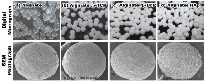
Digital photographs and SEM images of the prepared beads after drying.
Figure 1 illustrates the typical optical and SEM photographs of the prepared beads. There were no significant differences in the shape and size of the granules at macroscopic level. However, at higher magnification differences could be clearly observed in terms of roughness and shape. Alginate and alginate/HAp showed numerous cracks.
Figure 2 shows the XRD patterns of the prepared beads. Alginate showed a broad peak at 2𝜃 = 13.7°, consistent with its amorphous nature [26,27]. The other samples demonstrated peaks corresponding to each CaP (𝛼-TCP: JCPDS card 9–348, 𝛽-TCP: JCPDS card 9-169 and HAp: JCPDS card 9–432). There were no other foreign peaks in any of the samples.

XRD patterns of the prepared beads after drying.
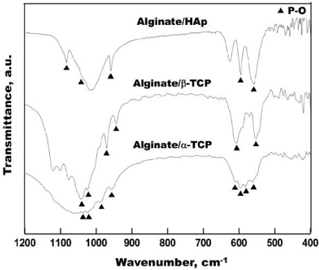
FT-IR spectra of alginate/𝛼-TCP, alginate/𝛽-TCP and alginate/HAp beads after drying.
Figure 3 presents the FT-IR spectra of alginate/𝛼-TCP, alginate/𝛽-TCP and alginate/HAp hybrid beads. Peaks of P-O characteristic of CaP were observed for all samples around 550–500 and 950–1050 cm−1 [28,29]. According to Ma et al., HAp should have a strong absorption band around 1035 cm−1 due to the stretching of PO4 3− [28]. However, it is well known that sodium alginate also has a strong absorption band of C–O–C at this wavenumber [30]. Thus, the observed P-O band of alginate/HAp around 1035 cm−1 would be very weak.
Swelling behaviour is a very important property of a drug delivery vehicle, because it has a great influence on the control of the drug release. Figure 4 describes the swelling ratio of the samples in ultrapure water at 37 °C. The hybrid beads reached their equilibrium swelling state at around 15 hours. In the first 12 hours, the swelling ratio decreased in the order: Alginate ∼ Alginate/𝛼-TCP > Alginate/HAp ∼ Alginate/𝛽-TCP. After 24 hours, it decreased in the order: Alginate > Alginate/HAp > Alginate/𝛼-TCP > Alginate/𝛽-TCP.
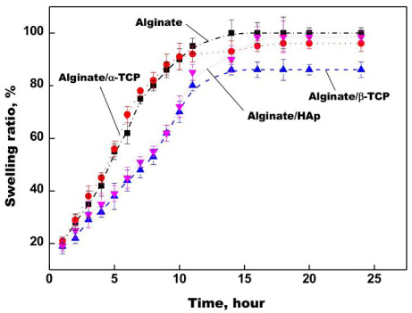
Swelling ratio of the prepared beads after immersion in ultrapure water for various periods (n = 3).
Figure 5 describes the density and porosity of the prepared samples. The density decreased in the order: Alginate/HAp > Alginate/𝛽-TCP > Alginate/𝛼-TCP > Alginate. The porosity of alginate was around 65%, while those of the other samples were around 30% irrespective of the kind of CaP. There were distinct differences in the density and porosity among the samples. The added CaP was assumed to fill the pores in the alginate matrix, thereby reducing the porosity.

Density and porosity of the prepared beads.
As a preliminary model of bioabsorption of the beads in an injection site, degradation was monitored in vitro by measuring the dry weight of the samples as a function of incubation time in saline at 37 °C (Fig. 6). Weight loss after 35 days decreased in the order: Alginate > Alginate/𝛽-TCP > Alginate/𝛼-TCP > Alginate/HAp. The SEM images for the beads before and after immersion in PBS are shown in Fig. 7. Although a lot of pores were formed in alginate after immersion, significant changes in size and shape were not observed. It was found that the addition of CaP to the alginate beads suppressed degradation in the aqueous environment, and this phenomenon would be induced by suppression of the swelling.
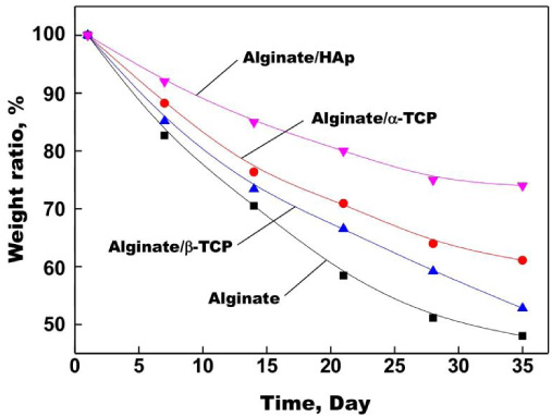
Degradation rate in vitro of the prepared beads in a saline solution at 37 °C for various time periods (n = 4).
Blocks fabricated from all types of beads were tested for their compressive strength (Fig. 8). Equal weights and volumes were maintained throughout the experiment to maintain the homogeneity of the system. The compressive strength of the blocks of alginate, alginate/𝛼-TCP, alginate/𝛽-TCP and alginate/HAp were 122.84 ± 4.39, 125.52 ± 11.2, 119.37 ± 7.84 and 129.26 ± 12.4 MPa respectively.
Figure 9 shows the release profiles of methylene blue from the beads. Alginate showed a burst release profile. The t 50 value (50% of drug release) for alginate was 55 minutes whereas the t 50 values of the composite beads were extended to 100–300 minutes.
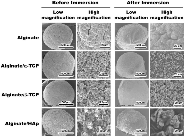
SEM photographs of the prepared beads before and after immersion in saline at 37 °C for 35 days.
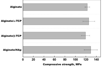
Compressive strength of the blocks fabricated by accumulation of the prepared beads (n = 8).
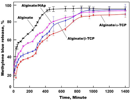
In vitro release profiles of methylene blue from the prepared beads.
Composite microspheres of alginate and various CaP were prepared through gelation by Ca2+. XRD results indicate that the amorphous nature of alginate is diminished by the accumulation of CaP particles (see Fig. 2). The crystallinity of each CaP was confirmed to remain intact in the ionically cross-linked polymer network, which may affect the mechanical properties of the hybrids.
Addition of HAp or 𝛽-TCP to the alginate microspheres decreased the swelling ratio in aqueous conditions (see Fig. 4). In pure alginate, the large number of hydrophilic groups and the void space in the cross-linked network would lead to the highest swelling ratio. The addition of HAp or 𝛽-TCP to alginate beads was found to suppress the initial swelling in the aqueous environment. Ionic interactions between alginate and the added CaP would block significant water penetration. Meanwhile, alginate/𝛼-TCP showed a similar swelling profile to alginate. 𝛼-TCP could easily react with the surrounding water to convert into low crystalline HAp [31]. Therefore, a large amount of water could presumably penetrate into alginate/𝛼-TCP. In the case of alginate/HAp, the swelling ratio significantly increased after 12 hours to resemble that of alginate. This could possibly be due to the degradation of the interface between alginate and HAp by the swelling, although detailed structural changes should be further analysed. Improvement of chemical bonding between alginate and HAp particles by silane coupling agents may suppress the significant swelling at the later stage.
Compressive strength of the blocks fabricated by accumulation of the microspheres was more than 100 MPa irrespective of inorganic phase (see Fig. 8). It is assumed that intergranular voids in the blocks are filled with polyacrylic acid even in pure alginate. The measured compressive strength of the hybrid blocks is comparable to that of typical glass ionomer cements [32]. Tanahashi et al. demonstrated that carboxyl groups in organic polymers form tight ion-ion interactions with Ca2+ [33]. Therefore, similar interactions may exist between CaP and the carboxyl groups in alginate and/or the polyacrylic acid in the present samples. Blocks fabricated from the microbeads are thus expected to be useful materials for bone substitutes. For this purpose, not only compressive but also bending strength is important and should be clarified in the future. In order to enhance the bending strength, reinforcement of the interface between the constituent phases is important. In this study, ionic polyacrylic acid was used as a binder for fabrication of the blocks. This would enhance bonding of each microsphere, since components of the microspheres also have ionic characteristics.
It was found that the incorporation of CaP into the alginate beads is effective for the slow release of methylene blue (see Fig. 9). This can be attributed to the suppression of swelling and biodegradation (see Figs 4 and 6). Moreover, CaP likely adsorbs methylene blue tightly onto their surfaces, having a larger quantity of ionic functional groups than alginate. Notably, the initial release rate of alginate/HAp was higher than that of alginate/𝛽-TCP. 𝛽-TCP is reportedly more negatively charged than HAp [34], thus the former adsorbs the positively charged methylene blue more tightly than the latter. This has implications in the selection of the appropriate kind of CaP according to the target drug to obtain the desired drug release profile.
In order to further understand the release behaviour, the release data were investigated by fitting the cumulative release data. Several mathematical equation models describe drug dissolution and/or release from DDS. In the modern era of controlled-release oral formulations, the Higuchi equation has become a prominent kinetic equation in its own right, as evidenced by employing drug dissolution studies that are recognized as an important element in drug delivery development [35]. After simplifying the above equation, the Higuchi equation can be represented in the simplified form:
The data obtained were plotted as cumulative percentage drug release versus square root of time. Therefore, the simple Higuchi model will result a linear Q versus t 1∕2 plot having a gradient, or slope, equal to K H and we say the matrix follows t 1∕2 kinetics.
For a detailed kinetic study, we selected the initial period drug release data from 5 to 120 minutes. The cumulative % drug release vs. square root of time graph has been plotted. As shown in Fig. 10, it is clear that all sets of prepared beads may follow the Higuchi square root model. It was found that a higher correlation coefficient was observed for alginate/𝛼-TCP and alginate/HAp (0.99), whereas for the alginate and alginate/𝛽-TCP sphere it was 0.98. Hence, we can say that the drug release profile of methylene blue as a model drug from the developed spheres mainly follows a diffusion-controlled release mechanism at the initial stage, although the mechanism may be changed later by partial degradation.
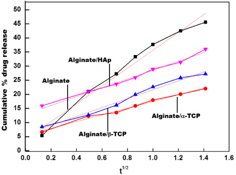
Higuchi model kinetic release of methylene blue from the prepared beads.
The obtained hybrid microspheres are expected to be applied for a drug delivery carrier. Many studies on in vitro and in vivo comparisons of drug release of polymer microspheres have been reported [36,37], but not many have studied inorganic materials. Wang et al. prepared microspheres with an average particle size of 14.3 μm in which the doxycycline was incorporated into poly(lactide-co-glycolide) (PLGA)-coated HAp microspheres, and compared their release behavior in vitro and in vivo [38]. The controlled release characteristics were improved by the PLGA coating. Furthermore, 40% of the contained doxorubicin was released in 150 h or more in vitro, and the steady concentration was maintained in mouse plasma. On the other hand, although the incorporated substances were different, the microspheres produced in this study released 40% of the methylene blue in 1 to 5 h. The main reason for these phenomena is thought to be the difference in hydrophobicity of the polymers. It is assumed that PLGA suppressed the penetration of the surrounding water into the microspheres due to its hydrophobicity and contributed to the controlled release. In future studies, control in the hydrophobicity of the polymers to optimize the drug release behavior is needed.
In this work, we performed a comparative study on alginate-based composite beads modified with various kinds of CaP. Particles with high sphericity were obtained irrespective of the kind of CaP. There were no significant differences in the mechanical behaviour of the blocks fabricated from the beads. The swelling properties and in vitro release profile of methylene blue differed between the beads of different CaP. The drug release profile from the developed spheres follows the diffusion-controlled release mechanism. The approach used in this study not only enhanced the bead strength substantially, but also prolonged the bead dissolution for the controlled release of drugs. We have successfully demonstrated that microbeads with a suitable release profile for the target drug can be obtained by fabrication from alginate and the appropriate kind of CaP.
Footnotes
Acknowledgements
Conflict of interest
None to report.