
Abstract
BACKGROUND:
The biologic scaffolds derived from decellularized tissues and organs have been successfully developed in a variety of preclinical and/or clinical studies.
OBJECTIVE:
The new decellularized liver-regenerative 3D printing biomaterials were designed and prepared for cell-based liver therapies.
METHODS:
An extraction process was employed to remove the tissue and cellular molecules from porcine liver via pretreatment of supercritical fluid of carbon dioxide (ScCO2). Varying porosities of the decellularized liver tissues were created using papain-containing reagent treatments after ScCO2.
RESULTS:
The resulting liver-regenerative 3D printing biomaterials of decellularized liver collagen scaffolds were characterized by Fourier transform infrared spectroscopy, thermo-gravimetric analysis, differential scanning calorimetry and scanning electron microscopy.
CONCLUSIONS:
The decellularized liver collagen scaffolds with good thermal stability (>150 °C) were obtained and employed as liver-regenerative 3D printing biomaterials for cell-based liver therapies.
Introduction
Genetically distinct types of collagen have been fractionated into types I, III, IV and V preparations using judicious salt precipitation from dilute acid and neutral pH solutions. The average distribution of collagen was 42.5, 39.5, 6.9 and 10.6 percent types I, III, IV and V collagen respectively in normal liver [1]. The biologic scaffolds derived from decellularized tissues and organs have been successfully developed in a variety of preclinical and clinical studies [1–3]. Decellularization was used to efficiently remove inhabiting cell contents from a tissue or organ while keeping components and functional proteins that constitute the extracellular matrix (ECM) [1–3]. Importantly, the decellularized organs provided a translucent biologic scaffold composed of an acellular naturally occurring matrix, which retained tissue-specific three-dimensional ultrastructure, intact vascular system, and mechanical integrity [1–3]. Hence, numerous biologic materials for scaffolds have been designed and used in various tissue engineering and medical applications [1–11]. With the advantages of biocompatibility, degradability and non-toxicity, natural materials such as collagen were considered in particular [7–10]. Collagen scaffolds may either be entirely derived from natural sources or hybridized with synthetic polymers. Naturally derived collagen possesses the potential advantages of specific cell interactions and hydrophilicity. Naturally cross-linked collagen scaffolds provide effective starting materials for the fabrication of collagen-based scaffolds. The purified scaffolds retain their biomechanical properties as long as collagen is not denatured by the extraction procedure. Insoluble collagen scaffolds have been used to produce biomaterials for ligament and tendon repair, ocular implants and dressings, organ-regenerative collagen biomembrane, drug delivery vehicles, and as scaffolds for tissue engineering [7–10].
In this study, a new decellularization procedure combined with supercritical fluid of carbon dioxide (ScCO2), alkaline and papain to prepare liver-regenerative biomembrane with collagen scaffolds, which show a good potential in several medical applications. This study describes an effective procedure for the decellularization of porcine liver. The resulting liver-regenerative biomembrane with decellularized collagen scaffolds via a designed procedure are combined with papain-containing reagent treatments after supercritical fluid. Collagen scaffolds were characterized by Fourier transform infrared spectroscopy (FTIR), thermo-gravimetric analysis (TGA), and scanning electron microscopy (SEM).
Materials and methods
Materials
All the reagents of acetic acid, alcohol, sodium hydroxide, TritonX-100, sodium dihydrogen phosphate, disodium hydrogen phosphate and papain were used and purchased from Sigma-Aldrich (USA).
A new decellularization procedure using ScCO2 before chemical and papain-containing reagents treatments
The porcine liver, which was treated with ScCO2, was soaked in a mixture of 2.0% aqueous sodium hydroxide solution and 3.0% aqueous TritonX-10 solution for 6 h at 25 °C [7]. The sample was washed with PBS (0.2 g potassium dihydrogen phosphate and 1.150 g disodium dihydrogen phosphate) at the intermediate interval under ultrasonic wave to remove residual fat, and organic matter. The resulting ScPLS1 was obtained (Table 1).
A new decellularization procedure by using alkaline and papain treatments after supercritical fluid of carbon dioxide
A new decellularization procedure by using alkaline and papain treatments after supercritical fluid of carbon dioxide
(a) ScCO2 was employed before decellularization reagent treatments. (b) ScPLS: scaffolds from porcine liver via ScCO2 treatments.
The porcine liver, which was treated with ScCO2, was soaked in a mixture of 2.0% aqueous sodium hydroxide solution and 3.0% aqueous TritonX-10 solution at 25 °C for 6 h with magnet mixer. The sample was further treated with 0.5 U/ml aqueous papain solution at 25 °C for 12 h and washed with PBS at the intermediate interval under ultrasonic wave to remove residual fat, and organic matter. The resulting liver collagen scaffold, ScPLS2, was obtained (Table 1). The resulting liver collagen scaffolds were analyzed and characterized by FTIR, SEM, TGA and DSC.
A new decellularization procedure using after papain-containing reagents treatments
The porcine liver, which was treated with ScCO2, was soaked in 0.5 U/ml aqueous papain solution at 25 °C for 12 h, followed by soaked a mixture of 2.0% aqueous sodium hydroxide solution and 3.0% aqueous TritonX-10 solution at 25 °C for 6 h with magnet mixer. The sample was further washed with PBS at the intermediate interval under ultrasonic wave to remove residual fat, and organic matter. The resulting liver collagen scaffold, ScPLS3, was obtained. The resulting liver collagen scaffolds were analyzed and characterized by FTIR, SEM, and TGA (Table 1).
Results and discussion
Fourier transform infrared spectroscopy of liver collagen scaffolds
The structural construction due to the decellularization of porcine liver was confirmed by FTIR spectroscopy, as depicted in Fig. 1. All the peaks found in the FTIR spectrum were corresponding to liver collagen scaffolds with ECM structure. The entire scaffolds showed characteristic amide I (∼1641 cm−1), amide II (∼1555 cm−1) and amide III (∼1234 cm−1), amide A (∼3289 cm−1), and amide B (∼3110 cm−1) absorption bands of collagen.
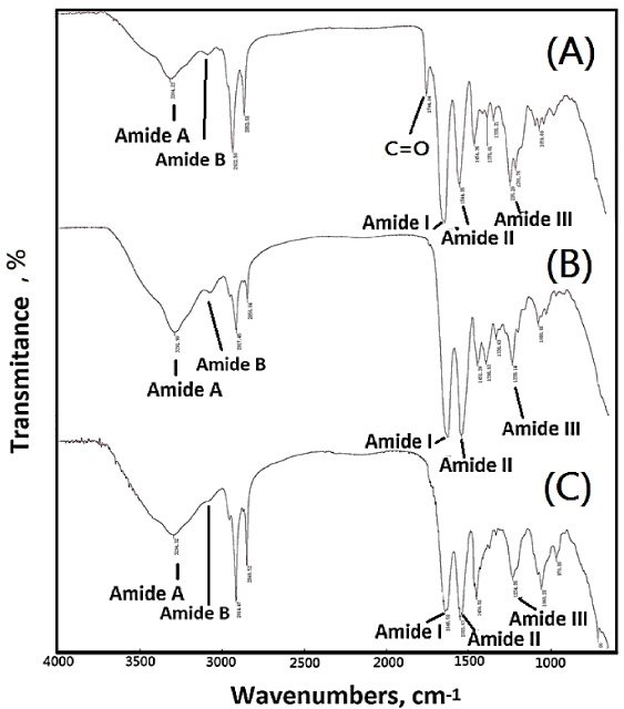
FTIR spectra of (A) ScPLS1, (B) ScPLS2, and (C) ScPLS3.
In Fig. 1(A), the liver collagen scaffold shows the characteristic absorption band of water bounded amide A (∼3304 cm−1) and exhibited a broad band associated with the N–H stretching vibration of hydrogen-bonded amide groups. The characteristic absorption band of amide B (∼3104 cm−1) exhibited a relative broad band associated with the N–H stretching vibration of protein groups. The characteristic absorption band of amide I (∼1643 cm−1) corresponds to the carbonyl group was present within the triple helix structure in the secondary structure of the collagen protein. The N–H bending and N–H stretching vibration were assigned to amide II (∼1544 cm−1) and amide III (1235 cm−1), respectively. All scaffolds exhibited C–H stretching absorption bands at 2922 cm−1 and 2852 cm−1, corresponding to the CH2 and CH3 in the collagen scaffolds and lipids. The IR spectral data exhibit all the characteristic peaks corresponding to the entire scaffolds. The absorption band at ca. 1746 cm−1 𝛿 (C=O) was attributed to the fatty acid.
The FTIR spectrum of collagen scaffold samples ScPLS2 and ScPLS3 showed the characteristic peaks corresponding to fatty acids was disappeared, which implied no lipid residue in the resulting liver collagen scaffold as shown in Figs 1(B) and 1(C). The introduction of papain-containing reagent treatments after supercritical fluid of carbon dioxide (ScCO2) into the procedures would enhance the efficiency of decellularization of liver collagen scaffolds no whether the papain-containing reagent treatment was carried out before and after NaOH/Triton X treatments (Table 1).
Microstructures of resulting scaffolds were characterized by SEM as shown in Fig. 2. A microstructure with pore shape was remarkably observed in the liver collagen scaffolds derived from porcine liver with ScCO2 treatment. Even papain was not further employed to move the residues of small lipid and protein molecules such as ScPLS1, the pore-containing microstructure can be observed in Fig. 2(A).

Scanning electron micrographs of (A) ScPLS1 with 50 μm scale, (B) ScPLS2 with 50 μm scale, and (C) ScPLS3 with 50 μm scale.
The additional papain treatment was considered to solve the lipid problems of porcine liver. Figure 2(B) shows the SEM of ScPLS2, which was decellularized by using an papain (aq) treatment after ScCO2. The mixtures of 2% NaOH (aq) and 3% Triton (aq) were further employed within the same procedures of ScPLS2. The porous microstructures could be remarkably observed and cleared in the SEM images of ScPLS2. The pore diameter of pore-containing microstructures could be observed in the range between 15 μm and 50 μm and separated by the boundary walls with a wide in the range between 3 μm and 5 μm (Fig. 2(B)). The mixtures of 2% NaOH (aq) and 3% Triton (aq) were employed before papain (aq) treatments and the pore-containing microstructures were still observed, as shown in Fig. 2(C). The pore diameter of pore-containing microstructures could be observed in the range between 3 μm and 10 μm and separated by the quite narrow boundary walls (ca. <1 μm) (Fig. 2(B)).
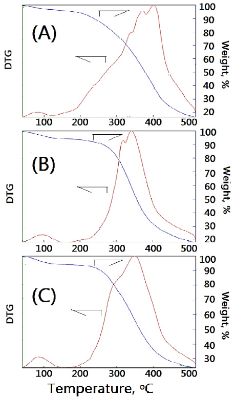
Thermogravimetric analysis of (A) ScPLS1, (B) ScPLS2 and (C) ScPLS3.
Thermal stability of resulting liver collagen scaffolds could be characterized by TGA. The main loss was presented in two different temperature ranges of area I (<150 °C) and area II (150–500 °C). The curve in area I corresponded to the loss of the physisorbed and chemical water in the resulting scaffold, which represented the 5–8 wt% of the scaffold, which occurred at 40–100 °C. The following main loss, occurring in the area II, for the scaffold was observed at 340 °C (Figs 3(B) and 3(C)). The losses would be related to the combustion of the resulting liver collagen scaffolds. In Fig. 3(A), the main loss was also presented in two different temperature ranges of area I and area II. The 5 wt% loss of the resulting liver collagen scaffold without papain (aq) treatments was observed at 40–100 °C. The following main loss, occurring in the range of 150 and 400 °C (area II in the thermogram), was related to the combustion of collagen scaffolds (Fig. 3(A)). The resulting liver collagen scaffold without papain (aq) treatments showed a relatively high thermal stability which might be the complicated associations among ECM and impurities. Also these complicated associations could be supported by the DTG results of ScPLS1. A broad band in the DTG curve with some shoulders and peaks was observed, as shown in Fig. 3(A). The relative simple profiles were observed in the DTG curves of ScPLS2 and ScPLS3.
Furthermore, differential scanning calorimetry (DSC) studies were performed to understand the behavior of resulting liver collagen scaffolds on the application of thermal energy. The DSC curves of the resulting decellularized liver collagen scaffolds with different treatments such as ScPLS1, ScPLS2 and ScPLS3 were at 100 °C, 110 °C, and 120 °C, respectively (Fig. 4). The relative remarkably transition temperatures suggest that the decellularized collagen scaffold had good stability at high-temperature environment. Thermal stability also had an influence on the durability of the resulting scaffolds. The thermal stability of ScPLS1 with lipids would show a board curve in the range of 80 °C and 240 °C, which might to due to complicated associations among ECM and impurities. This specific phenomenon was observed in TGA and SEM results, as shown in Figs 2(A) and 3(A).
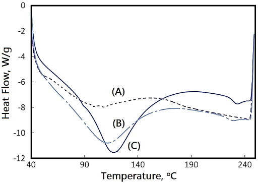
DSC analyses of (A) ScPLS1, (B) ScPLS2 and (C) ScPLS3.
In this study, decellularized liver collagen scaffolds with porous microstructures were successfully prepared from porcine liver by using papain (aq) treatments after ScCO2. The retain ECM and integrity scaffold-structure was observed. The decellularized liver collagen scaffolds with good thermal stability were observed. The resulting scaffolds could be employed in some medical applications such as liver-regenerative 3D printing biomaterials for cell-based liver therapies.
Footnotes
Acknowledgement
The author would like to acknowledge the PARSD Pharmaceutical Technology Consultants Ltd Co. for financial and technical support. The author also thanks Ciao-Yi Syu and Yu-Lin Shen for their technical assistance.
Conflict of interest
None to report.