
Abstract
BACKGROUND:
In stem cell therapy, due to the lack of an effective carrier, a large number of transplanted stem cells are lost and die. Therefore, finding a suitable carrier has become a further direction of stem cell therapy.
OBJECTIVE:
In research on the co-culture of polycaprolactone (PCL) with 1,1 ′ -Dioctadecyl-3,3,3 ′ ,3 ′ - tetramethylindocarbocyanine perchlorate (DiI) labeled bone marrow mesenchymal stem cells (BMSCs), we observe the effect of materials on the growth and proliferation of DiI labeled stem cells, and the effect of DiI labeling on patch preparation, so as to find a kind of biomaterial suitable for the growth and proliferation of BMSCs, and find a suitable cell carrier for stem cell therapy of myocardial infarction and in vivo tracing.
METHODS:
Clean grade Sprague Dawley rats were selected as experimental objects, BMSCs were isolated and cultured, and the surface markers were identified by flow cytometry. After the BMSCs were cultured for 3 passages, the BMSCs were stained with DiI dye, and the BMSCs DiI and PCL biomaterial film were co-cultured. After 24 hours, the cell growth was observed under fluorescence microscope, and fixed for scanning under electron microscope. The cell proliferation was detected by CCK-8 at 1, 4, 7, 10 days of culture. The measurement data conforming to normal distribution are expressed in the form of mean ± standard deviation (
RESULTS:
BMSCs were strongly positive for CD90, CD44H, but negative for CD11b/c, CD45. Under fluorescence microscope, BMSCs DiI showed red light, fusiform or polygonal. Under the scanning electron microscope, the cell patch formed by co-culture of PCL film and DiI-BMSCs had a large number of cells on the surface and normal cell state. CCK-8 assay showed that the OD value on the first day was 0.330 ± 0.025; The OD value was 0.620 ± 0.012 on the 4th day, 1.033 ± 0.144 on the 7th day and 1.223 ± 0.133 on the 10th day. There was significant difference among the time points (P < 0.05).
CONCLUSIONS:
The cell patch made of PCL film and DiI labeled BMSCs can survive and proliferate on the surface, so it can be used as a scaffold material for stem cell therapy in vivo.
Keywords
Introduction
Stem cell therapy is a method with great potential to treat a variety of diseases. It is the research hotspot and focus in recent years [1]. Through stem cell transplantation, the differentiation potential of stem cells can be used to replace damaged cardiomyocytes and improve some functions of damaged heart, so as to treat myocardial infarction [2]. Bone marrow mesenchymal stem cells (BMSCs) have the advantages of easy material selection, no immune rejection, no ethical problems and multi-directional differentiation potential [3–6], which are suitable for stem cell therapy. However, due to the lack of effective vector, a large number of transplanted stem cells lose and die, resulting in poor treatment results. Therefore, finding a suitable carrier has become a further direction of stem cell therapy.
Polycaprolactone (PCL) is a polyester obtained by ring opening polymerization of caprolactone monomer. It has good biocompatibility, absorbability, excellent processability, diverse synthetic processes and low production cost [7]. It has been widely used in medical fields such as tissue engineering [8]. In this study, rat BMSCs were labeled with 1,1 ′ - dioctadecyl-3,3,3 ′ ,3 ′ -tetramethylindocyanine perchlorate (DiI) and co-cultured with PCL film to observe the effect of PCL film on the adhesion and proliferation of BMSCs, so as to lay a foundation for further in vivo tracing experiment and further application.
Materials and methods
Materials
SPF grade Sprague Dawley (SD) rats aged 4–5 weeks, with a body weight range of (120 ± 20) g, were purchased from SBF (Beijing) Biotechnology Co., Ltd. [animal certificate no. SCXK (Beijing) 2019-0010] (China); Fetal bovine serum was purchased from Gibco (USA); Penicillin streptomycin double antibody, trypsin, cell counting kit-8 (CCK-8) reagent and DiI dye were purchased from Shanghai Yisheng (China); Dulbecco’s modified Eagle medium (DMEM) was purchased from Hyclone (USA); Flow cytometry antibody was purchased from Abcam (USA); and PCL films were obtained from the Department of Chemical Engineering of Tsinghua University and Suzhou Institute of Environmental Innovation of Tsinghua University (China). Films were prepared by electrospinning: in the environment of 25–30 °C and humidity below 50%, the fibers were prepared using prepared PCL solution by electrospinning device and deposited on the collection device. The average fiber elastic modulus of the material were 0.5 GPa.
Isolation, culture and passage of BMSCs and Identification of surface antigens
Two 5-week-old SD rats were killed after neck amputation. The femur and tibia were taken out. The bone marrow cavity was washed with high sugar DMEM medium. The washing solution was collected and centrifuged (centrifugation radius 10 cm, rotating speed 1000 r/min, time 5 min). The cells were resuspended, inoculated in 6-well plates, cultured in 37 °C and 5% CO2 incubator. Take P3 generation BMSCs cells for standby.
Take P3 generation BMSCs cells, digest, centrifuge (centrifugation radius 10 cm, rotating speed 1000 r/min, time 5 min), count cells, and adjust the cell concentration to 50000/ml. Take 5 tributary tubes, and add monoclonal fluorescent antibodies to each tube successively: p-phycoerythrin (PE) anti-CD90 antibody, fluorescein isothiocyanate (FITC) anti-CD44 antibody, allophycocyanin (APC) anti-CD11b/c antibody and PE/cyanine7 anti-CD45 antibody. Set a blank group and test on the computer.
DiI fluorescence labeling of BMSCs
Take P3 generation BMSCs cells and add 1 ml to each well at a concentration of 5 μmol of DiI dye, after staining for 15 min, rinse, digest, centrifuge (centrifugation radius 10 cm, rotating speed 1000 r/min, time 5 min), resuspend, count, adjust the cell concentration to 50000/ml, and set aside. Take 2 ml to 6-well plates were cultured for 24 h, and the cell morphology was observed under fluorescence microscope.
Fabrication and observation of DiI labeled cell patches
Cut the PCL film into a circular piece with a diameter of 5 mm and a thickness of 0.5 mm with a punch, sterilize it by ultraviolet irradiation for 1 h, and then put it into a 96 well plate. Add 200 to each hole μl prepared DiI labeled cell suspension and cultured. After 24 hours, the adhesion and growth of cells were observed by fluorescence microscope.
Scanning electron microscopy of DiI labeled cell patches
After the DiI labeled cell patch was cultured for 24 hours, it was observed and photographed under scanning electron microscope.
DiI labeled cell patch proliferation measured with CCK-8
The DiI labeled cell patch prepared according to the appeal method was cultured for 1d, 4d, 7d and 10d. The cell proliferation was measured by CCK-8. Four multiple wells were set at each time point, and the optical density (OD) at 450 nm was measured by microplate reader.
Statistical analysis
The experimental data were statistically processed by SPSS 20.0 (IBM Corp., Armonk, NY, USA). The measurement data conforming to normal distribution are expressed in the form of mean ± standard deviation (X ± s). One way ANOVA was used for comparison among multiple groups, LSD analysis was used for pairwise comparison, and the difference was statistically significant (P < 0.05).
Results
Isolation, culture and passage of rat bone marrow mesenchymal stem cells
After primary culture for 6–8 hours, the cells began to adhere to the wall. After 3–4 days, the cells were fusiform or polygonal, mainly polygonal (Fig. 1a). After the third passage, the cells became smaller and the shape tended to be uniform, fusiform, like fibrous cells, forming cell clusters (Fig. 1b).
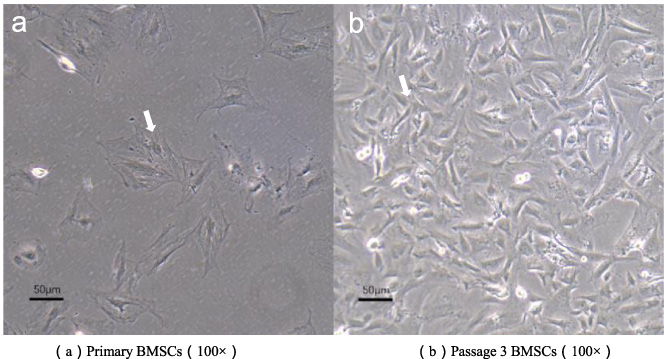
Microscope picture of BMSCs.
CD90 and CD44H are characteristic surface antigens of BMSCs. The positive rate of CD90 was 99.16% (Fig. 2a), the positive rate of CD44H was 99.72% (Fig. 2b), which were strongly positive; CD11b/c was the characteristic surface antigen of leukocytes, and its expression rate was 2.32% (Fig. 2c), which was negative; CD45 was the surface antigen of hematopoietic cells, and its expression rate was 3.10% (Fig. 2d). This result was consistent with the immunophenotypic characteristics of bone marrow mesenchymal stem cells.
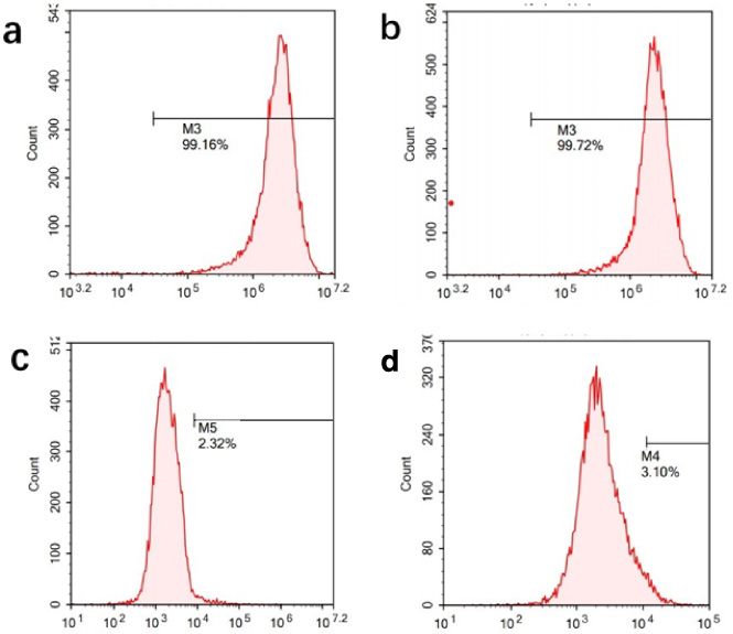
FCM of BMSCs.
DiI is a kind of lipophilic fluorescent dye, which can stain the whole cell membrane and emit red light under the excitation spectrum of 550 nm. Under the fluorescence microscope, BMSCs were spindle shaped or polygonal. The shape is clear and recognizable, and the staining is uniform (Fig. 3).
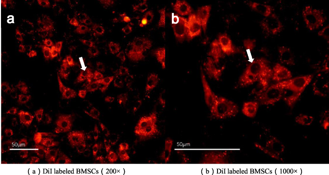
Fluorescence microscope photographs of DiI labeled BMSCs.
Under the fluorescence microscope, DiI labeled BMSCs grow well on the material, emit red light, and the cell morphology is clear and recognizable (Fig. 4).
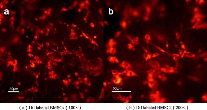
Fluorescence microscope photographs of the cell patch.
The morphology of BMSCs on the surface of PCL films was observed by electron microscopy, and a large number of BMSCs were located on the surface of the material and grew inside the pores, which were tightly attached to the material. The cells were mostly spindle shaped, grew in a cuboidal shape, and secreted extracellular matrix surrounded the cell periphery, connected in sheets (Fig. 5).
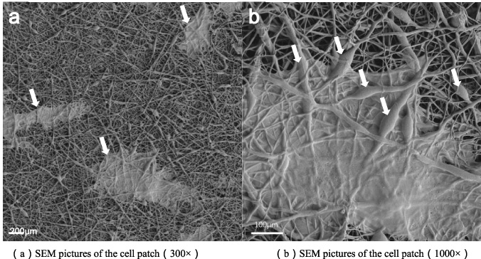
SEM pictures of DiI labeled cell patch.
The cell proliferation was measured by CCK-8 (Fig. 6). The OD value on the first day was 0.330 ± 0.025. On the fourth day, the OD value was 0.620 ± 0.012 and the value-added rate was 181.250%. On the 7th day, the OD value was 1.033 ± 0.144 and the value-added rate was 439.375%. On the 10th day, the OD value was 1.223 ± 0.133 and the value-added rate was 558.125%. There was a significant difference among the time points (P < 0.05). With the extension of time, the number of cells increased gradually, which proved that DiI labeled BMSCs on PCL film could proliferate.
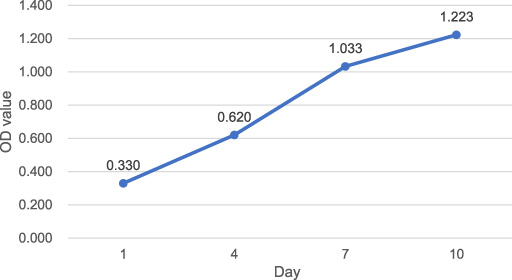
Proliferation curve of cell patch.
Myocardial infarction has attracted much attention because of its rapid onset, severe condition and poor prognosis. At present, the main treatment methods, namely conservative thrombolysis and anticoagulation in internal medicine and surgical coronary artery bypass grafting, cannot fundamentally solve the problem of cardiomyocyte reduction. With the deepening of cell and tissue engineering research, stem cells have been gradually used in the treatment of myocardial infarction and show broad application prospects [9,10].
Bone marrow mesenchymal stem cells can improve cardiac function through paracrine, immunosuppression, intercellular interaction and other mechanisms. Paracrine effect is the main mechanism for bone marrow mesenchymal stem cells to play a therapeutic role, including promoting angiogenesis, anti-apoptosis, reducing inflammation and so on. The exosomes secreted by bone marrow mesenchymal stem cells can regulate the survival of myocardial cells after infarction and help to improve myocardial revascularization. Sun et al. [11] Showed that miR-486-5p carried by BMSC-exo plays a pivotal role in the regulatory process by suppressing PTEN expression, activating the PI3K/AKT signaling pathway, and subsequently inhibiting the apoptosis of injured cardiomyocytes.
However, in stem cell therapy, transplanted cells are easy to lose and die due to blood flow impact and mechanical action of the heart itself. Therefore, it is particularly important to find a suitable carrier to ensure that transplanted cells can adhere and grow on the surface of the heart [12]. PCL has good biodegradability and biocompatibility, good shape memory, excellent processability and low production cost. It is one of the ideal carrier materials [7]. Based on the above reasons, I chose PCL as the carrier material to make cell patch, and co-cultured BMSCs with PCL film.
In the experiment, because the light transmittance of PCL film is poor, the cell morphology cannot be observed by light microscope when the cells are directly inoculated on its surface. DiI dye is a cell membrane dye that is non-toxic to living cells and has no effect on cell growth and proliferation [13]. Therefore, we stained the cell membrane with DiI in advance, so that the morphology of cells attached to PCL membrane can be observed directly under fluorescence microscope, which lays a foundation for further experiment and application of tracing in vivo.
The surface morphology of PCL film and the attachment of cells can be more clearly observed by scanning electron microscope. The cells are closely attached to the surface of material scaffold, and the secreted extracellular matrix covers the material and cell surface in sheets. Studies have shown that Pu and P (3HB-co-4HB) materials can also adsorb and grow BMSCs [14]. However, compared with this experiment, it can be found that the number of cell attachment is less, and there is no coverage of extracellular matrix. Therefore, the biocompatibility of the two materials may not be as good as PCL, and the cells cannot secrete extracellular matrix and adhere to the surface of the material. Therefore, PCL is more suitable as a carrier of cell patch. The surface of PCL film prepared by electrospinning is rough, which can promote the attachment of cells. In addition, the pores on the surface of the membrane also enable the cells to obtain more space and three-dimensional support. After attachment, the cells produce extracellular matrix and adhere to the material, which provides a supporting site for the continuous adhesion and growth of cells. In addition, the detection results of CCK-8 showed that BMSCs could proliferate on PCL film. Therefore, PCL can be used as a carrier for DiI labeled cell patch.
In the next stage of in vivo application, we plan to cut the PCL film into smaller sheets and transplant it to the surface of infarcted myocardium after planting cells, so as to make the cell patch play its role on the surface of myocardium and avoid falling off of patch transplantation due to excessive volume.
Although this experiment proved that BMSCs had good compatibility with PCL film, we did not know the degradation rate and the effect of degradation products after cell patch transplantation in vivo, and what factors are secreted by cell patch to contribute to revascularization of myocardium. Next, we will further study whether the cell patch with PCL film as carrier can degrade in animals, and the factors secreted by cell patches, so as to lay a foundation for its further clinical application.
Conclusions
Within the limits of the present study, we conclude that DiI stained BMSCs can normally adhere to PCL film, and PCL is suitable as the carrier of cell patch.
Footnotes
Conflict of interest
No potential conflict of interest was reported by the authors.
Funding
This study was partially supported by the National Natural Science Foundation of China (81870181).