
Abstract
BACKGROUND:
Hydrogel is a three-dimensional structure that has the potential to absorb and retain water within the mesh of its porous network structure. Currently hydrogels made from natural biopolymers are preferred in the discipline of biomedical applications because of their blood compatibility, adhesion of platelets and protein binding, ease of administration and delivery of ingredients to the place of action.
OBJECTIVE:
The aim of this work was to prepare a hydrogel from natural biopolymers and evaluate its blood compatibility, swelling nature, prolonged degradation and morphological features in order to further recommend its clinical use.
METHODS:
To prepare hydrogels, different combinations of gelatin, dialyzed SF, curcumin and N, N methylene bisacrylamide (MBA) were evenly mixed on a magnetic stirrer. After an hour of the gelation process it was kept in a refrigerator at 4 °C. For the characterization and biocompatibility studies of hydrogel, the swelling test and biodegradation analysis, SEM, FTIR, in vitro coagulation tests, total serum albumin and cholesterol level analysis were applied.
RESULTS:
Injectable hydrogels were successfully made with significantly correlated combinations of polymers. The analysis of physiochemical biocompatibility studies and morphological characterization were done effectively.
CONCLUSION:
The results of the study indicate that hydrogels made from natural biopolymers are a potential source and suitable matrices with excellent biocompatible nature acting as a useful device in delivering drugs.
Introduction
Hydrogel is a biological fluid or structure with a three-dimensional network that is widely used in various biomedical applications and has unique properties, such as the potential to absorb large amounts of water and other biological fluids (due to the formation of a hydrophilic polymer network), easy flexibility, and a high degree of biocompatibility [1]. The biodegradability, swelling property, and biocompatibility of the hydrogel can be controlled and modified by the type of polymers used and the preparation procedures [2]. The rapid hydrogel formation as well as the gelling ability are controlled by the association of the polymer network and the functional groups of the polymers used [3].
Hydrogels as biopolymers exceptionally form a network in which the crosslinked chains are either covalently linked [4] or form a non-covalent interaction based on chemical crosslinkers and physical actions as well as enzymatic processes [5]. The formed crosslinkers of the hydrogels form a permeable network that allows the movement of molecules like loaded drugs, nutrients and oxygen through the intertwined structure [6].
Hydrogels, which are in situ injectable gelling systems ideal for drug delivery, are nowadays preferred in biomedicine due to their biocompatibility, biodegradability, flexibility [7] and ease of administration [8]. They provide a potential solution to current treatment complications, especially preventable neural retinal diseases such as age-related macular degeneration and glaucoma, which are prevalent worldwide. There are several treatment options available, but many of them have a number of problems and limitations. For all these problems use of hydrogels as drug delivery material offers a solution.
Naturally obtained biopolymers like silk fibroin (SF), gelatin (GE) and curcumin (CU) have countless application in the area of regenerative medicine including tissue engineering due to their biodegradability and biocompatibility [9], more remarkably hydrogels made from natural polymers exhibit high potential as drug delivery units [10].
Silk fibroin (SF) is appropriately used to prepare biomaterials for biomedical applications in the form of hydrogels [11], scaffolds, films, particles, and sponges [12], all of which have been experimentally confirmed to have remarkable properties of blood compatibility, interaction with cells, biodegradability, biocompatibility, and low potential cytotoxicity and inflammatory response in surrounding tissues [13], water absorbability, and low immunogenicity [14], they can be used for the purpose of better drug delivery devices.
Curcumin (1E,6E)-1,7-bis(4-hydroxy-3-methoxyphenyl) hepta-1,6-diene-3,5-dione), a turmeric root, is a pale yellow chemical produced naturally as Curcuma longa and consists of various chemicals that exhibit antimicrobial, antioxidant, anti-inflammatory, antineoplastic, wound-healing, and potentially chemotherapeutic properties, and is best suited for biomedical applications [15].
Gelatin (C102H151N31O39) is obtained and produced by the process of partial hydrolysis of natural collagen [16]. It has exceptional gelling behavior and also shares functional properties with proteins, which is why it is widely used in many areas of the processing industry, such as food processing, cosmetics, pharmaceuticals, and biomedical device manufacturing [17,18].
Timolol maleate (C17H28N4O7S) is a beta-adrenoceptor blocker currently used to treat glaucoma. It lowers high pressure in the eye and helps prevent blindness by reducing the amount of fluid in the eye [19].
In this study, an injectable hydrogel was prepared from a combination of natural biopolymers (SF/CU/GE with N,N’ methylenebisacrylamide (MBA) as a crosslinker) and its blood compatibility, swelling ability, prolonged degradation and morphological properties were investigated under in vitro conditions to recommend it for further clinical use.
Materials and methods
Materials
Bombxy mori cocoons and Curcuma longa (Turmeric) rhizome were obtained from a local market in North Cyprus. Gelatin, Tween 80, Calcium chloride (CaCl2), Sodium triphosphate Pentabasic (Na5O10P3), Ethanol (C2H5OH, 98%) were all purchased from Sigma Aldrich. SnakeSkin® Dialysis Tubing of 3500 molecular weight cut out membranes was purchased from Thermo Scientific USA. Anhydrous sodium carbonate (Na2CO3) and anhydrous calcium chloride (CaCl2) obtained from EMSURE® Merck chemicals in Darmstadt, Germany, Timolol maleate (C17H28N4O7S) purchased from a local pharmacy in North Cyprus.
Silk fibroin purification
The purification process was performed by cutting the SF cocoons into smaller pieces and mixing them with 0.1 M Na2CO3 solution in a flask using an electro-magnetic stirrer at a speed of 1.5 rpm for three sessions at 70 °C. After each degumming, SF was washed with ultrapure water and dried overnight at room temperature [13], then the solution was dissolved with a strong electrolyte solution (molar ratio of nH2O:nCaC12:nCH3CH20H;8:2:1) with continuous stirring at 70 °C. Finally, the solution was dialyzed with a snakeskin dialysis membrane against deionized water to remove all the strong electrolyte in the silk fibroin solution [6].
Composition used for hydrogel formation
Composition used for hydrogel formation
The percolation extraction method was used by adding acetone to 144 g of Curcuma longa powder. After completion of curcuminoid extraction, rotary evaporator was used to concentrate the extract, column chromatography was used to purify the concentrated curcumin extract, using silica gel (SiO2) and dichloromethane as adsorbents. After purification by heating, dichloromethane was removed and finally the by-product was collected in the form of powder and subjected to thin layer chromatography analysis. Column chromatography setup was performed to isolate the pure curcumin sample.
Preparation of hydrogels
To prepare hydrogels, gelatin (GE), dialyzed silk fibroin (SF), curcumin (CU), and N, N methylene bisacrylamide (MBA) (as a cross linker) were used in the following protocols as it is indicated in Table 1.
SF/GEL/MBA (sample coded by IVA): 1.5 ml of 3% SF, 5 ml of 10% GE and 5 μl MBA were mixed evenly on a magnetic stirrer and left to gelate then the hydrogel stored under refrigerator at 4 °C. SF/GEL/CU/MBA (sample coded by IVB): 2 ml of 3% silk fibroin, 0.25 ml of CU and 5 μl MBA were added to 5 ml of 10% GE and mixed properly on a magnetic stirrer later left to gelate and kept overnight at 4 °C. SF/GEL/CU/MBA (sample coded by IVC): 2 ml of 3% silk fibroin, 0.25 ml of CU and 5 μl MBA were added to 3 ml of 10% GE which were all mixed on a magnetic stirrer. After an hour of the gelation process it has been kept under refrigerator at 4 °C and the samples were freeze dried into a balloon at −20 °C Table 1.
Swelling tests were carried out by immersing the samples in 20 ml of PBS (pH 7.4) solution and ABS solution at 37 °C at predetermined time intervals the hydrogel was carefully taken and blotted with filter paper and weighed and returned back to the same medium so that measurement was made to the maximum swelling point. To reduce the error, the procedure was repeated for three times. The range of pH was chosen to mimic the human physiological conditions and swelling ratio was calculated by the formula:
In vitro coagulation and fibrinogen activity test analysis
For in vitro anti-coagulation test analysis STA compact was used. Fresh blood sample collected from the healthy donor in sample collection tubes containing anticoagulant trisodium citrate 0.109 M (3.2%) according to the CLSI guidelines and centrifuged at 850 RCF for 10 min’ blood plasma were taken and sample of hydrogel dropped inside and incubated under static condition of 37 °C for about 15 min. Prothrombin time, Activated partial thromboplastin time and fibrinogen analyses were performed. APTT results and PT analysis results were reported in seconds and International Normalized Ratio INR and fibrinogen results were reported as mg/dL.
Parameters of complete blood count
Parameters of complete blood count
Fresh human blood samples were collected in tubes with citrate to bind the calcium and stopping the coagulation. The intrinsic pathway activated by adding calcium to inverse the anticoagulant effect of the citrate and an activator were mixed into the plasma sample the time was measured up to the point of a clot formation this test is called partial because the reaction mixture is free of the tissue factor.
Prothrombin time (PT)
The prothrombin time was measured using the plasma. Blood was drawn into a test tube containing liquid citrate by binding the calcium it acted as an anticoagulant. The blood was mixed and centrifuged to isolate the plasma from the blood cells [13]. Factor II (tissue factor) was added and the time that the sample takes to clot was measured.
International normalized ratio (INR)
International normalized ratio (INR) is a calculation made to normalize prothrombin time INR is based on the ratio of the patient’s prothrombin time and the normal mean prothrombin time. Prothrombin time is a test to know how fast the blood clots in patients getting oral anticoagulant treatment [13]. The INR uses the ISI to equate all thromboplastins to the reference thromboplastin through the following
Fibrinogen activity test
Fibrinogen is a soluble protein in the blood plays a role in clotting and wound healing process which provides a conducive environment for supporting the development of new tissues. 150–400 mg/dl is the normal range of fibrinogen concentration in the human blood [20], even though the range may vary in different laboratories and depending the method they follow.
Total serum albumin and cholesterol level
Fresh blood collected from the healthy donor and sample hydrogel were mixed and incubated for 30 min under 37 °C then centrifuged by the speed 1500 RCF for 10 min, serum albumin and cholesterol level were measured. serum albumin is a protein in the blood plasma represents almost half of the total proteins ranging in between 3.5 g/dL to 5 g/dL in healthy persons protein [10,21]. Cholesterol is a natural substance made by the body around 75% of it is made by liver and is important for normal physiological activities in optimal conditions. A total cholesterol score of 200 mg/dl or lower is considered to be optimal [22,23].

Swelling % hydrogels of sample IVA, IVB, IVC in PBS pH 7.4.

Swelling % of hydrogels of sample IVA, IVB, IVC in ABS pH 4.7.

Swelling % of hydrogels of sample IVA, IVB, IVC in ABS pH 1.
Automated hematology analyzer was used for complete blood count analysis (Cell Dyn Ruby; Abbott Laboratories, Diagnostic Division, Abbott Park, IL, USA). Fresh blood collected and sample (hydrogel with drug and without drug) immersed K2 EDTA was used as anticoagulant. The samples were mixed by shaker (300 revolution/minutes) which facilitates the interaction of sample surface and blood cell, for 20 min and then the following parameters were analyzed Table 2.
The biodegradation test of hydrogels in lysozyme solution at 37 °C
The biodegradation test of hydrogels in lysozyme solution at 37 °C
Coagulation analysis, fibrinogen, total cholesterol and serum albumin analysis of hydrogels (IVA, IVB, IVC)
PT = prothrombin, INR = international normalized ratio, APTT = activated partial thromboplastin time, IVA, IVB and IVC are coded samples of hydrogels, HK = control blood plasma.
Fresh human blood from the healthy donor was collected and then centrifuged using 100 RCF for 15 min, platelet rich plasma prepared and sample of hydrogel were immersed into it for about 15 min under human physiological condition 37 °C. Peripheral smear test was used to determine the adhesion morphology, micro-particle platelet formation on the surface of hydrogel by using light microscope with low (100×) and high (400×) magnification power.
Scanning electron microscope (SEM) analysis
Morphological and structural features of injectable hydrogels prepared at static condition were carried out by using scanning electron microscope (SEM, at TUBITAK-MAN using a JEOL/JSM-6510LVF). After the samples were lyophilized the micro-structure were imaged at ×100 and ×200 magnification at 20 kv of acceleration voltage.
Fourier transform infrared (FTIR) analysis
The Fourier transform infrared (FTIR) spectra of tested samples were carried out in the laboratory of Eastern Mediterranean University, Famagusta, Northern Cyprus using a Perkin Elmer Spectrum 65 FTIR.
X-ray diffraction
X-ray diffraction of freeze-dried samples of the hydrogel was carried out at a central laboratory at Middle East Technical University Turkey, using a Shimadzu XRD-6000 model diffractometer with Cu X-ray tube ( = 1.5405 Å (10−10 m). The crystallinity index is calculated and determined by the technique based on the methods formulated by Kim and his colleagues [24] it consists of measuring the maximum intensity, I110, at 2𝜃 =16°. The analysis of crystallinity index was calculated by using Eq. (1):
All data obtained were presented as mean ± standard deviation. Significance difference were performed by a student’s t test at a probability level of 0.05 and one-way (ANOVA) used for determining the difference among the groups using Graphpad prism version 8.0.2 software.
Total blood count analysis of hydrogel samples coded by IVA, IVB and IVC
Total blood count analysis of hydrogel samples coded by IVA, IVB and IVC
Swelling kinetics
The equilibrium swelling nature of prepared injectable hydrogel were tested by using acidic phosphate saline (ABS, pH 1.2 and pH 4.7) and basic medium phosphate buffer saline (PBS, pH 7.4). The swelling behavior of the samples coded with IVB and IVC swells uniformly with time and accordingly reached its maximum equilibrium ratio. Meanwhile, the equilibrium swelling ratio of the sample encoded with IVB was highest among the groups within 180 min and that of IVC within 45 min in PBS solution with pH 7.4. Whereas the same sample in ABS of pH 4.7 reaches its maximum equilibrium within 90 min for IVB and 45 min for IVC as well as in ABS solution of pH 1.2 sample IVB within 35 min and IVC within 20 min, however one of the samples coded by IVA quickly swells up in both ABS and PBS within 10 min then degrade totally.
The % swelling result in PBS pH 7.4 for IVA 98%, IVB 1469.2% the highest of all among the groups (P < 0.0001) and IVC 1393% shown in Fig. 1 respectively. While Figs 2 and 3 gives the swelling % in acid buffer of pH 4.7 for the same sample IVA 145%, IVB 700.4% and IVC 1242.6% respectively and in ABS of pH 1.2 for the same samples were IVA 125%, IVB 1440.9% and IVC 1092.1% respectively.
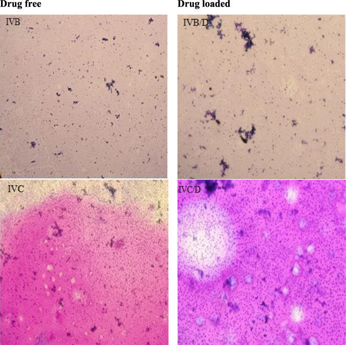
Peripheral blood smears for platelet adhesion analysis of IVB, IVB/D, IVC & IVC/D at 400× magnification.

Wright–Giemsa stains of IVB, IVB/D, IVC, IVC/D at 400× magnification for erythrocyte morphology analysis.

SEM micrograph of sample IVA, IVB, IVC 200 μm.
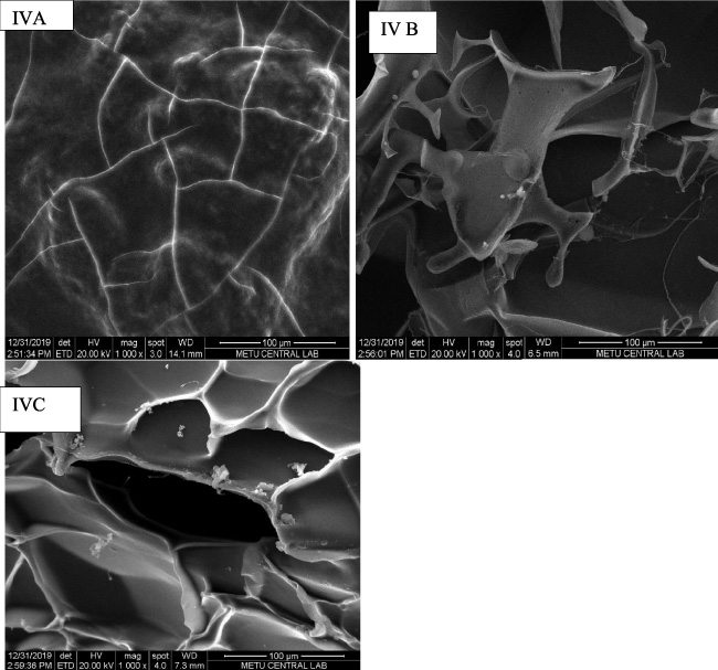
SEM micrograph of sample IVA, IVB, IVC 100 μm.

FTIR spectra of samples (a) IVA, (b) IVB and (c) IVC hydrogels.
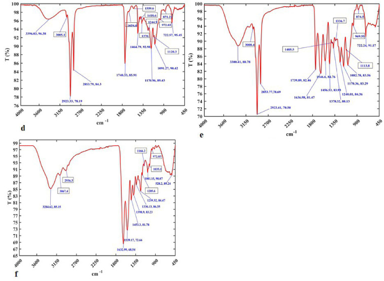
FTIR spectra of samples (d) IVA-D, (e) IVB-D and (f) IVC-D hydrogels.

XRD pattern of hydrogel with drug and without drug coded by IVA, IVB, IVA/D and IVB/D containing SF/Gel/Curcumin in different compositions.
The result confirmed that the swelling of hydrogels occurs due to the uptake of large amounts of liquid at the beginning of the experiment and gradually becomes saturated as the network of the hydrogel expands when more liquid is taken up, confirming the hydrophilic nature of the hydrogel due to the free hydroxyl and amino groups within the polymer matrix [25]. However, the IVA sample swells immediately in both acidic and basic buffer solutions within 10 min because it is more amorphous and contains a smaller amount of silk fibroin compared to the other samples, which is consistent with the study by Terada and his colleagues [26].
The hydrogels were tested against the lysozyme solution (0.2 mg/100 mL H2O) at 37 °C. The experiment was repeated three times. The results are shown in Table 3. The hydrogels were synthesized, with a varying amount of silk fibroin, gelatin, curcumin and a cross linker MBA. As the amount of gelatin increases with respect to silk fibroin, it enhances the crosslinking and affects the rate of biodegradation. These results indicate that the biodegradation rate may be controlled by gelatin.
In vitro coagulation, total cholesterol and serum albumin analysis of hydrogels
The anticoagulant effect of the hydrogel was assessed by (APTT) and (PT) measurements, which were used to evaluate secondary hemostasis. The values of PT and APTT activation were measured for citrated control plasma and after contact with samples (IVA, IVB & IVC) at a temperature of 37 ± 1 °C.
The results of the test for APTT, PT and (INR) are in the standard range without significant statistical difference from the control, confirming that the prepared injectable hydrogels are biocompatible in terms of coagulation activity. Total cholesterol level and serum albumin are also within the desirable limits according to the National Cholesterol Education Program (NCEP) standard Table 4.
In vitro coagulation analysis and fibrinogen activity test of hydrogels with drug (combinations IVA/D, IVB/D and IVC/D)
The anticoagulant effect of the hydrogel loaded with the drug (timolol maleate) was evaluated by the APTT, PT, and INR values displayed in the selected unit (seconds, INR, % ratio). The following results were obtained for samples (IVA/D, IVB/D & IVC/D) at a temperature of 37 ± 1 °C. The results obtained are in agreement with the test values of the standards, which ensures that the hydrogels produced are in the best position in terms of biocompatibility. A test of fibrinogen activity was also performed for the samples with and without drugs, and they were found to be compatible according to the standard value without significant difference.
Complete blood count analysis
Hydrogels were immersed in fresh blood with K2 EDTA as an anticoagulant. No significant difference was observed in all samples compared to the control, as shown in Table 5, all results were below the limit range. This experiment confirms the fact that the polymers used for the preparation of the hydrogels are biocompatible and have no negative effect on the total of erythrocytes, leukocytes and platelets in normal blood.
Peripheric smear test for in vitro platelet adhesion analyses
For evaluating hemostatic condition of the hydrogel, in vitro peripheric smear test was applied as it is shown in the Figs 4 and 5 microscope micrographs of both hydrogels with drug and hydrogel without drug there is no platelet adhere formation on the surface of hydrogel which makes the hydrogel to be promising blood compatible biomedical device.
Scanning electron microscopy analysis
The SEM micrographs shown in Fig. 6 illustrate the morphology of IVA, IVB, and IVC encoded hydrogels prepared from various combinations of silk fibroin, gelatin, curcumin, and the crosslinker MBA. In the sample of IVA (Fig. 6), the number of pores was small and their morphology was rough, IVB and IVC contain visible microspheres with open pores that facilitate drug transport. The increased amount of SF with decreasing gelatin and the presence of curcumin might be responsible for the morphological characteristics of IVC, which have better pore structure and good interconnectivity (Figs 6 and 7 IVC).
Fourier transform infrared spectroscopy analysis
In the hydrogel, the specific groups of SF/GE/CU/MBA are characterized by the absorption band in Fourier transform infrared spectroscopy (FTIR).
In (Fig. 8a), the FTIR spectra showed the absorption band region between 1630.48 cm−1 and 1382.6 cm−1 assigned to amide I, and in (Fig. 8b), the band region assigned between 1537.41 cm−1 and 1451.4 cm−1 the amide II absorption mainly from the NH bending in plane bending vibrations and CN stretching in protein backbone of silk fibroin, which is in agreement with the work of Adali and colleagues, the bands secure the 𝛽-sheet confirmation of the samples [27]. (Fig. 9) confirms that the 2923.10 cm−1 and 2853.42 cm−1 peaks are due to CH3 stretching, the curcumin loaded sample (Fig. 9f) shows an absorption band at 1556.4 cm−1 for amide I and 1456.5 cm−1 for amide II, which is consistent with previous studies [17]. Since curcumin was completely mixed into the mixture, the stretching vibration could be very limited and the bands disappeared in the complexes of SF [28]. Based on the presence of CO and CN vibrational stretching of amide I and amide II in the gelatin, the absorption bands were reported to be 1636 and 1558, which is in agreement with the study by Khade and colleagues [29]. The hydrogel showed an absorption band of 3300.41 cm−1, 3283.94 cm−1 and 3284.10 cm−1, which were attributed to OH and NH stretching vibrations in all hydrogels.
X-ray diffraction analyses
X-ray diffraction pattern of SF/Gel/curcumin hydrogel gives three characteristic crystallinity peaks at 2𝜃 = 11.62°, 20.56° and 10.46° of high intensity since there is more periodicity because of the preferred crystal orientation, however in the drug-loaded sample new weaker crystalline peaks appear at 2𝜃 = 12.58°, 20.56° and 9.12°. As indicated in the result, the degree of crystallinity and crystalline structures are affected by the drug loaded hydrogel as shown in the Fig. 10.
Conclusion
In the present study, an injectable hydrogel was successfully prepared from naturally derived biopolymers and the physiochemical biocompatibility and morphological properties were determined. The overall result of the analysis confirmed that the samples encoded with IVB and IVC were suitable for biomedical applications in vitro.
The morphological structure of the samples from scanning electron microscopy analysis also showed that the samples labeled IVA have low and rough pores, while the samples labeled IVB and IVC contain miscible microspheres with open pores, which are important for drug loading and facilitate drug transport. The presence of curcumin in the sample provides a better pore structure and good interconnectivity that promotes optimal drug loading. Therefore, the result of the study shows that hydrogels made from natural biopolymers are a potential source and suitable matrices with excellent biocompatibility that serve as a useful tool for drug delivery.
Footnotes
Acknowledgements
The authors acknowledge the participants in this research.
Conflict of interest
The authors declare no potential conflicts of interest concerning the research, authorship, and/or publication of this work.