
Abstract
BACKGROUND:
Magnesium (Mg) enhances the bone regeneration, mineralization and attachment at the tissue/biomaterial interface.
OBJECTIVE:
In this study, the effect of Mg on mineralization/osseointegration was determined using (Ti,Mg)N thin film coated Ti6Al4V based plates and screws in vivo.
METHODS:
TiN and (Ti,Mg)N coated Ti6Al4V plates and screws were prepared using arc-PVD technique and used to fix rabbit femur fractures for 6 weeks. Then, mineralization/osseointegration was assessed by surface analysis including cell attachment, mineralization, and hydroxyapatite deposition on concave and convex sides of the plates along with the attachment between the screw and the bone.
RESULTS:
According to Scanning Electron Microscopy (SEM) and Energy Dispersive Spectroscopy (EDS) analyses; cell attachment and mineralization were higher on the concave sides of the plates from both groups in comparison to the convex sides. However, mineralization was significantly higher on Mg-containing ones. The mean gray value indicating mineralized area after von Kossa staining was found as 0.48 ± 0.01 and 0.41 ± 0.04 on Mg containing and free ones respectively. Similarly, Fourier Transform Infrared Spectroscopy (FTIR) and X-ray diffraction (XRD) analyses showed that hydroxyapatite growth was abundant on the Mg-containing and concave sides of the plates. Enhanced mineralization and strong attachment to bone were also detected in EDS and SEM analyses of Mg-containing screws.
CONCLUSION:
These findings indicated that (Ti,Mg)N coatings can be used to increase attachment at the implant tissue interface due to accelerated mineralization, cell attachment, and hydroxyapatite growth.
Introduction
Titanium (Ti) and its alloys have been extensively used as metallic biomaterials in hard tissue applications. Despite their unique properties such as relatively low elastic modulus, high strength and biocompatibility, there are several reported cases for failed long term applications due to low osseointegration performance and wear resistance [1,2]. Functional surface coatings are commonly used to tailor the surface properties of the Ti based implant materials facilitating osseointegration. For instance, Ca-P coatings are widely used to improve the performance of hard tissue implants. Porous structure of Ca-P coatings let the nutrient flow and cell migration towards the implant surface and leads to better osseointegration between the implant and surrounding tissues [3–6]. Magnesium (Mg) is also considered as one of the best choices for surface modifications of Ti-based biomaterials in the last decades to overcome these issues [7–9].
When compared to non-degradable conventional metallic materials that are widely used in surface coating of hard tissue implants, Mg has various advantages such as biodegradability; biocompatibility, mechanical specifications matching the bone tissue and better tissue interactions [10–12]. Furthermore, Mg is the most abundant cation in the human body and it also exists in natural bone structure [13,14]. These properties make Mg based implant materials highly desirable for long-term implantation by preventing revision surgeries leading to additional cost and complications.
Pure Mg usage in hard tissue applications has some limitations due to its corrosion behavior in aqueous solutions or physiological media. Therefore, different methods have been used to improve both the intrinsic and surface properties of the Mg based materials to control corrosion. Purifying, alloying, grain refinement, and heat treatment techniques have been proposed to improve intrinsic properties of the Mg materials [15–18]. The mechanical properties and corrosion behavior of binary Ca-Mg alloys, for example, can be adjusted by controlling the Ca content and process parameters [19]. Corrosion rate and mechanical properties of the alloys such as ultimate tensile strength and elongation were largely improved after hot rolling and hot extrusion. Functional coatings are deposited onto Mg materials with various techniques such as chemical conversion, plasma electrolytic oxidation, by using inorganic and organic materials to reduce the corrosion rate and maintain the bulk mechanical properties [20,21]. MgF2 coating using vacuum evaporation deposition method significantly reduced corrosion of Mg-1Ca alloy [22]. Similarly, more favorable corrosion resistance was obtained when 1 wt % nanosized HA particles were coated onto Mg–2.9Zn–0.7Zr in comparison with uncoated one [23].
Mg containing coatings have the potency to overcome the limitations of the Mg-based implant materials as well as the methods used to improve the intrinsic and surface properties of the Mg materials. Mg containing coatings deposited onto the hard tissue implants limit the biocorrosion while maintaining the positive effects of Mg on cells and mineralization. Mg incorporated titanium nanotube arrays on Ti surface had been shown to accelerate bone mesenchymal stem cell proliferation rate by Mg release from the surfaces [24]. In another study, titanium implants coated with mesoporous
In the present study, we investigated the surface properties of the (Ti,Mg)N thin film coated bone fixation devices that are in contact with two different tissues (bone and muscle) to assess the effect of Mg on mineralization in vivo. For this purpose; (i) convex and concave surfaces of the (Ti,Mg)N and TiN coated bone fixation devices were characterized following the 6th week of bone fracture fixation on femur of the rabbits and (ii) cell attachment and mineralization on these surfaces were evaluated.
Materials and methods
Coating and characterization of the plates and screws
Ti6Al4V based plates (2 × 4 × 60 mm) and screws (diameter: 2 mm) were used to fix bone fractures formed on the femur of rabbits. (Ti,Mg)N and TiN thin film coatings were deposited onto the plates and screws with arc PVD technique. Coating parameters optimized in our previous studies were used during the coating process [26,30]. Briefly, two Ti and one Mg target were placed in the system as shown in Fig. 1 and target currents were set to 90A and 15A respectively to obtain <10% Mg content in coatings. Surfaces of plates and screws were first cleaned with ion bombardment and to homogenously coat samples, plates and screws were rotated both along their axes and along an axis passing through the center of the vacuum chamber while applying bias voltage (−150 V). Whole process was carried out in the nitrogen atmosphere (pressure: 1 atm).
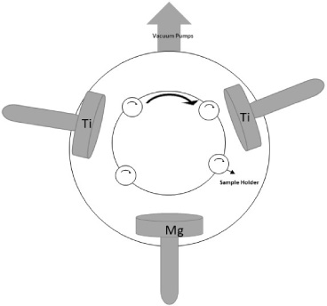
Schematic of the coatings system.
Scanning electron microscopy (SEM) was used to observe the homogeneity and thickness of the deposited coatings, Energy dispersive X-ray spectroscopy (EDS) to determine the elemental composition of the coatings and X-ray diffractometer to assess the phases formed in the coatings were used as shown in our previous study [30].
Coated plates/screws were used to fix these fractures formed on the femur of the rabbits whose weights were around 3.1 ± 0.5 kg. Sixteen healthy male rabbits taken from “ABDEHAM Experimental Animal Center” were operated at Istanbul University Faculty of Medicine in accordance with animal protocols that were approved by the Animal Experiments Ethical Committee of Istanbul University. All surgical procedures were done as described in our paper [30]. Basically, a 50–60 mm incision cut was made with an osteotome and a fracture site was formed in the middle of the femur (Fig. 2).
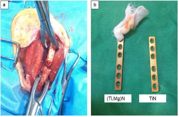
(a) Fracture formed in the middle of the femur and (b) thin film coated plates.
Coated plates and screws were autoclaved and sonicated in pure acetone and ethanol before the surgical operations. Rabbits were randomly divided into two groups for TiN (Group 1) and (Ti,Mg)N (Group 2) thin film coated plate fixation (n = 8 per group). They were all euthanized after 6 weeks of bone healing with an overdose of Propofol® injection and plates were removed and their surfaces were analyzed to determine the performance of two groups after in vivo implantation.
Calcium deposition on implanted plates were analyzed by von Kossa staining (n = 4) while the remaining four samples were used for Fourier Transform Infrared Spectroscopy (FTIR), Scanning Electron Microscopy (SEM), Energy dispersive x-ray spectroscopy (EDS) and XRD analyses. Characterization studies were carried out on concave sides that were in contact with bone and convex sides that were in contact with muscles and body fluids.
SEM and EDS analyses
SEM (EVO 40, Carl ZEISS, Germany) and EDS (Noran, Inc. 606M 1FSS, USA) were used to determine mineralization and cell attachment over the TiN and (Ti,Mg)N coated titanium substrates. A thin gold coating (ca. 15 nm) was deposited on the samples and analyses were carried out on both concave and convex sides of the two plates and screws from each group. Surfaces of the coated substrates were also checked for corrosion with SEM. EDS elemental analyses were carried out from 3 different places on both sides along the plates and on the screws using iridium ultra software. Presence and amounts of mineralized structures on the surfaces were determined. Point analyses were also done to check if the mineralization was distributed on surfaces homogeneously.
von Kossa staining and image J analyses
von Kossa staining was performed to visualize mineralization on surfaces and to assess osseointegration. First, TiN and (Ti,Mg)N plates were incubated in 4% formaldehyde solution at 4°C, washed with dH2O and treated with silver nitrate solution (2 mL, 5%). Samples were exposed to UV light in a dark chamber for 45 min until calcium deposits were visible. Plates were then washed with dH2O for 3 times, and sodium thiosulphate (5%) was applied on plates for 3 min. Samples were washed again with dH2O for 3 times and nuclear fast red stain was applied for 5 min. Image J program was used to determine the size and distribution of stained area. Whole plate surface that was in contact with the bone was chosen for image J analysis and the mean gray area where no staining occurred because of the lack of mineralization was quantified.
FTIR and XRD analyses
FTIR and XRD analyses were performed to determine the mineralization and phases formed on two plates that were randomly selected from each group. Plates were kept in 4% formaldehyde solution at 4°C until FTIR analyses.
Statistical analysis
Statistical analysis was done to compare the mineralization on concave and convex sides of the plates after von Kossa staining. Statistical analysis was performed using a student’s t-test in Microsoft Office Excel. The p values <0.05 were considered as statistically significant.
Results
SEM analysis
SEM micrographs of two randomly selected plates coated with TiN thin films are shown in Fig. 3. Cell type, densities of cells and amounts of matrix deposited on convex and concave sides of the TiN coated plates were found to be different. Enhanced red blood cell attachment was observed on the convex side of the plates (Fig. 3a) whereas bone cell density was higher on the concave sides of the TiN coated plates (Fig. 3c). Moreover, plate surfaces on both sides were homogeneously covered with the mineralized structures (Figs 3b and 3d).
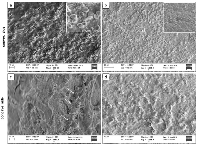
SEM micrographs of convex (a,b) and concave (c,d) sides of the two different plates coated with TiN thin films after bone fracture fixation. Arrows shows bone cells, inlets (a,b) shows red blood cells and mineralized coatings, respectively.
SEM micrographs of the two randomly selected plates coated with (Ti,Mg)N thin films are shown in Fig. 4. Attached bone cell density was higher on the concave sides of the Mg containing plates (Figs 4c and 4d). Thick mineralized structures were also seen on the convex side of the plates (Figs 4a and 4b) similar to TiN coatings (Figs 3a and 3b).
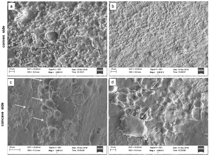
SEM micrographs of convex (a,b) and concave (c,d) sides of the two different plates coated with (Ti,Mg)N thin films after bone fracture fixation. Arrows show bone cells.
SEM micrographs of the TiN and (Ti,Mg)N coated screws also provided information about effects of local Mg presence on the cell attachment and mineralization (Fig. 5). High amounts of mineralized structures were observed in almost all threads of the Mg containing screws (Fig. 5a). However, the amount of mineralized structures on TiN thin film coated screws was significantly low (Fig. 5b, Supplementary Figure S1). Furthermore, no corrosion was detected on the coated surfaces in SEM micrographs.

SEM micrographs of the (a) (Ti,Mg)N and (b) TiN coated screws after 6 weeks of bone fixation. Inlets show mineralized structures, numbers show the points where EDS analysis were carried out.
Calcium and phosphate presence indicating mineralization on the surface of plates was determined by EDS analysis and summarized in Table 1. Although there is difference in calcium and phosphate amounts accumulated on convex and concave sides of Mg coated plates, both were found higher with respect to the Mg free ones. Moreover, mineralization was higher on the concave sides of the plates in all groups. Point EDS analysis that was carried out on the plate surfaces showed that mineralization was not homogeneously distributed on whole surfaces (Supplementary Figure S2).
Elemental composition of the plate surfaces after 6 weeks of implantation
Elemental composition of the plate surfaces after 6 weeks of implantation
Higher amounts of calcium and phosphate accumulation on Mg containing screws detected in EDS analysis was in accordance with SEM observations. For instance, calcium and phosphate amounts were 3.62% ± 0.48% and 13.95% ± 1.58% for Mg containing surfaces, respectively. However, these amounts reduced to 0.46% ± 0.14% and 5.96% ± 1.80% on TiN coated surface (Fig. 5b). Point analysis was also carried out on screws from two different points (1 and 2 in Fig. 5) and an increase in calcium and phosphate amounts were observed in mineralized structures. As expected, Ca and P amounts were found low for point 1 (0.27% and 1.18%, respectively) while they were significantly higher for point 2 (10.70% and 33.64%, respectively) on Mg containing surfaces (Fig. 5a). Similarly, lower Ca and P amounts were detected for point 1 when compared with point 2 on TiN coatings observed in Fig. 5b (0.14% and 3.62% versus 0.54% and 17.76%).
Mineralized structures on the surfaces of the plates were visualized by von Kossa staining (Fig. 6). Concave side image of one plate from each group (Figs 6a and 6e) was used to show the surface color before the staining (control group). Dark areas on the concave sides of TiN (Figs 6b and 6c) and (Ti,Mg)N (Figs 6f and 6g) coated plates represent the calcium deposits (CD) stained by the replacement of calcium ions with silver after the procedure. Calcium deposits were more apparent on Mg containing surfaces in comparison with Mg-free ones. Calcium deposition was also higher on the convex side of the Mg containing surfaces (Figs 6d and 6h).
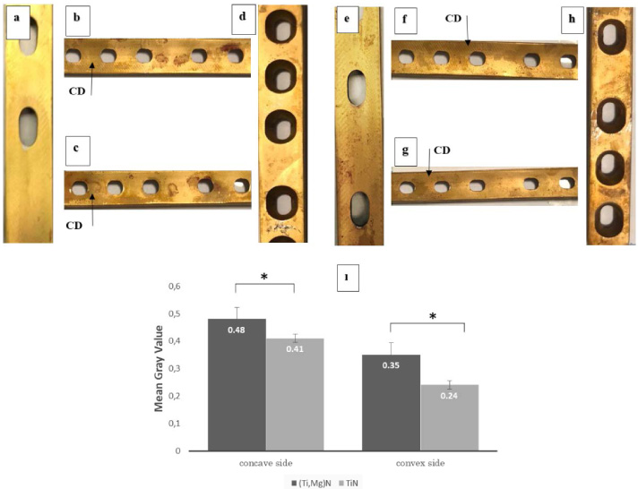
von Kossa staining of coated plate surfaces after 6 weeks of fracture fixation. Concave sides of the TiN (a) and (Ti,Mg)N (e) coated surfaces before the staining ( control groups). Concave sides of the TiN (b,c) and (Ti,Mg)N (f,g) coated plates after the staining. Convex sides of the TiN (d) and (Ti,Mg)N (h) coated plates after the staining. Mean gray area of the both sides of the plates (i). Calcium Deposits (CD), −∗ on the graph shows a statistically significant difference (p < 0.05, n = 4).
Image J analysis provided a semi quantitative data about the calcium deposition on the surfaces (Fig. 6i). Mean gray value that decreased with the increased mineralization on surfaces was significantly higher on both sides of the TiN coated plates when compared to Mg containing TiN coated plates.
FTIR analysis was used to provide information about mineralization on the TiN and (Ti,Mg)N coated plates (Fig. 7). PO4-3 is one of the characteristic groups of the HA deposition and peaks at 564, 577, 947, 997, 1090, 1102, 1113 ve 1114 cm−1 indicates the presence of this group in a crystalline HA structure 28. Intensity of these peaks was higher on the convex site of the (Ti,Mg)N thin film coated plates, but a substantial decrease was observed on concave sides (Fig. 7b). Calcium carbonate or carbonate apatite (CO3-2) is another group used to assess HA deposition and characterized by peaks observed at 1456 (Fig. 7b), 1460 (Fig. 7b) and 1574 (Fig. 7a) cm−1 in FTIR analysis. These peaks were more obvious on the convex side of the (Ti,Mg)N coated plates. HPO4-2 group at 864 ve 866 cm−1, (P2O7)-4 group at 719 and 979 cm−1 also indicate HA deposition 26 on surfaces and was found on the convex side of the Mg doped TiN coated surfaces. Briefly, calcium phosphate deposition density was found higher on convex/concave sides of the (Ti,Mg)N thin film coated plates (Fig. 7b) in comparison with convex/concave sides of the TiN thin film coated plates (Fig. 7a).

Surface FTIR (a,b) and XRD (c,d) analyses of the TiN and (Ti,Mg)N coated plates after 6 weeks of bone fracture fixation.
Characteristic Ti, TiN and Mg peaks were detected on concave and convex sides of the coated plates before the surgery (as coated ones) and after the implantation (Figs 7c and 7d). Furthermore, new peaks that are attributed to HA accumulation also appeared in XRD patterns both on TiN and (Ti,Mg)N coated plates. HA peaks were observed on both sides of the plate surfaces from each group.
Discussion
In the present study, we assessed the in vivo performance of Mg doped TiN thin film coatings that were deposited on fixation plates and screws. TiN coatings have been commonly used to improve the wear resistance of orthopedic and prosthetic devices in clinical applications [31,32]. However, they have a limited function to form strong bonds with the surrounding tissues due to their bio inertness [33]. New generation biomaterials are expected to induce tissue regeneration [34], so we hypothesized that Mg doping into the TiN coatings would promote the osseointegration in implant/bone interface due to positive effects of Mg on mineral accumulation and bone mineral density [7,35,36].
In order to observe the effect of Mg on in vivo mineralization, surface properties of (Ti,Mg)N thin film coated plates and screws were investigated after fracture fixation on femurs of rabbits following the encouraging results obtained for accelerated HA formation [26], enhanced cell attachment/proliferation, mineralization [28,29], and effective rBMSC differentiation [29] after Mg doping in vitro. After 6 weeks of fracture fixation, significantly higher mineralization on both surfaces of the (Ti,Mg)N coated plates were observed in von Kossa staining when compared to Mg free ones (Fig. 6) which is consistent with previous in vivo studies conducted in the intramedullary space of guinea pig femora [37]. Moreover, mineralization was higher on concave sides of the plates in each group (Fig. 6i) where periosteal tissue layer was present. Mg-ion release from the coating was thought to be the reason for osteoblastic cell differentiation from this tissue layer and enhanced mineralization at implant/bone interface. This hypothesis is consistent with the previous work by Chaya et al., in which Mg plates and screws were used to fix rabbit ulna fracture [38]. In their study, degradation in Mg devices led to enhanced bone formation suggesting that Mg release from degrading devices stimulated stem cells within the periosteum and initiated bone formation over them. Cells such as osteoblasts and pluripotent mesenchymal stem cells within the periosteal tissue are known to facilitate bone growth and repair [39,40]. Another reason for the higher mineralization on the concave sides of the plates may be due to the cell density. As shown by SEM micrographs, it was higher on concave sides of the plates in comparison with the convex sides for both groups (Figs 3 and 4). Previously, we demonstrated the effects of cell presence on in vitro mineralization by using hFOB cells. Therefore, mineralization might also be positively affected from the enhanced cell density on the surfaces. When we examined the SEM micrographs (Figs 3 and 4) and Von Kossa staining (Fig. 6), density of bone cell attachment and mineralization were higher on concave sides of the (Ti,Mg)N coated surfaces.
On the convex sides of the plates (Fig. 6), lower mineralization (Table 1) and bone cell attachment (Figs 3 and 4) were detected. These surfaces were in contact with the muscle tissues that have higher water content and blood flow compared to bone [41]. Therefore, absence of the cells that may differentiate/migrate to substrate surfaces to initiate bio mineralisation might led to decreased mineral formation on these surfaces. However, mineralization was still higher on convex sides of Mg containing surfaces when compared to Mg free ones. It was previously shown that Mg in the coatings might act as nucleation center and increase the calcium phosphate deposition on the surfaces and crystalline HA formed only on Mg containing coatings incubated in Mg free simulated body fluid (SBF) [26]. These coatings were also shown to promote calcium and phosphate accumulation when incubated in more complex environment like cell culture medium [28]. In the present study, convex sides of the plates that were in contact with body fluids and mostly muscle cells showed similar behavior supporting the in vitro experiments. The mechanism of cell attachment on the convex sides of the TiN coated surfaces is not fully understood (Fig. 3a) but the reason of presence a higher amount of red blood cells on coatings may be due to the damages formed on the blood vessels during the surgical operations.
Higher amounts of calcium and phosphate deposition on Mg containing plate surfaces were also confirmed by EDS analysis (Table 1). Although local differences arising due to the variances in the size of mineralized structures were observed in point analysis, their surface density was higher on the concave sides of the plates for both groups. Moreover, the Ca/P ratio was very low on the surfaces of all plates. On Mg containing surfaces, Mg ions act as the nucleation center for the hydroxyapatite deposition along with the Ca and bind phosphate ions in the mineralized structure. This may enhance the phosphate accumulation on the surfaces. Moreover, released Mg from the coating replaces the Ca ions in the hydroxyapatite, and Mg doped hydroxyapatite grew on the surfaces [26]. These factors can reduce the Ca/P ratio in the mineralized structures. If the (Ca + Mg)/P ratio was analyzed on the accumulated HA coatings, this ratio can increase. On the Mg free surfaces, abundance in local potassium concentration or enhanced potassium attraction due to the partial positive charges following the protein adsorption onto the surfaces from body fluids may be the causes of the low Ca/P ratio. Nevertheless, a reduction in the difference between calcium and phosphate concentrations was determined at concave surfaces due to the presence of osteoblasts. Enhanced calcium phosphate presence on biomaterial surfaces contributes to acceleration of the HA formation and in turn, bone formation at implant/bone interface. Wen et al. [42] and Doe et al. [43] showed the accelerated HA deposition on the Ti implants following the Ca treatment on the surfaces. HA coated titanium implants had been shown to accelerate bone formation in rabbit and porcine models [44,45]. More pronounced HA formation on Mg containing surfaces was also demonstrated by FTIR (Fig. 7) and XRD analysis, supporting the previous findings on the positive role of Mg on HA deposition. Not only the surface density but also strong binding to the surface was important for better performance, therefore screws used for the fixation were observed by SEM micrographs and high amounts of mineralized tissue was observed in threads of the (Ti,Mg)N coated screws in comparison with the Mg free ones (Fig. 5a, Supplementary Figure S2). This might be the indication of the stronger binding at the screw/bone interface. These results indicate that Mg doping into TiN coatings led to better osseointegration in vivo.
Conclusion
In the present study, we assessed the surface properties of the Mg doped TiN thin films deposited on plates and screws in a rabbit model. We demonstrated that Mg doping into TiN coatings enhanced the mineralization on the devices and led to strong bond formation at implant/tissue interface. Furthermore, density of the mineralization was different for the concave and convex sites of the fixation plates. It was higher on concave sites of the Mg containing surfaces probably due to the migrated cells from bone toward surfaces, showing the positive effect of Mg on cell proliferation and attachment. To our knowledge, this is the first study to investigate osseointegration properties of Mg doped TiN coatings on a hard tissue material in vivo, and the results showed that Mg doped TiN thin films are good candidates to be used as coating on Ti based materials due to their high osseointegration potency.