
Abstract
BACKGROUND:
At present, surgical resection and chemotherapy are still the main treatments for hepatocellular carcinoma and other cancers, but the curative effect and survival rate are not ideal.
OBJECTIVE:
In this study, we aim to prepare a carrier with low toxicity, high biocompatibility and targeted transport for the treatment of hepatocellular carcinoma.
METHODS:
CdSe quantum dots (QDs) modified with oleic acid were synthesized. Then hydrophobic CdSe QDs and hydrophilic super-paramagnetic Fe3O4 particles were encapsulated into different layers of liposomes to form magnetic fluorescent liposomes (MFLs). MFLs in the aqueous would quickly drift towards the external magnet and the entire process was clearly observed with fluorescence microscope. The fluorescence spectra revealed that the fluorescence properties of MFLs were similar to that of CdSe QDs.
RESULTS:
QDs had an average size of 3.32 nm with good fluorescence properties. The size of MFLs was about 100 nm (transmission electron microscopy (TEM) analysis showed the average size of MFLs was about 82.8 nm and dynamic light scattering (DLS) detection showed 111.9 nm). After being cultured with MFLs for 8 h, HepG2 cells were labeled by MFLs, and good fluorescence images were obtained. MTT analysis also expressed their good biocompatibility.
CONCLUSION:
The prepared MFLs had multi-function and could be used as ideal drug carriers.
Introduction
Cancer is one of the leading causes of human death in modern society [1]. At present, the treatment of cancer such as hepatocellular carcinoma (HCC) is still based on the comprehensive treatment of surgical resection. The survival rate of surgical resection is unsatisfactory, and many patients require chemotherapy. However, toxicity and side effects, as well as lack of targeting, severely impede the effects of chemotherapy [2–4]. Selectively transporting to the target with low toxicity and high biocompatibility is one of the most critical issues for the treatment of hepatocellular carcinoma.
Liposomes are ideal carriers for drug delivery. And magneto liposomes (MLs, i.e., magnetic nanoparticles encapsulated within liposomes) were kind of multifunctional hybrid liposome/nanoparticle assemblies that have received considerable attention since their introduction in 1988 [5]. Magnetic nanocrystals are loaded into the interior of hydrophilic liposomes, and magnets nanoparticle can be used to guide MLs in vivo [6,7]. According to previous studies, a sufficient amount of magnetic nanoparticles can be accumulated in the target lesion area by controlling an external magnetic field in vitro [8–10].
The magnetically guided drug delivery lacks direct observability, which leads to the problems such as inaccurate positioning, real time monitoring. The introduce of fluorescent probes may be a solution. Quantum dots (QDs) are sorts of semiconductor nanocrystals and were studied as novel luminescence probes in biological imaging, fluorescence labeling and detection which can be used to solve the above problems [11–13]. They have fascinating optical properties such as broad absorption spectra, narrow and symmetrical emission spectra, high photostability and long fluorescence lifetime, all of which are superior to those of traditional organic dyes [14–17]. At present, group II–VI semiconductor nanocrystals have been studied, especially CdSe quantum dots (CdSe QDs). A wildly used synthesis route of CdSe QDs is organometallic precursor method, which was first proposed in 1993 [18]. In 2005, Deng et al. developed a cheap and green route to prepare a high-quality CdSe QDs [19].
Magnetic fluorescent liposome (MFLs) is a new type of physical targeting liposome [20–22]. Strijkers et al. reviewed the research progress of MFLs and proposed the design and Application Strategies of multichannel targeting liposomes [23]. Ding et al. reported that by embedding transferrin on the surface of liposome, under the joint action of magnetic force and transferrin receptor, the uptake of cells increased and the transport of blood-brain barrier increased accordingly [24]. It suggested that there might be a bright future in drug delivery to the brain. Chen et al. Modified the surface of magnetic fluorescent liposome with alpha fetoprotein (AFP) and successfully constructed a multifunctional magnetic fluorescent liposome probe with double targets (magnetic target and antibody target) [25]. Although magnetic nanoparticles have achieved some success in preclinical trials, at present, the products of liposomes related to magnetic fluorescence only exist in the laboratory. This is a direction worth exploring.
According to others’ studies and our negative experiments [26,27], fluorescence quenching usually occurred when QDs were in contact with magnetic nanoparticles directly. So we propose a kind of carrier, in which QDs and nano-Fe3O4 located in different layers inside liposome. On the one hand, this model can help real-time monitoring of targeted transport, on the other hand, it can monitor the leakage of liposomes and so on. In addition, if the drug is added to the liposome, the drug release can be monitored by fluorescence quenching.
In this work, the hydrophobic CdSe QDs were dispersed in phospholipid bilayer and hydrophilic Fe3O4 nanoparticles were encapsulated into interior vesicle of liposome. The CdSe QDs and Fe3O4 nanoparticles were isolated by phospholipid bilayer to avoid fluorescence quenching. Phospholipid bilayer could lock CdSe QDs firmly in the liposome membrane and avoid QDs leakage. We aim to prepare MFLs with good magnetic response, biocompatibility and fluorescence imaging ability, and use them as drug carriers in the biomedical field.
Experimental section
Materials
Cadmium oxide (CdO, 99.0%), paraffin liquid (99.0%), oleic acid (OA, 99.0%), selenium powder (Se, 99.999%), ethanol (99.5%), ethyl acetate (99.7%), chloroform (99.0%), polyethylene glycol (PEG1000), and diethyl ether (99.0%) were obtained from Kelong chemical Co., Ltd. (Chengdu, China). Cholesterol (Chol, 95%) and egg phosphatidylcholine (EPC) were purchased from TCI (Shanghai) Development Co., Ltd. Dulbecco’s modified Eagle’s medium (DMEM) and fetal bovine serum (FBS) were obtained from HyClone (Logan, USA). Penicillin-streptomycin, 3-(4,5-dimethyl-2-thiazolyl)-2,5-diphenyl-2-H-tetrazolium bromide (MTT) were purchased from biosharp (Hefei, China). Dimethyl sulfoxide (DMSO) was purchased from Sigma-Aldrich (St. Louis., MO, USA). HepG2 cells were obtained from the National Engineering Research Center for Biomaterials (Sichuan University). The nano-Fe3O4 magnetic fluids were provided by our laboratory [28].
Instruments
Ultraviolet and visible spectrophotometry (UV-Vis) absorption spectra were recorded with a UV-Vis spectrophotometer (UV1901PC, China) at room temperature. High-resolution transmission electron microscopy (HRTEM) images were detected with FEI Tecnai GF20S-TWIN equipment. Diffraction of X-rays (XRD) measurements were performed at X’ Pertpro MPD instrument. Fourier transform infrared (FTIR) spectra were recorded with a Nicolet 170SX FTIR spectrometer. Energy dispersive spectrometer (EDS) measurements were performed at an FEI INSPECT F scanning electron microscopy. The particles sizes were determined by using a Mastersizer 2000 (Malvern Instruments Ltd., UK) laser particle size analyzer. Fluorescence spectra were recorded on a F97-Pro fluorospectrophotometer (Lengguang tech. Ltd., China). All fluorescence microscopy images were obtained by using Shun Yu XD30-RFL instrument. Laser scanning confocal microscopy (LSCM) images were detected with Leica TCS SP5 equipment. The optical density (OD) data were measured by micro-plate reader (KHB ST-360, China).
Synthesis of CdSe QDs
CdSe QDs modified with oleic acid were prepared by “green chemical synthesis” and the synthetic procedure was briefly described as following [19]. Firstly, the Cd-OA precursor was synthesized: 2 mmol of CdO, 8 mmol of OA, and 20 mL of paraffin liquid were loaded in a three-neck flask and heated at 150 °C until the reddish CdO completely dissolved and generated a light yellowish homogeneous solution. Secondly, the Se precursor was prepared: 0.4 mmol of Se powder in 20 mL of paraffin liquid was heated to 220 °C in another three-necked flask, which was heated until the solution became orange. Then 12 mL of Cd-OA precursor solution was swiftly injected into Se precursor solution while being rapidly stirred. The final temperature was maintained at 220 °C for 25 min and then the reaction mixture was cooled to room temperature. The entire processes were carried out under argon atmosphere. Finally ethyl acetate and ethanol were added into the mixture to precipitate CdSe QDs. The precipitate CdSe QDs were separated by centrifugation, and then washed with ethyl acetate for several times, and dissolved in chloroform for further experiments. The final concentration of CdSe QDs was 1.6 mg/mL.
Preparation of magnetic fluorescent liposomes
The MFLs were prepared by using the reverse phase evaporation (REV) method [29]. EPC, Chol and PEG1000 in a weight ratio of 30:5:3 were dissolved in CdSe QDs chloroform solution in a round-bottomed flask and the mixture formed into a thin film in the rotary evaporation apparatus (RE-2000, China) under vacuum at 50 °C. Then nano-Fe3O4 magnetic fluids (3.8 mg/mL), diethyl ether and phosphate buffered saline (PBS, pH = 7.4) were added in turn. Subsequently, the dispersion was sonicated in the ice-water bath to produce a homogeneous emulsion. A viscous gel was visualized by using rotary evaporation apparatus to remove diethyl ether at room temperature. Extra PBS was added with continuation of evaporation under reduced pressure. Eventually, a homogeneous solution was obtained by sonication.
Cell culture
HepG2 is an immortalized cell line consisting of human liver carcinoma cells, derived from well-differentiated hepatocellular carcinoma, which have adherent properties and grow as monolayers in small aggregates. They are non-tumorigenic and have high proliferation rates. So the HepG2 cell line is commonly used in drug metabolism and hepatoxicity studies.
The HepG2 cells were cultured in H-DMEM supplemented with 10% (v/v) FBS and 1% (v/v) penicillin-streptomycin at 37 °C in a 5% CO2 humidified environment incubator.
MTT assay
HepG2 cell lines were seeded at 6000 cells per well onto 96-well plates and incubated for 24 h. The growth medium was replaced with a fresh one containing MFLs at different concentrations (0, 0.08, 0.2, 0.4, 0.8, 2, 4, 8 mg/mL, respectively). HepG2 cells were cultured for 48 h and then washed with PBS. Subsequently, growth medium containing MTT (5 mg/mL) was added to each well. After 4 h incubation, DMSO was added, followed by gentle shaking for 15 min to dissolve purple formazan crystals in darkness. The optical density (OD) of the solution in each well was detected by using a micro-plate reader at 492 nm. The cell viability was calculated by the formula:
In this case, the cell viability of control group (concentration of MFLs was 0 mg/mL) was set to be 100%.
Characterization of CdSe QDs
As shown in Fig. 1a, the maximum absorption peak of CdSe QDs was 541 nm. According to the empirical formula [30]:
The diameter of the CdSe QDs could be calculated as 3.14 nm. (D was the diameter of QDs and 𝜆 was the wavelength of the UV-Vis absorption.)
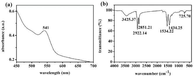
(a) UV-Vis spectrum of CdSe QDs and (b) FTIR spectrum of CdSe QDs.
The broad absorption peak of FTIR spectrum in Fig. 1b at 3425.37 cm−1 illustrated that O-H stretching vibration originated from the surrounding water, which was absorbed by CdSe QDs [31]. The intense absorption peaked at 2922.14 cm−1 and 2851.21 cm−1 ascribed to -CH2- stretching vibrations. Absorption peaks at 1534.22 cm−1 and 1434.25 cm−1 were characteristic absorption peaks of -COO−, which belonged to OA [32]. Furthermore, the Cd-Se stretching vibration peak at 725.7 cm−1 was observed [33]. These observations demonstrated that the CdSe QDs capped with OA were successfully prepared.
EDS was used to further confirm the elemental compositions of CdSe QDs. The results indicated the presence of C, O, Se and Cd of CdSe QDs. The C and O elements came from capping agent of OA and absorbed water. So the average atomic ratio of C to O (76.51:13.70) was lower than the stoichiometric ratio in OA (9:1). Besides, the atomic ratio of Se to Cd (4.73:5.05) was close to the theoretical value in CdSe (1:1).
The XRD spectrum was used to confirm the crystal structures of CdSe QDs. From Fig. 2, the diffraction features appearing at 25.4°, 42.1°, 49.7° corresponded to the (111), (220), (311) planes of the zinc-blende phase of CdSe crystal (JCPDS NO.19-0191), which was consistent with the fact that zinc-blende phase was the most stable form at lower temperature [19].

XRD spectrum of CdSe QDs.
The HRTEM image (Fig. 3a) showed that the obtained CdSe QDs with excellent monodispersity were near-spherical. The distinct lattice fringes on the HRTEM confirmed the good crystallinity of the QDs and the lattice distance of 3.49 Å corresponded to the (111) plane of CdSe QDs. As shown in Fig. 3b, the average size of CdSe QDs was about 3.32 nm (measured by Nano Measurer software) which matched the calculation results from UV-Vis spectrum.

(a) HRTEM image of CdSe QDs and (b) particle size distribution of CdSe QDs.
An orthogonal experimental design was applied to optimize the preparation condition of blank liposome. The results of orthogonal test and range analysis were shown in Table 1. According to the R values, the factors that affected the particle size could be sequenced as: ratio of EPC to PEG (C) > concentration of lipid (A) > ratio of EPC to Chol (B) > ratio of organic phase to aqueous phase (D). The minimum size of blank liposome was obtained when ratio of EPC to PEG, concentration of lipid, ratio of EPC to Chol, ratio of organic phase to aqueous phase were 10:1, 4 mg/mL, 6:1, 5:1, respectively (A3B2C1D3).
Design and directive analysis of orthogonal test
Design and directive analysis of orthogonal test
A: concentration of lipid (mg/mL); B: ratio of EPC to Chol (w/w); C: ratio of EPC to PEG (w/w); D: ratio of organic phase to aqueous phase (v/v); “R” refers to the result of range analysis.
Based on the optimal preparation condition of blank liposome, we made some adjustments to determine the preparation condition of MFLs. The final synthetic condition of MFLs was: ultrasonic time = 15 min, the amount of Fe3O4 = 20 μL, the amount of QDs = 2 mL.
The size and morphology of MFLs prepared on the optimal synthetic condition were determined by laser particle size analyzer (DLS) and HRTEM. The MFLs had an average hydrodynamic size of 111.9 nm and narrow size distribution, which indicated that the prepared MFLs were uneasy to be phagocytized by cells of the mononuclear phagocytic system, therefore ensuring the prolonged circulation effect [34,35]. From the HRTEM image (Fig. 4), the morphology of MFLs was near-spherical and the average size of them was about 82.8 nm, which was smaller than their hydrodynamic size, due to the different principle of detection methods [36]. In the HRTEM image, the darker area should be nano-Fe3O4, whose diameter met the expected size. SEM-EDS analysis of MFL indicated the existence of Fe, Cd and Se. QDs were oil-soluble while nano-Fe3O4 can disperse stably in the water. The reasonable explanation about the MFLs structure was that QDs and nano-Fe3O4 located in different layers inside liposome just like the structure of a Ferrero Rocher chocolate.
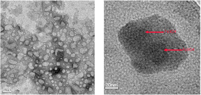
HRTEM images of MFLs.
As shown in Fig. 5, the CdSe QDs had a broad absorption range from 300 to 500 nm and the strongest excitation peak appeared at 468 nm (see inset image). So 468 nm was selected as the excitation wavelength (Ex) to measure the emission spectrum of QDs. The CdSe QDs had a narrow symmetrical emission peak at 570 nm (Ex = 468 nm). Meanwhile, MFLs reserved most of fluorescence properties of CdSe QDs, suggesting the presence of QDs in liposome.
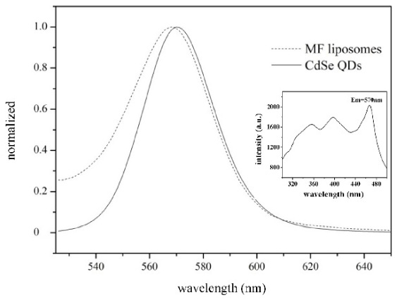
Fluorescence spectra of CdSe nanocrystals and MFLs (inset was the excitation spectrum of CdSe).
A droplet of MFLs solution was dropped on the glass slide and it showed uniform distribution without external magnetic field (Fig. 6a). And then a permanent magnet (N35, 850 Gs) was placed on one side of glass slide to verify the magnetic response of MFLs. The MFLs moved toward magnet as soon as the magnet was placed and then aggregated at the edge of droplet. The entire process was recorded with CCD camera.
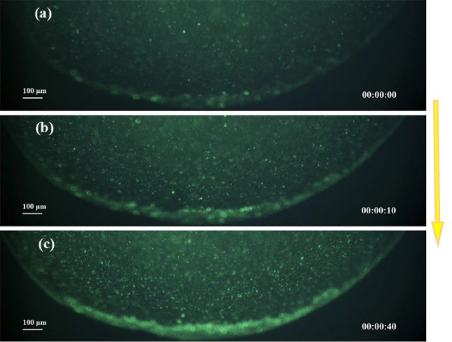
Fluorescence CCD images of the MFLs moving toward the external magnetic field (the yellow arrow indicates the direction of the external magnetic field).
Gaëlle Béalle mentioned that the magnetic liposomes they prepared only carrying magnetic fluid had certain magnetic mobility under the action of magnetic field [37]. The magnetic liposome could accumulate on the target tissue surface and increase the concentration at the target site. It could be concluded that the liposome loaded with Fe3O4 and QDs had a quick magnetic response and good luminescence property, which exhibited great potential for targeting and visual detecting.
To verify the biocompatibility of MFLs, we systematically tested in vitro cytotoxicity through an established MTT assay. As shown in Fig. 7, the cell viability remained at 97.8% when the concentration of MFLs was 0.08 mg/mL. And the cell viability was 82.2% when the concentration of MFLs was 0.8 mg/mL. As the concentration of MFLs increased from 0.08 to 0.8 mg/mL, the cell viability fell sharply. While the concentration of MFLs increased from 0.8 to 8 mg/mL, the cell viability fell gently. The cell viability still reached 60.1% when the concentration of MFLs was up to 8 mg/mL, indicating that MFLs had subtle cytotoxicity and owned good biocompatibility.

Cytotoxicity of MFLs in HepG2 cells.
The MFLs were incubated at 37 °C with HepG2 cells for 8 h to study the cellular internalization of MFLs with cells. The labeling effect of MFLs was investigated by using fluorescence microscope and LSCM. Figure 8 was the microscopy image of HepG2 after being incubated with MFLs. Figure 8a was detected by using optical microscopy. Figure 8b was obtained with fluorescence microscope, and Fig. 8c was the overlay of a and b. The adherent cell morphology showed a spindle or angular shape, which indicated that the cells grew normally. The MFLs aggregated at where the cells were, indicating that MFLs could label the cells with the interaction between MFLs and cells. LSCM results further confirmed the labeling ability of MFLs. As shown in Fig. 9, the MFLs entered into HepG2 cells, which might be the result of the endocytosis of cells. In addition, it was mentioned in Xie’s paper that by detecting the accumulation image of magnetic nanocrystals in 4T1 cells, it showed that it had the ability to bind the target tissue specifically [38]. According to the toxicity test of hepatoma cells shown in Fig. 8 and Fig. 9, it can be concluded that MFLs had minimal cytotoxicity and good biocompatibility, and they successfully labeled the cells.
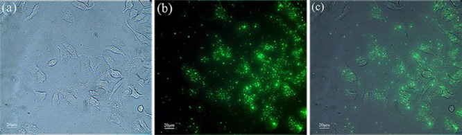
Microscopy images of HepG2 after incubating with MFLs.
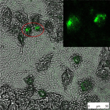
LSCM images of HepG2 (inset was fluorescence image of the cells in red circle).
The zinc-blende CdSe QDs modified with OA showed a narrow symmetrical emission peak at 570 nm and had uniform particle size distribution. The MFLs were successfully synthesized by using REV method. The prepared MFLs exhibited good fluorescence properties and quick magnetic response under an external magnetic field. Cell experiments showed that the MFLs owned good biocompatibility and cell labeling ability. All these results suggest that the MFLs we prepared have great potential for targeting and labeling in biomedicine.
Footnotes
Acknowledgements
This work was supported by the National Key Research and Development Program of China (grant no. 2016YFC1100900 and 2016YFC1100903). The authors thank the Instrumental Analysis Center of Sichuan University for helping with characterization.
Conflict of interest
None to report.