
Abstract
BACKGROUND:
Biomaterials must allow revascularization for a successful tissue regeneration process. Biomaterials formulated from the extracellular matrix (ECM) have gained popularity in tissue engineering because of their superior biocompatibility, and due to their rheological properties, ECM-hydrogels can be easily applied in damaged areas, allowing cell colonization and integration into the host tissue. Porcine urinary bladder ECM (pUBM) retains functional signaling and structural proteins, being an excellent option in regenerative medicine. Even some small molecules, such as the antimicrobial cathelicidin-derived LL-37 peptide have proven angiogenic properties.
OBJECTIVE:
The objective of this study was to evaluate the biocompatibility and angiogenic potential of an ECM-hydrogel derived from the porcine urinary bladder (pUBMh) biofunctionalized with the LL-37 peptide (pUBMh/LL37).
METHODS:
Macrophages, fibroblasts, and adipose tissue-derived mesenchymal stem cells (AD-MSC) were exposed pUBMh/LL37, and the effect on cell proliferation was evaluated by MTT assay, cytotoxicity by quantification of lactate dehydrogenase release and the Live/Dead Cell Imaging assays. Moreover, macrophage production of IL-6, IL-10, IL-12p70, MCP-1, INF-γ, and TNF-α cytokines was quantified using a bead-based cytometric array. pUBMh/LL37 was implanted directly by dorsal subcutaneous injection in Wistar rats for 24 h to evaluate biocompatibility, and pUBMh/LL37-loaded angioreactors were implanted for 21 days for evaluation of angiogenesis.
RESULTS:
We found that pUBMh/LL37 did not affect cell proliferation and is cytocompatible to all tested cell lines but induces the production of TNF-α and MCP-1 in macrophages. In vivo, this ECM-hydrogel induces fibroblast-like cell recruitment within the material, without tissue damage or inflammation at 48 h. Interestingly, tissue remodeling with vasculature inside angioreactors was seen at 21 days.
CONCLUSIONS:
Our results showed that pUBMh/LL37 is cytologically compatible, and induces angiogenesis in vivo, showing potential for tissue regeneration therapies.
Keywords
Introduction
Nowadays there is an increasing clinical demand to obtain specific organs or tissues for regenerative medicine, therefore, the need to manufacture biomaterials with improved biocompatibility and biodegradability capabilities has arisen in tissue engineering [1]. To achieve functional restoration of injured tissues and organs, three factors are required: cells, scaffolds, and biosignals [2]. Scaffolds based on natural extracellular matrix (ECM) confer mechanical properties and stimulate migration, proliferation, and differentiation of various cell populations, including mesenchymal stem cells (MSC), promoting its growth and differentiation [3–5]. The ECM has structural and signaling proteins such as collagen, elastin, fibronectin, vitronectin, glycosaminoglycans, cytokines, and growth factors [6]. ECM scaffolds can be obtained from different tissues, such as skin, liver, pancreas, bone, small intestine, and urinary bladder, among others [7], and formulated as aerogels, pastes, creams, and hydrogels. Porcine urinary bladder ECM (pUBM) retains functional signaling and structural proteins that make it an excellent option in regenerative medicine of the larynx, esophagus, stomach, intestine, urethra, and heart [8–13]. ECM-hydrogels have become an excellent alternative in tissue regeneration because can be easily applied in critical defects with minimal clinical intervention, either directly or injection [14].
The formation of blood vessels from pre-existing vessels (angiogenesis) is a determining step in the repair and regeneration of damaged tissues, such as skin, bones, nerves, muscles, and organs such as the heart and lungs. The importance of new blood vessel formation is to provide nutrients and oxygen to cells located in deep tissue, inducing waste distribution and allowing cell vitality in the host tissue [15,16]. There are several functionalized hydrogels with bioactive molecules such as proteins or growth factors and cells or their derivatives, showing angiogenic properties, including commercials AlgiMatrix, Geltrex, and Matrigel, as well as some free manufacture, such as collagen, alginate or ECM, among others [17,18]. Moreover, many synthetic materials can induce vascularization and allow rapid and orderly bone healing [19].
Hydrogels could incorporate various compounds, including small molecules such as peptides, expanding their use for the regeneration of various tissues. Peptides are small molecules that can perform a variety of functions, transmitting signals, and interacting in essential functions of cellular and molecular biological processes. The main function of proangiogenic peptides is to act as a mimetic signal recognized by growth factor receptors that induce angiogenesis [20,21]. Some of these angiogenic peptides are LL-37, QK, RoY, PBA2-1c, and Exendin-4 [22–24]. The antimicrobial peptide LL-37, the only cathelicidin present in humans, is effective in killing several drug-resistant pathogens [25]. LL-37 peptide-encapsulated chitosan hydrogels have been investigated to assess deep tissue wound healing in a mouse model, which demonstrated that these scaffolds induce the synthesis of proangiogenic factors such as HIF-1α and VEGF-A at 21 days after implantation [26], supported by the fact that LL-37 activates ERK and Akt signaling pathways, promoting lymphangiogenesis in lymphatic endothelial cells [27].
Despite that several biomaterials for revascularization of damaged tissues shows promising potential, there is an urgent need for new angiogenic products, therefore, in this work we evaluated, for the first time, the angiogenic potential of an ECM-hydrogel derived from the porcine urinary bladder (pUBMh) biofunctionalized with the LL-37 peptide (pUBMh/LL37).
Materials and methods
Preparation of porcine urinary bladder (pUBMh) biofunctionalized with the LL-37 peptide (pUBMh/LL37)
The porcine urinary bladders were donated by the Chester White market (Fapsa y Asociados, Sinaloa, Mexico), ISO 9001-2008 certified quality. The decellularization protocol was the one established by Silva et al., with minimal modifications [28], briefly, bladders were decontaminated by transporting them in phosphate-buffered saline (PBS) solution containing 100 IU/ml penicillin and 100 mg/ml streptomycin. Urothelium was separated from the muscle by mechanical delamination and submerged in a shaking 0.25% trypsin solution overnight, frozen at −80 °C, lyophilized, pulverized followed by pepsin-activated enzymatic digestion to obtain a pregel or pUBM (20 mg/ml). Before and after the decellularization, samples were taken, fixed in 10% formalin, embedded in paraffin, cut into 7 μm sections on RM2125 RTS microtome (Leica Biosystems), and stained with hematoxylin and eosin to demonstrate the complete removal of residual cells. Finally, pUBM was biofunctionalized with the LL-37 peptide (10 μg/ml. Tocris Bioscience a Bio-Techne Brand), mixed with 1X PBS and NaOH (1 M), allowing jellification for 30 min at 37 °C to form the pUBMh/LL37.
Cell culture
Macrophages RAW 264.7 (TIB-71, ATCC), Fibroblast NIH/3T3 (CCL-92, ATCC), and human adipose-derived mesenchymal stem cells (AD-MSCs, PCS-500-011-ATCC) were cultured in Dulbecco Modified Eagle’s medium (DMEM, GIBCO, USA) supplemented with 10% fetal bovine (FBS, GIBCO) and an antibiotic cocktail containing penicillin-streptomycin (10,000 U/mL) (Gibco) at 37 °C with 5% CO2, the medium was changed every third day. All cell lines were used in the third passage upon reaching 80% cell confluence for the experiments. Cells were harvested using 0.25% trypsin-EDTA (GIBCO).
Soluble extracts from hydrogels
Soluble extracts of pUBMh/LL37 were prepared as follows: 1 ml of the hydrogel and 9 ml of DMEM were plated with 0.05 mm glass microbeads in a tissue disruptor for 1 min at full speed (Disruptor Genie, Scientific Industries, USA) and incubated at 37 °C with constant slow orbital shaking for 24 h. Finally, the samples were centrifuged at 13,000 rpm, the recovered supernatants were filtered through a 0.22 μm membrane (Cytiva Whatman, Uniflo Syringe Filters, USA) and then used for all in vitro assays described below.
Analysis of mitogenic activity
Methylthiazoletetrazolium colorimetric assay (MTT CellTiter 96 NON-Radioctive Cell Proliferation Assay, PROMEGA, USA) was used to measure proliferation. 7.5 × 10³ cells/well were exposed to the soluble extracts of pUBMh/LL37 for 24, 48, and 72 h, and measurements were made. Briefly, the media was removed, and the cells were incubated with 3 4,5-dimethylthiazol-2-yl) 2,5-diphenyltetrazolium bromide. 15 μl of MTT solution was added to each well over 4 h. The formazan salts were then dissolved by adding 100 μl of stop solution. The optical density (OD) of the samples was measured at 490 nm in a microplate reader.
Analysis of cytotoxicity
To evaluate cytotoxicity, 15 × 103 cells/well were exposed to the soluble extracts pUBMh/LL37 for 48 h, and measurements were made quantifying lactate dehydrogenase (LDH) release in supernatants by colorimetric assay following the manufacturer’s instructions (LDH Cytotoxicity assay, Sigma-Aldrich, USA). OD was measured at 490 nm in a microplate reader. Total cell lysis with 0.9% Triton X-100 was used as a control for comparison.
Evaluation of toxic effects by Live/Dead assay
Live/Dead assay was performed to assess cell membrane integrity and morphology using thiazole orange and propidium iodide. 10 × 104 cells/well were exposed to the soluble extracts of pUBMh/LL37 for 48 h, and then incubated with 84 nM thiazole orange (Sigma-Aldrich) and 4.3 μM propidium iodide (Sigma Aldrich) for 5 min at room temperature. The samples were then examined using a TCS SP8 Confocal microscope system (Leica, Germany) at 40×. Relative fluorescence units (RFU) were measured, and cell viability percentages were obtained. Cell damage induced with 100 μM hydrogen peroxide was used as a control for comparison.
Expression analysis of cytokines
For evaluation of the inflammatory response, 5 × 10⁵ macrophages/well were exposed to the soluble extracts of pUBMh/LL37 for 48 h, and IL-6, IL-10, IL-12p70, MCP-1, IFN-γ, and TNF-α were measured in the supernatants using the Mouse Inflammation CBA Kit according to the manufacturer’s instructions (BD Bioscience, USA). Data were acquired using the Accuri C6 flow cytometer and quantification with FCAP Array v3.0 software (BD Bioscience, USA). LPS were used to treat the cells as a positive control (Lipopolysaccharide from Salmonella enterica serotype Typhimurium, Sigma-Aldrich).
In vivo biocompatibility
Wistar rats of 250--300 g were maintained at 24 ± 2 °C, 50% humidity, 12 h light-dark cycles, and provided with water and diet ad libitum, according to the Mexican Official Norm NOM-062-ZOO-1999 and the guide for the care and use of laboratory animals [29]. Biocompatibility was assessed by subcutaneous injection of the ECM-hydrogel. Briefly, the sedation technique began by placing 1 μL of sedative cocktail (70% Ketamine, 15% Xylazine, 15% Injectable Water) per mg of weight, intramuscularly. Trichotomy and disinfection with 70% alcohol were performed, and the dorse was divided into quadrants taking the transverse midline and sagittal midline as a reference, then 200 μL of pUBMh/LL37 was infiltrated by injection in quadrant 1, 200 μL of chloroform (positive control) in quadrant 2 and 200 μL of 1X PBS (negative control) quadrant 3, using 5 rats for each group. Specimens were sacrificed at 48 h of exposure. Then, euthanasia was performed with a gas chamber, to remove tissue biopsies, which were fixed in 10% formalin for histological evaluations.
In vivo angiogenesis
The angiogenic potential was assessed using subcutaneous micropocket implants. Briefly, after trichotomy and disinfection of the dorse, a pocket was made in the subcutaneous stroma using an N15 scalpel with immediate implantation of the angioreactor the angioreactors (10 × 5 mm) loaded with 200 μL of pUBMh/LL37 (quadrant 1) or empty as a control (quadrant 2) were implanted and keep for 21 days, using 5 rats for each group. After euthanasia, angioreactors were removed and the material inside was recovered for histological evaluations.
Statistical analysis
Experiments were performed three times in triplicate, and results were represented as mean ± SD. ANOVA and Dunnett’s post hoc test were used for 3 or more parametric variables. p < 0.05 was considered statistically significant.
Results and discussion
Obtained pUBMh is a cell-free scaffold rich in collagen fibers
Following the methodology of Silva et al. [28], we obtained an ECM scaffold with large gaps or spaces within and free of cells after trypsin treatment (Figs 1A and B). In addition, a great disposition of collagen content is observed even after mechanical manipulation and contact with the enzymatic solution, this result is of great interest since it is mentioned that an ECM scaffold should retain high content of collagen fibers and signaling molecules after its formulation as hydrogels [30,31]. Then, decellularized material was (20 mg/mg) digested with pepsin to form a pregel, which was biofunctionalized with the LL-37 peptide (10 μg/ml) and jellified to obtain pUBMh/LL37. The ECM-hydrogel showed a clear and whitish appearance with soft but resistant consistency (Fig. 1C), ideal for direct application or being loaded inside angioreactors. Gels were used for the in vivo assays, and soluble extracts were used for the in vivo analyses.

Preparation and characterization of pUBMh/LL37. pUBMh pregel was obtained from the decellularization of porcine bladders. Samples were taken before and after trypsin/EDTA treatment, processed for histology, and observed at 40× magnification. Initial bladder tissue (A) showed normal connective tissue seed with fibroblast (black arrow), surrounded by residual muscular tissue with abundant cells (red arrow). After the decellularization process (B), only cell-free regions and acellular lacunae were seen (black arrow) and dispersed collagen bundles (red arrow). pUBMh/LL37 had a clear and whitish appearance with soft but resistant consistency (C). Scale bar 20 μm.
Macrophage, fibroblast, and AD-MSCs were exposed to the soluble extracts of pUBMh/LL37 for 24, 48, and 72 h to assess its effects on mitogenic activity (Fig. 2). There were no significant changes in proliferation for any cell line during the exposure times (p > 0.05) in comparison to the control group (DMEM). Therefore, this material promotes normal cell proliferation, maybe for the presence of remaining ECM components. Following other reports, it is known that fibronectin, collagen, hyaluronic acid, and proteoglycans, can promote greater cell proliferation [32–34]), and also LL-37 peptide stimulates the migration and proliferation functions of stem cells derived from adipose tissue through the early growth response 1 and the MAPK pathway [35,36]. Our findings also agree with the results reported by Ayala-Ham et al., since the behavior of cell proliferation and metabolic activity of murine macrophages exposed to ECM-hydrogel derived from bovine bone, increased at 48 and 72 h [37].
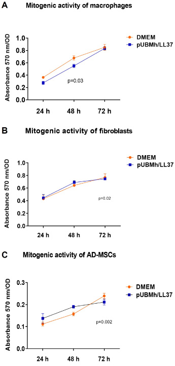
Metabolic activity induced by pUBMh/LL37. Proliferation kinetics were performed in macrophages (A), fibroblasts (B), and AD-MSC (C) exposed to pUBMh/LL37 soluble extracts up to 72 h. MTT assay showed that the ECM-hydrogel did not interfere with proliferation in any cell line during the exposure times (p > 0.05).
Cytotoxicity was also evaluated at 48 h measuring the percentage of LDH release (Fig. 3) in the exposed and control group (DMEM). In general, there were no differences in cell viability between the groups (p > 0.05). In addition, the live/dead assay was used to evaluate cell membrane integrity and morphology. We found similar percentages (Fig. 4A, B, and C) of live cells for macrophages (92.8 ± 2.17), fibroblasts (92.3 ± 4.93), and AD-MSCs (96.9 ± 1.86). In addition, cell morphology was intact after exposure to the soluble extract (Fig. 4D, E, and F) in all groups for the three cell lines. This result agrees with a report by Hussey et al., where demonstrated that ECM-hydrogels prepared from the dermis (dECM), bladder (UBM), or small intestinal submucosa (SIS) are not cytotoxic, since they performed in vitro metabolic and cytocompatibility assays on NIH-3T3 fibroblasts and primary equine mesenchymal stem cells showed almost 100% viability over 24 h [38]. It also corresponds to what was reported by Crum et al., since they demonstrated that UBM with extracellular nanovesicles attached to the matrix are not cytotoxic and do not harm cell proliferation in vitro [39]. Altogether, these results confirmed the cytocompatibility of the porcine urinary bladder (pUBMh) alone, as reported by Jiménez-Gastelum et al. [40], as well as the functionalized hydrogel with the LL-37 peptide (pUBMh/LL37) evaluated in this work.

pUBMh/LL37 is cytocompatible. LDH release assays were performed in macrophages (A), fibroblasts (B), and AD-MSC (C) exposed to pUBMh/LL37 soluble extracts for 48 h. We found that LDH release patterns were similar (p > 0.05) between the different cell lines compared to the DMEM group.

Cell viability and morphology. Macrophages (A, D), fibroblasts (B, E), and AD-MSC (C, F) were exposed to pUBMh/LL37 soluble extracts for 48 h and evaluated by the Live/Death assay and Confocal microscopy. Cell viability was not compromised in any cell line (A, B, and C) (p > 0.5), demonstrating that hUBM-LL37 is biocompatible (p > 0.05). Furthermore, cell morphology retained its integrity (D, E, and F). In green: living cells and in red: dead/dying cells. Positive H2O2 treatment. Scale bar 50 μm.
We analyzed the cytokine release profile of macrophages when in contact with soluble extracts of pUBMh/LL37 for 48 h, to understand the inflammatory effect and the synthesis of interleukins for cell recruitment. We observed that the expression of some cytokines increased significantly (Fig. 5) in the exposed group in comparison to the control (DMEM), for TNF-α (1283.22 ± 96.35 vs 247.37 ± 18.91 pg/ml, p = 0.0001) and MCP-1 (493.96 ± 49.25 vs 51.67 ± 24.21 pg/ml, p = 0.01). There were no changes of consideration for IL-6 (84.85 ± 25.97 vs 106.10 ± 6.45), IL-10 (43.22 ± 2.76 vs 40.39 ± 4.55), IL-12p70 (7.74 ± 1.35 vs 16.74 ± 2.57), and INF-γ (0.00 ± 0.00 vs 1.51 ± 0.31) expression between the groups. Damage or injury induces inflammation, under physiological conditions which increases blood flow, leading to the accumulation of interstitial fluid, accumulation of leukocytes, recruitment of inflammatory mediators, and upregulation of proinflammatory cytokines [41,42]. Reports of liver tissue regeneration mention a dose-dependent increase in the mitotic rate after exposure to TNF-α [43]. Besides, TNFα is required for optimal VEGF production and angiogenesis in aortic cultures, being this pathway of angiogenic induction dependent on macrophages which produce TNF-α and mediate its proangiogenic effect [44]. On the other hand, it has been shown that MCP-1 is a chemotactic cytokine that actively stimulates angiogenesis activating the transcription factor ETS-1 [45] to upregulate VEGF and HIF-α, key molecules in the angiogenic process [46,47]. Also, MCP-1 mediates TGF-β–stimulated angiogenesis by enhancing the migration of mural cells toward endothelial cells and thus promoting the maturation of new blood vessels [48]. Our finding is in agreement with the report by Jiménez-Gastelum et al. for pUBMh alone [40], and suggests that these overexpressed cytokine are involved in the initial stage of inflammation leading to tissue regeneration and revascularization.

Cytokine expression analysis. Using the cytometric bead array assay, we found an increase in the proinflammatory cytokines TNF-α (A) and the chemotactic cytokine MCP-1 (B) in macrophages exposed to hUBM/LL37 soluble extracts for 48 h (p < 0.05).
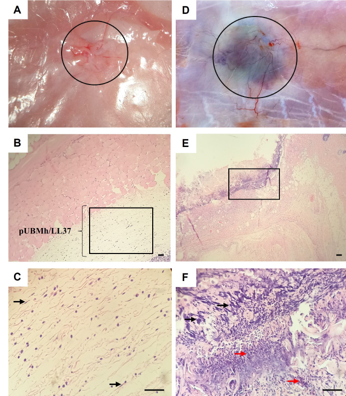
In vivo biocompatibility of pUBMh/LL37. After 48 h of subcutaneous injection of pUBMh/LL37 and chloroform (as damage control) into the dorsal region, anatomy and tissue reaction was observed. pUBMh/LL37 remains stable in a well-defined area (black circle), with no subdermal abnormalities (A), with low cellularity within the material (black square) (B), and under magnification (C), fibrillar structure, few polymorphonuclear and fibroblast-like cells (black arrows) were seen. In contrast, chloroform injection caused severe damage by inducing a hematoma (D), with severe inflammatory infiltrate as well as dense connective tissue (black square) (E) and under magnification (F), epithelioid cells interacting with fibroblastoid-type cells (black arrows) inflammatory infiltrates (red arrows) are seen Scale bar 50 μm.
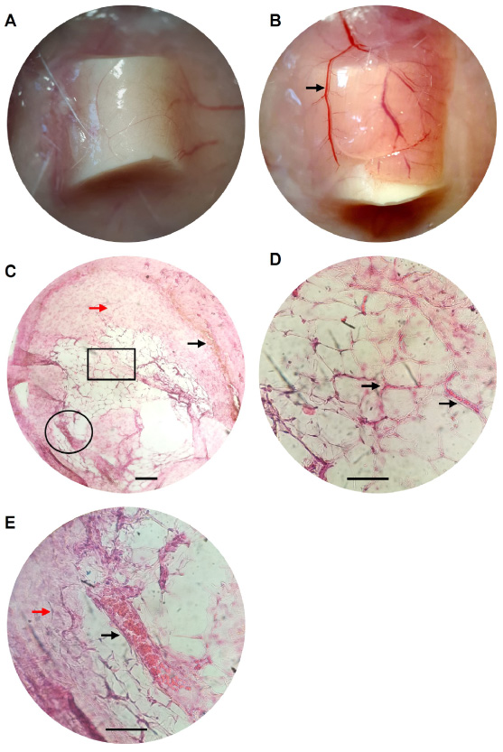
In vivo vasculogenesis. After 21 days of dorsal subcutaneous implantation of empty (negative control) or pUBMh/LL37-loaded angioreactors, revascularization was evaluated. Both empty (A) and loaded (B) angioreactors were well integrated into the host tissue, but the loaded angioreactor showed bigger and more numerous blood vessels in the external area (black arrow). Complete transverse section histology of the recovered material inside angioreactor (C) showed loose fibrous connective tissue (red arrow) and scattered and trabecular networks (black square), a region with hemosiderin deposition (black arrow), and neoformation of blood vessels containing abundant erythrocytes (black circle). Under magnification, vascular primordium (black arrows) (D), as well as the presence of fibroblast-like cells (red arrow) and a longitudinal section of a perfunded vein (black arrow) can be observed (E). Scale bar 50 μm.
Subcutaneous infiltration of pUBMh/LL37 in the dorsal region or rats was performed to assess biocompatibility in vivo for 48 h. Gross examination showed a normal aspect of the dermis, and that ECM-hydrogel has an excellent integration into the host tissue, resists reabsorption, and retained its 3D structure, appearance, and softness (Fig. 6A). In the histological evaluation, a fibrillar structure with dispersed collagen bundles can be observed, populated by fibroblastoid-type cells with few polymorphonuclear cells (Fig. 6B and C). In contrast, the chloroform injection caused severe damage and hematoma (Fig. 6D) with severe inflammatory infiltrate (Fig 6, E and F). Výborný et al., observed a similar behavior of increased colonization of fibroblast cells when using an umbilical cord ECM-derived hydrogel, confirming our findings [49], on the other hand, ECM-derived hydrogels have shown potential advantages as they are included as potent bioactive materials that provide a viable microenvironment for therapeutic applications, are easy to apply to fill critical defects and without risk of immunological rejection [50]. In the case of pUBMh, it is known that when applied to wounds it strongly stimulates healing and this is since it preserves growth factors and intrinsic signaling molecules such as FGF, VEGF, PGF, TGF-β, and BMP4, among others [51].
Implantation of pUBMh/LL37 loaded angioreactors induces angiogenesis in vivo
Subcutaneous angioreactors loaded with pUBMh/LL37 were implanted in the dorsal area of rats to assess their biocompatibility in vivo. After 21 days, euthanasia was performed and angioreactors were recovered for analysis. By gross inspection, we found a complete integration of the implanted angioreactors in the transepithelial basement membrane, both empty and loaded with the ECM-hydrogel (Fig. 7, A and B). Complete transverse section histology of the recovered material inside the pUBMh/LL37-loaded angioreactor (Fig. 7C), showed a loose fibrous connective tissue stroma with scattered and trabecular networks (Fig. 7D), with the presence of spindle cells consistent with fibroblasts, and neoformation of blood vessels containing abundant erythrocytes (Fig. 7E). No type of tissue or residual material was found inside the empty angioreactor.
These findings allow us to confirm that our scaffolds have chemotactic and angiogenic capacity. This agrees with what was reported by Yang, Xu, et al. since they evaluated the behavior of a hyaluronic acid hydrogel loaded with LL-37 in pressure ulcers, finding an increase in epithelial thickness and great capillary density, as well as an increase in VEGF-A and HIF-1α from day 11 of interaction [26]. These data agree with what was reported by Pike. et al., since they demonstrated rapid and prolonged in vivo angiogenic response of implanted hyaluronan hydrogels loaded with VEGF and bFGF [52]. It also agrees with what was reported by Dissanaca et al., since they evaluated the formation of blood vessels in vivo in a murine model for 14 days, on a PURAMATRIX + VEFG scaffold loaded with HUVECs and DPSCs [53].
Conclusions
Our results show that the ECM-hydrogel derived from porcine urinary bladder biofunctionalized with the LL-37 peptide (pUBMh/LL37) is cytocompatible and allows the proliferation of macrophages, fibroblast, and mesenchymal stem cells, and induces the synthesis of TNF-α and MCP-1 in macrophages, key molecules for inflammation, recruitment, and cell differentiation during tissue repair. Moreover, this ECM-hydrogel is biocompatible and induces angiogenesis in vivo. All these properties have potential in regenerative medicine since this functional construct could be used to restore, maintain, or improve damaged tissues or even whole organs.
Footnotes
Acknowledgements
The authors thank the Mexican company Fapsa y Asociados S.A. De C.V. for donating the necessary bladders to carry out this important project. This research was funded by PROFAPI-UAS grant PRO/A2/012.
Author contribution
JLG, AAH, GJG, RRP conducted laboratory tests, JGRQ, AAH, YCS, MB collected the data, MAM, RRP, HCU, and MB analyzed the results. MAM and RRP conceived and designed the study. JGRQ, AAH, JLG, and RRP wrote the manuscript. YCS and HCU contributed to the final version of the manuscript. All authors read and approved the final manuscript.
Conflict of interest
No potential conflict of interest relevant to this article was reported.