
Abstract
BACKGROUND:
Ureteral stents are commonly used in urology. However, complications such as encrustation and infection on the surface of the stent, and injury to the ureteral mucosa can occur after implantation, causing discomfort for patients.
OBJECTIVE:
We intend to confirm the biosafety of polyvinylpyrrolidone (PVP) hydrophilic coating and its lubrication properties for surface modification of ureteral stents to reduce friction and improve patient comfort.
METHODS:
Based on our previous studies, we have developed a PVP hydrophilic coating for surface modification of ureteral stents. We firstly investigated the cytotoxicity, intradermal irritation, delayed type hypersensitivity, and acute systemic reactions of stent coating extracts. We further characterized the break strength, retention strength, and dynamic friction of the stent.
RESULTS:
The cell survival rate of all experimental groups was greater than 70%. No hypersensitivity reaction, systemic toxicity reaction, or obvious intradermal reaction were observed. The above results indicate that the test results of the modified stent meet the requirements of ISO 10993-5: 2009 (Cytotoxicity); ISO 10993-10:2021 (Sensitization and Irritation); ISO 10993-11:2017 (Acute Systemic Toxicity). After soaking in artificial urine for an extended period, there was no obvious change in its super-slip performance.
CONCLUSION:
Our results confirm the safety and lubrication characteristics of PVP hydrophilic coating for ureteral stent surface modification. The performance of this coating has the potential to reduce complications after stent implantation, thereby improving patient comfort, reducing medical burden, and has a good clinical application prospect.
Keywords
Introduction
Ureteral stents are integral to endourologic practice, commonly used in in the perioperative management of urolithiasis, relief of ureteral obstruction, and management of ureteral reconstruction [1]. By supporting the ureter, they relieve upper urinary tract obstruction, prevent stricture formation, drain urinary leakage, and reduce postoperative complications [2]. However, long-term retention of ureteral stents can lead to complications such as infection, encrustation, and damage to the ureteral mucosa. In severe cases, these complications may impair drainage and cause ureteral obstruction, renal insufficiency, or even renal failure. This can inflict significant pain and financial burden on patients while also increasing the strain on limited medical resources [3,4].
Each year, a considerable number of patients are affected by the serious clinical problem of encrustation [5]. Encrustation occurs when minerals from the urine deposit on the surface of an indwelling stent. Risk factors include the duration of stent indwelling time, bacterial colonization, patient-specific factors, and physical characteristics of the stent [6]. Encrusted stents become ossified (calcified) and fragile, losing their durability and increasing the chances of fracture of the stent or avulsion of the ureter during withdrawal [7]. As such, an ideal stent material should not only possess good biocompatibility and mechanical properties to alleviate intra- and extra-luminal obstruction, but also reduce encrustation, infection, and patient discomfort [8].
The most commonly used materials for ureteral stents are polyethylene, polyurethane and silicone, which exhibit different tendencies for encrustation [9]. Polyurethane combines the softness of silicone with the hardness of polyethylene and its excellent blood and tissue compatibility make it a popular choice in the medical field. However, polyurethane is not as smooth as other materials such as silicone, which increases surface friction and mineral deposition incidence making the stent more difficult to place and remove, which can potentially cause discomfort to the patient [10]. Research shows that stent surface modification is a promising method to reduce ureteral stent-related complications and make it suitable for clinical application [11]. Hydrophilic coating has the advantages of enhanced hydrophilicity, improved biocompatibility and reduced protein adsorption [12]. Hydrophilic hydrogels can lubricate the stent surface and reduce friction, helping to inhibit or prevent encrustation and biofilm formation [13–15]. Nevertheless, current ureteral stents often have issues such as low bonding strength between surface coatings and substrates, easy shedding, insufficient super lubricity, and unsatisfactory antibacterial and anti-encrustation performance.
Polyvinyl Pyrrolidone (PVP) has good physiological inertness and biocompatibility and is widely used as a lubricating ingredient in hydrophilic lubricating coatings [16,17]. The hydrophilic nature of PVP hydrogel coatings facilitates stent placement by significantly reducing friction between the catheter and ureteral tissue, which is crucial for the implant of stent into the human body and potentially increasing patient comfort [18]. Furthermore, the PVP coating also helps avoid the deposition of urinary crystals that trigger encrustation. However, PVP has poor adhesion and must be combined with adhesive components. Polyurethane prepolymer and polyol are commonly used as adhesive components in hydrophilic lubricating coatings [19].
In this study, we utilized a hydrophilic lubricating coating composed of PVP, polyurethane prepolymer, and polyol as the primary components. The ureteral stent loaded with a zwitterionic polymer synthesized in previous research reports, and a hydrophilic-coated ureteral stent was prepared by thermal curing [20]. We expect that the stent we have prepared will have good biocompatibility and can use its hydrophilic properties to reduce the friction force on the surface of the ureteral stent, thereby reducing the incidence and severity of complications during ureteral stent placement and improving the treatment effect and comfort of patients. Therefore, we tested the cytotoxicity, systemic toxicity reaction and hypersensitivity reaction of the stent to detect its biosafety. And further characterized the physical properties of the stent to verify its improvement in stent performance.
Methods and materials
Preparation of polyvinylpyrrolidone hydrogel coating ureteral stent
The preparation of the membrane biomimetic ureteral stent was completed in cooperation with Chengdu Daxan Innovative Medical Tech. Co., Ltd [20,21].
The stent, characterized by an outer diameter of 4.8 Fr and an effective length of 30 cm, possesses a coating thickness that varies between 1.41 and 1.97 μm. The coating, thinnest at the stent’s bend bearing the Daxan logo and thickest at another bend, exhibits a non-uniform distribution attributed to the dip coating process. Notably, the coating is denser at the lower end. The stent undergoes sterilization via ethylene oxide.
Biocompatibility test
The experimental animals and procedures were approved by the Ethics Committee of the First Affiliated Hospital of Chongqing Medical University (permit number: 2022-K44). Throughout the experimental process, animal welfare concerns were well monitored.
Cytotoxicity
Preparation of extract solution
0.25% ZDBC polyurethane film and high-density polyethylene film were selected as positive and negative controls, respectively. Both films were autoclaved at 121 °C for 15 minutes and purchased from Hatano Research (lot numbers: B-213K, C-212). Membrane biomimetic ureteral stents were leached in complete MEM (minimal essential medium) (pH 7.2) containing 10% fetal bovine serum (FBS) at a rate of 3 cm2/mL in an incubator at 37 °C ± 1 °C for 72 hours. Negative and positive controls were leached under the same conditions at rates of 3 cm2/mL and 6 cm2/mL, respectively. Stent extracts were diluted with cell culture medium to four concentrations: 100%, 75%, 50%, and 25%.
Experiment procedure
L-929 cells (ATCC cell line CCL-1) were used to assess the cytotoxicity of the samples. The cells were inoculated in a 96-well culture plate at a concentration of 1 × 10⁵/mL, with 100 μL of cell suspension added to each well. The cells were then cultured in a 37 °C incubator with high humidity and 5% CO2 for 24 hours. The medium used for cell culture was MEM containing 10% FBS, supplemented with 4 mM L-glutamine, 100 units/mL penicillin, and 100 μg/mL streptomycin.
Negative control (high-density polyethylene film extract), medium control (medium), positive control (0.25% ZDBC polyurethane film extract), and experimental groups (100%, 75%, 50%, 25% sample extract) were set up. When a near-confluent cell layer formed, the supernatant medium was discarded. 100 μL of different extracts were added to each group, and 6 wells were reseeded in the positive control, negative control, and experimental groups, respectively. After culturing for 24 hours, cell morphology was observed and the culture supernatant of each well was removed. 50 μL of MTT (1 mg/mL) was added to each well and incubated for 2 hours. The MTT solution was then removed and 100 μL of isopropanol was added to each well. The plate was shaken at low speed for 10–30 minutes to fully dissolve the MTT crystals. The absorbance OD570 value of the culture plate was measured at a measurement wavelength of 570 nm and a reference wavelength of 650 nm.
Cell viability calculation
Cell viability (relative proliferation rate) = absorbance value of the experimental group/absorbance value of the medium control group × 100%.
Intradermal irritation
Preparation of extraction solution
The membrane-modified stents were leached with saline and corn oil at a ratio of 3 cm2/mL for 72 hours in an incubator at 50 °C ± 2 °C to prepare the extract.
Experimental animals and feeding conditions
Three male New Zealand white rabbits, approximately 5 months old and weighing between 2.9–3.4 kg, were used as experimental animals. The rabbits were fed standard feed (Beijing Keao Xieli Feed Co., Ltd., batch number: 22034113) and had free access to food and water. During the experiment, the relative humidity in the animal room was maintained at 47.3%–68.4%, the temperature at 19.7 °C–21.7 °C, and the ventilation rate at 10–20 times per hour. The cages measured 146 ∗ 66 ∗ 45 cm3 and were well lit with a 12-hour light/dark cycle.
Experimental procedure
Before the experiment, all hair on the backs of rabbits was removed. 10 pairs of injection points were evenly selected on both sides of each rabbit’s spine, with a minimum interval of 1 cm between each point. On the left side, 0.2 mL of saline or corn oil extract was injected at 5 sites. On the right side, 0.2 mL of the corresponding control solution was injected at the same sites. Skin reactions were observed and scored immediately, and at 24, 48, and 72 hours after injection. The scoring standard of intradermal reaction is shown in Table 1. Any other clinical symptoms in the animals, apart from skin reactions, were recorded at each time point.
Visual assessment of skin reactions
Visual assessment of skin reactions
The sum of the erythema and edema scores at the injection sites of the experimental and control groups were calculated at 24, 48, and 78 hours. The sum was divided by 15 to obtain the score for each animal. The scores for the experimental or control groups of the three animals were added and divided by 3 to obtain their respective overall mean scores.
Delayed type hypersensitivity test in guinea pigs
Preparation of extract
Prepare the extraction solution as described in Section 2.2.2.1.
Experimental animals and grouping
40 male guinea pigs, approximately 4–6 weeks old and weighing between 335.20–481.11 grams, were purchased from Pizhou Dongfang Breeding Co., Ltd. (Experimental animal quality certificate number: 202221248). The guinea pigs were randomly divided into 4 groups: the control group (saline or corn oil) had 7 animals per group, and the experimental group (saline or corn oil extract) had 13 animals per group.
Experimental procedure
2.2.3.3.1. Intradermal injection phase
Before the experiment, hair was removed from an area of approximately 4 cm * 6 cm on the backs of the guinea pigs. Three pairs of intradermal injection sites were selected for each animal: sites a and b were near the head end, while site c was located near the tail. Site a was injected with a saline and Freund’s Complete Adjuvant (FCA), while site b was injected with extraction solution or medium control. Site c was injected with saline, FCA and extract solution or medium control, with a ratio of 1:1 between the saline and FCA. The prepared preparations were injected in pairs on both sides of the midline of each animal’s back, with 0.1 ml injected intradermally at each site.
2.2.3.3.2. Topical application phase
After 7 days of intradermal induction, all injection sites were shaved again and smeared with approximately 0.5 mL of 10% sodium dodecyl sulfate (SDS) paraffin solution per animal. On the 8th day, filter paper measuring approximately 2 cm * 4 cm was saturated with saline or corn oil extract for each experimental group and applied locally to cover the injection site on each animal’s back and as much surrounding skin as possible. The area was then wrapped in retractable wrapping material for 48 hours and secured with tape. The control group was treated in the same way as the experimental group using the extraction medium. The patch was removed after 48 hours.
2.2.3.3.3. Excitation
14 days after topical induction, the upper ventral side (uninduced area) of the guinea pigs was shaved. Filter paper measuring approximately 2 cm * 2 cm and soaked with sample extract was applied to the shaved area on the right side of each animal. Filter paper of the same size soaked in control medium and was applied to the shaved area on the left side of each animal. The dosing site for each animal was then wrapped with stretchable wrap and taped for 24 hours.
2.2.3.3.4. Observation
Throughout the experiment, the animals were clinically observed daily and weighed before the first administration and on the final day of the experiment. Approximately 24 and 48 hours after the removal of the patch, skin reactions at the excitation site were observed and scored according to the Magnusson and Kligman grading criteria (Table 2).
Magnusson and Kligman scale
Magnusson and Kligman scale
Preparation of extraction solution
The extract was prepared as described in Section 2.2.2.1.
Experimental animals and grouping
20 female mice, approximately 4–6 weeks old at the beginning of the experiment, were purchased from Beijing Weitong Lihua Experimental Animal Technology Co., Ltd. (certificate number: 110011221104524318). The mice weighed between 19.67–24.45 grams and were randomly divided into 4 groups of 5 animals each. Two control groups (saline or corn oil) and two experimental groups (saline or corn oil extract) were set up.
Experimental procedure
Animals in groups 1 and 2 were given saline extract and saline by intravenous injection, while animals in groups 3 and 4 were given corn oil extract and corn oil by intraperitoneal injection. The administration volume for each animal was 50 mL/kg, with an intravenous injection rate of approximately ≤2 mL/min. The animals were observed for clinical toxicity immediately after injection and at 4, 24, 48, and 72 hours later. The animals were also weighed before and after administration for three consecutive days.
Characterization of stent performance
Preparation before mechanical test
Based on previously reported concentrations, the contents of each component in artificial urine are as follows: NaCl is 6.17 g/dm3, NaH2PO4 is 4.59 g/dm3, Na3C6H5O7 is 0.944 g/dm3, MgSO4 is 0.463 g/dm3, Na2SO4 is 2.408 g/dm3, Na2SO4 is 4.75 KCl g/dm3, CaCl2 is 0.638 g/dm3, and Na2C2O4 is 0.043 g/dm3 [22]. The pH of the artificial urine solution is between 5.5 and 6.5. The stent was soaked in artificial urine at 37 ± 1 °C.
Break strength
Static tensile tests were conducted according to the recommendations of ASTM F 1828-97 standard [23]. Tests were performed at a distance of 35 mm from the stent body before and after soaking for 35 days. The test section was clamped in the fixture of a microcomputer-controlled electronic universal testing machine, with 5 mm clamped at each end and a gauge length set at 25 mm, at a tensile rate of 200 mm/min. Three stents were randomly selected for testing.
Retention strength
The ureteral stent fixation strength test was conducted according to the recommendations of ASTM F 1828-97 [23]. Tests were performed at a distance of 35 mm from the stent body before and after soaking for 35 days. A polyoxymethylene resin funnel block was prepared for testing. After the stent reached thermal equilibrium at 37 ± 3 °C, it was pulled upward at a rate of 500 mm/min to pass through the bell mouth. The maximum pulling force required for the stent to completely pass through the bell mouth was recorded. Three stents were randomly selected for testing.
Dynamic frictional force
The ureteral stent dynamic friction test was conducted according to the recommendations of ASTM F 1828-97 [23]. Before starting the test, the stent was soaked in distilled water for at least one minute. Then the stent was passed through the upper part of the test ring and completely immersed in the water column. A microcomputer-controlled universal testing machine was used to pull out at a constant speed of 500 mm/min, and at least 100 data points were collected for each test, and the average value was calculated as the dynamic friction detection value. Polyurethane and coated stents were tested before and after soaking for 35 days, with three samples taken for each type.
Statistical analysis
The results are expressed as mean ± standard deviation and the differences between data sets were evaluated using one way ANOVA test and pairwise comparisons were evaluated used using t-test. P-value less than 0.05 was considered statistically significant.
Results
Cytotoxicity
After the culture period, normal cell morphology was observed in the experimental group. Figure 1a displays the relative cell viability rate calculated based on the OD570 values for each group by using the left medium control group as a reference. There was no significant difference in cell viability between the left and right medium control groups (P > 0.05). The survival rate of the negative control was 101.86%, while the positive control had a survival rate of 1.04%, indicating significant cytotoxicity. The cell viability gradually decreased with the increase of the concentration of stent extracts.

Results of biocompatibility test. (a) The relative cell viability rate calculated based on the OD570 values for each group by using the left medium control group as a reference, resulting from the MTT assay results. (b) Results of cell delayed type hypersensitivity reaction test. The score was calculated based on the Magnusson and Kligman sensitization grading criteria. (c) The erythema and edema scores of intradermal irritation test. (d) Weight changes at different time points in various animals, results from systemic toxicity experiments. Data were presented as mean ± SD. MC, medium control, NC, negetive control, E.25, extract resolution, PC, positive control. (*P < 0.05, **P < 0.01, ***P < 0.001).
During the experiment, no abnormal clinical reactions were observed and their body weight changes were normal. According to the Magnusson and Kligman sensitization grading criteria, the reaction scores of the control group were 0 at both 24 and 48 hours after challenge, with a 0% positive rate of sensitization, indicating that saline and corn oil did not cause sensitization. Similarly, the reaction scores of the experimental group at 24 and 48 hours after challenge were also 0, with a 0% positive incidence of sensitization, proving that the sample did not have the potential to cause skin sensitization (Fig. 1b).
Intracutaneous irritation
During the experiment, no abnormal clinical symptoms were observed at each time point except for skin reactions. The body weight changes of the animals were normal. In the saline group, no skin irritation symptoms appeared at the injection points 24, 48, and 72 hours after local intradermal injection. No erythema or edema appeared in any of the animals in each group and their erythema and edema scores were all 0 (Fig. 1c). The average score of the corn oil control group was 1.93 and the average score of the corn oil experimental group was 2, indicating that corn oil can cause local reactions (Fig. 1c). However, the difference between the two groups at each time point was not more than 1, proving that this sample did not have the potential to cause intradermal irritation.
Acute systemic toxicity
After injection of the extraction solution at 0, 24, 48, and 72 hours, the mice were in good general condition. They had stable breathing, free movement, and no deaths. Their fur was smooth and their diet was normal. There were no abnormal secretions from their mouth, nose or eyes and their urine and stool were normal. Their body weight increased as shown in Fig. 1d. None of the mice in the experimental group lost more than 10% of their body weight. This indicates that the health status of the experimental group may be similar to that of the negative control group.
Break strength
Figure 2a shows the results of the breaking strength. It can be seen that there was a slight change in the physical properties of the stent after soaking. Before soaking, the breaking force of the stent was 40.10 ± 0.79 N, and after soaking, its strength decreased slightly to 37.03 ± 4.45 N.
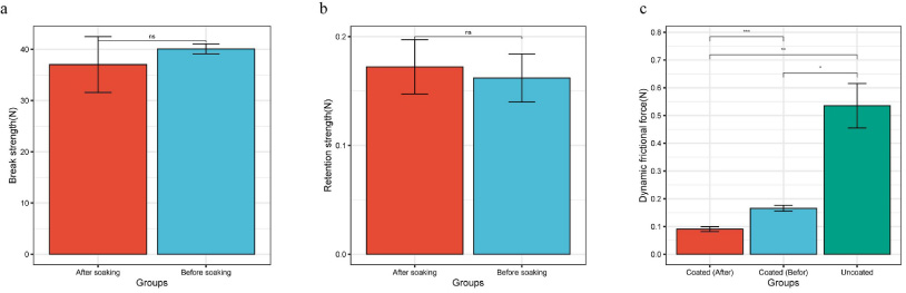
Results of stent performance characterization. (a) Break strength test results of stent before and after soaking. (b) Retention strength test results of stent before and after soaking. (c) An unmodified polyurethane stent was used as a control to compare dynamic frictional force.
There is no statistically significant difference in retention strength between the stent before and after soaking (P > 0.05) (Fig. 2b). And the retention strength of both is greater than 0.1 N.
Dynamic frictional force
Based on the results of dynamic friction force, it can be seen that the coating applied on the surface of the stent significantly reduces the friction force (p < 0.05). The average value of dynamic friction force of the polyurethane stent is 0.535 ± 0.065 N, while the the coated stent is 0.166 ± 0.009 N, which is about one-third of that before modification of stent surface. And after soaking for 35 days, the friction force of this stent is even lower than initial state (p < 0.05) (Fig. 2c).
Discussion
Encrustations of ureteral stent surface can foster bacterial biofilms, leading to infections and promoting crystal precipitation [24]. Preventing urinary tract infections in patients with long-term indwelling stents hinges on disrupting biofilm formation. Stent modification can effectively inhibit infection in patients with implanted ureteral stents. Various stents and stent coatings have been investigated to combat infection, with common experimental methods including surface treatment with repelling coatings, antimicrobial agents, surfactants, or selected biomolecules such as heparin or albumin [25,26]. Relevant anti-infection strategies are usually based on the following strategies: (1) Modify the stent surface to obtain anti-adhesion properties; (2) Add antimicrobial drugs to the stent material; (3) Combine the above two strategies when making ureteral stents. Surface modification of ureteral stents has been an active area of research in recent years [27].
Aleksandra Kuźmińska et al. prepared a modified polyurethane stent with a PVP coating and experimentally verified its biosafety and excellent anti-adhesion properties [28]. Our research builds on these existing studies and develops a hydrophilic lubricating coating with PVP, polyurethane prepolymer, and polyol as the main components to reduce friction on the stent surface, prevent encrustation on the stent surface, and improve patient comfort after implantation.
Good biocompatibility is the first requirement for a qualified ureteral stent. The MTT assay is a commonly used and convenient experiment to detect cell activity and evaluate biological toxicity [29]. L-929 fibroblasts are commonly used in cytotoxicity experiments due to their reproducible growth and biological response [30]. Our result suggested that the survival rate of stent extracts at each concentration exceeded 70%, meeting the standard for qualified materials [31]. Therefore, it can be assumed that the coating used on the ureteral stent surface will not cause significant cytotoxic reactions. Ureteral stents generally stay in the body for a long time. Therefore, contact of the stent surface with the ureteral mucosa may cause inflammation and hypersensitivity reactions, which will cause discomfort and even serious complications to patients. Delayed type hypersensitivity is a type of immune response that is characterized by antigen-specific T cell infiltration. It usually taking 24–72 hours to develop after the body is re-exposed to the same antigen stimulus [32]. We found that Magnusson and Kligman sensitization grading score were 0 for each, indicating that the coating does not cause significant sensitization. Further intradermal irritative reactions showed that at each observation time point, no other abnormal clinical symptoms were found in all animals except skin reactions. And no obvious skin reaction was observed in the saline group. Although skin reactions were observed in the corn oil group, the difference between the experimental group and the control group was less than 1, which may be due to the irritation caused by the corn oil. The above results show that the coating meets the requirements and has no potential intradermal irritation [33].
A stent that has become encrusted will calcify and become brittle, losing its durability and increasing the chance of stent fracture or ureteral avulsion during removal [34]. A ureteral stent with a break strength of >10 N, a retention strength of >0.05 N, and a dynamic friction force of <0.5 N can ensure its safety and effectiveness during clinical practice. In our study, the lubricating stent with a hydrophilic coating has a break strength and retention strength that fully meet the clinical use requirements. After soaking in artificial urine to simulate clinical use scenarios, there is no significant change in performance, and it is far higher than the clinical use requirements. This indicates that our coating can meet the basic requirements for stent retention while ensuring a reduction in friction, avoiding displacement during retention. The friction force of the coated ureteral stent is much lower than that of the uncoated stent. After soaking in simulated urine for 35 days, it still has excellent lubrication performance. Our research has demonstrated the excellent hydrophilic and ultra-smooth properties of the coating, which can help prevent encrustation on the surface of the stent and reduce complications from stent placement. In addition, the low friction on the surface of the stent can also reduce discomfort for patients during placement and removal, which ensuring the safety and effectiveness of the ureteral stent during long-term clinical placement.
In this study, we developed an effective hydrophilic coating to give the stent super-slip performance. This performance has the potential to prevent the adhesion of bacteria and proteins, reducing complications such as encrustation and infection on the surface of the ureteral stent after implantation in the body, thereby improving patient comfort and reducing medical burden. Our research has confirmed the biosafety and excellent super-slip performance of the stent, but further research is needed to test and improve our coating and evaluate its effectiveness in preventing adhesion, scabbing and infection.
Conclusion
In summary, our research on developing a hydrophilic lubricating coating for the surface modification of ureteral stents has the potential to significantly contribute to the field and improve patient outcomes in clinical practice. We initially conducted tests on the biocompatibility of the coating, including cytotoxicity, delayed hypersensitivity reactions, acute systemic reactions, and intradermal irritative reactions of stent coating extracts. The results confirmed that the coated stent has good biocompatibility and can be used in the body for an extended period. Furthermore, its physical properties remain stable after long-term soaking in urine, meeting clinical requirements. Additionally, this stent exhibits excellent lubrication performance, ensuring the safety and effectiveness of the ureteral stent during long-term clinical placement. Good tissue compatibility and low friction characteristics can reduce the urethral epithelial reaction after stent placement, prevent biofilm formation, and improve the efficiency of long-term drainage. The above results indicate that the coated stent we developed has the potential to be applied clinically to benefit a large number of patients who need to implant ureteral stents.
Footnotes
Acknowledgements
The authors have no acknowledgments.
Conflict of interest
The authors declare that they have no conflict of interest.
Funding
This work was supported by the Senior Medical Talents Program of Chongqing for Early and Mid Career Researchers supported by Chongqing Health Commission (No. 2022GDRC014).