



Abstract
BACKGROUND:
The ligament is the soft tissue that connects bone to bone and, in case of severe injury or rupture, it cannot heal itself mainly because of its poor vascularity and dynamic nature. Tissue engineering carries the potential to restore the injured tissue functions by utilization of scaffolds mimicking the structure of native ligament. Collagen fibrils in the anterior cruciate ligament (ACL) have a diameter ranging from 20 to 300 nm, which defines the physical and mechanical properties of the tissue. Also, the ACL tissue exhibited a bimodal distribution of collagen fibrils. Currently, the ability to fabricate scaffolds replicating this structure is a significant challenge.
OBJECTIVE:
This work aims at i) measuring the diameter of collagens of bovine ACL tissue, ii) investigating the fabrication of sub-100 nm fibers, and iii) fabricating aligned scaffolds with bimodal diameter distribution (with two peaks) resembling the healthy ACL structure. It is hypothesized that such scaffolds can be produced by electrospinning polycaprolactone (PCL) solutions.
METHODS:
To test the hypothesis, various PCL solutions were formulated in acetone and formic acid in combination with pyridine, and electrospun to generate sub-100 nm fibers. Next, this formulation was adjusted to produce nanofibers with a diameter between 100 nm and 200 nm. Finally, these solutions were combined in the co-electrospinning process, i.e., two-spinneret electrospinning, to fabricate biomimetic scaffolds with a bimodal distribution.
RESULTS:
Electrospinning of 8% and 15% PCL solutions, respectively, resulted in the production of fibers with diameters below and above 100 nm. The combined scaffold exhibited a bimodal distribution of aligned fibers with peaks around 80 and 180 nm, thus mimicking the collagen fibrils of healthy ACL tissue.
CONCLUSION:
This research is expected to have a society-wide impact because it aims to enhance the health condition and life quality of a wide range of patients.
Introduction
Ligament tissue consists of cells and extracellular matrix (ECM), which includes collagen, proteoglycans, glycoproteins, and elastin [1,2]. Ligament cells, i.e., ligamentocytes and ligamentoblasts, constitute nearly 90–95% of the cellular content [2], and play a key role in collagen synthesis, tissue growth, and remodeling [3]. Elastin accounts for 2% of the ligament dry weight and participates in tissue regeneration after stretching. These components assist the ECM in providing the safe gliding of fibers during mechanical loading, to afford adhesion sites for the cells and bind growth factors [1]. The main component of ACL is collagen making up about 60% of the total ligament dry mass. Collagen is mostly represented by collagen type I, nearly 95% of the total collagen content, and about 60–85% of the ECM dry weight, together with types III and V (∼5%) [2–4]. Collagen is the basic unit forming the hierarchical structure of the ligament. The structure of the ligament is based on a tropocollagen molecule, produced within a cell and then secreted as procollagen into an ECM [5]. These molecules aggregate to form the main nanostructural unit called fibrils, with a diameter distribution in the range from 20 to 150 nm [6–8]. A collagen fiber of around 1 μm in diameter is formed from a bunch of fibrils. A bunch of fibers constitute a primary bundle called a sub-fascicle, which in turn forms a secondary bundle called a fascicle. Further, a tertiary bundle forms the ligament with a diameter varying from 500 to 1000 μm. The hierarchically structured collagen units in the ligament tissue are aligned in the direction of the length of the ligament.
Ligaments were naturally designed to withstand large tensile loads exerted on the joint to prevent joint trauma. Such ability is possible owing to mechanical adaptation [9]. The main function of the ligament is to stabilize the joints by preventing and blockage of physically abnormal movement of the joint to minimize the effects of trauma. Also, the ligament provides the joints with stability and contributes to mobility. The ligament exhibits high mechanical strength and flexibility due to the rope-like structure of hierarchically arranged parallel collagen fibers [2,7].
Ligament injury is one of the most common reasons in orthopedic surgery of soft tissues, especially in the elderly population and athletes. The health problems related to ligaments affect around 100 million people worldwide annually [3,10]. The structure and function of the healed ligament are not comparable to its healthy form because of the intense synthesis of collagen type III, the disorganized matrix, and the randomly aligned fibers seen upon (scar) healing. Compared to collagen type I, collagen type III fibers are smaller and thinner, with inferior mechanical strength [9]. So, the weakened ligament brings the patient to a state of limited mobility and an increased risk of repeated rupture.
Different clinical approaches are used to remedy ligament injuries. The most widespread is ligament attachment to bone with surgical repair, which is highly susceptible to re-rupture resulting in high failure rates of 20–90% [1,7,11]. Therefore, tissue engineering using biodegradable grafts remains a plausible option [3]. Graft properties such as mechanical, structural, and biochemical depend on the choice of biomaterial and production techniques. They should be biocompatible to encourage cell growth and proliferation and not cause rejection, toxic, and allergic reactions by the donor [5]. Biodegradability is important to allow cells to synthesize collagen and other ECM components and replace the gradually degrading biomaterial [7]. Synthetic materials have more tailorable properties and are often used. They are relatively inexpensive, can be manufactured in large volumes, and their degradation rate is controllable. Polycaprolactone, polyglycolic acid, and poly-L-lactic acid are commonly used among synthetic polymers. Here, PCL was used as the material for constructing scaffolds due to its biocompatibility (approved by the FDA), biodegradability, and affordability. Scaffolds of PCL were fabricated using electrospinning technology because it allows the production of ultra-thin, aligned nanofibers and control of the fiber characteristic by optimization of process parameters.
Structurally, the grafts should mimic the properties of the native tissues. Since the ligament is mainly constituted by collagen fibrils on the nanometer scale, a graft fabrication method yielding nanofibers should be utilized in ligament tissue engineering. Electrospinning is a method used to fabricate ultrathin fibers from different polymers using an electric force [12]. However, both process and material properties should be tightly controlled to obtain fibers in the desired range or distribution. The frequently utilized solvents and their properties are reported in Table 1 [13–15]. The solvents studied were acetone, acetic acid, chloroform, dichloromethane (DCM), dimethyl-formamide (DMF), ethanol, formic acid, pyridine, and water. The parameters that affect the quality of PCL fibers in electrospinning are solubility in PCL, dielectric constant, boiling point, conductivity, surface tension, and viscosity. We were interested in a solvent that easily dissolves PCL, possessing high conductivity and dielectric constant, with optimal boiling point values, surface tension, and viscosity. A parametric study was performed to determine fiber diameter distribution that matched with that of the collagen diameter distribution of the bovine ACL tissue. Researchers in the field of electrospinning prepared solutions with these solvents and fabricated scaffolds with diameters around 100 nm (Supplementary Table S1). However, to our knowledge, they have not specifically targeted a narrow range of diameter distribution or a bimodal diameter distribution seen in native ligaments. Based on the properties of solvents reported in Table 1 as well as our findings in our preliminary experiments, PCL is highly soluble in acetic acid (AA), chloroform (CF), dichloromethane (DCM), and pyridine, so they were selected as candidate solvents in this study. This manuscript was derived from a thesis study of the lead author.
NA: Not available.
Here, a bovine ACL fibril diameter distribution model was selected to demonstrate the feasibility of our grafts fabricated from ultrafine fibers. Bovine ACL was selected as the reference tissue because it exhibits similarities to the human ACL tissue in collagen fibril diameter distribution. There are two peaks in the diameter distribution of ACL collagens for both species. The smaller peak is at about 50 nm for human [16] and about 60 nm for bovine [17]. The larger diameter peak appears between 120–150 nm for human ACL tissue and around 125 nm for bovine.
We hypothesized that the diameter distribution of native ACL tissue harvested from bovine can be replicated in a nanofiber structure. To test this hypothesis, this study aims at i) determining the distribution of collagen fibrils in bovine ACL, ii) investigating the conditions for the fabrication of sub-100 nm fibers, and iii) fabricating aligned scaffolds with bimodal diameter distribution resembling the ligament tissue structure.
The following materials were used in this study: Polycaprolactone MW = 80000 g/mol (#440744), acetic acid (#695092), formic acid (#1.10854), acetone (#270725), pyridine (#270970), dichloromethane (#210997), chloroform (#132950), N-N-Dimethylformamide (#319937), triton X-100 (#T9284), sodium dodecyl sulfate (#L3771). All chemicals were supplied by Sigma-Aldrich (Germany).
Harvesting, imaging, and collagen fibril diameter distribution of bovine ACL tissue
The bovine knee joints were obtained from a local abattoir right after the animal was sacrificed (n = 3). Collagen fibril diameter distribution was obtained from Transmission Electron Microscopy (TEM) images of sectioned tissues as described in our previous publication [18]. For TEM characterization, the location of ACL was identified, and specimens with dimensions of 1 mm × 1 mm × 1 mm were obtained from the midsection of the tissue. The specimens were fixed with a 2.5% solution of glutaraldehyde and gradually cooled down to 4 °C. A secondary fixation was carried out using 1% osmium tetroxide for 2 hours. After fixation, specimens were washed with PBS two times, each for 10 minutes. The specimens were, then, dehydrated through a graded series of 50% ethanol for 40 minutes, 70% ethanol for 12 hours, 96% ethanol for 20 minutes two times, 100% ethanol for 15 minutes two times, a mixture of 100% ethanol and propylene oxide for 10 minutes, and propylene oxide for 15 minutes two times, which was used as a transitional solvent. Then, the dehydrated specimens were infiltrated using different mixtures of resin and propylene oxide as recommended by the manufacturer. Finally, thin sections were cut perpendicular to the ligament’s longitudinal axis using ultramicrotome (Boeckeler PT-PC PowerTome Ultramicrotome, USA). Images were taken using a Transmission Electron Microscope (JEOL JEM-1400 Plus 120 kV TEM) and the diameter of fibrils was measured using Image J.
Fabrication of ultra-thin nanofibers with diameters below 100 nm
The solvents were used in various combinations and ratios to formulate a solution that will form bead-free and uniform nanofibers with diameters below 100 nm. Different solutions of PCL in acetone (Ace), formic acid (FA), and the mixtures of acetic acid (AA):(FA), Ace:FA, Ace:DMF (dimethyl formamide), (chloroform) CF: DMF, (dichloromethane) DCM: DMF were prepared. The concentration of PCL varied from 5 to 30%, the flow rate varied from 0.005 to 0.1 ml/h, the voltage changed between 5 and 30 kV, and spinning distance from 5 to 30 cm. Different needles of 21, 23, 26, and 30 G sizes were utilized. All the parameters tested are provided in Supplementary Table S2. Based on our initial qualitative (presence/absence of bead, formation of fibers or droplets, etc.) and quantitative (diameter) observations, a formulation of PCL dissolved in acetone and formic acid with the addition of pyridine was selected as the polymer-solvent combination and was further optimized in terms of processing parameters. Upon mixing, the polymer solution was transferred to the syringe with a 21 G needle (inner diameter about 0.514 mm) and attached to the syringe pump (210 Legato, CER201366), with a tip-to-collector distance of 7 cm. Next, the flow rate and voltage values were set at 0.03 ml/hr and 9 kV, respectively. The fibers were collected on a drum (diameter: 10 cm) to obtain random and aligned fibers. The rotation speed of the drum was fixed at 2000 rpm to collect aligned fibers, while it was kept stationary for random fibers.
Fabrication of nanofibers with diameters greater than 100 nm
Fibers with diameters larger than 100 nm were produced using a solution with 15% PCL concentration. The volume of pyridine was also increased to 2%. All other parameters were kept the same as described above.
As only a single voltage generator was used in the existing electrospinning setup, the applied voltage was set to 9 kV. The distance from the needle to the collector and needle size were left unchanged. The flow rate was increased to 0.06 ml/h, and the aligned fibers were collected on the drum at 2000 rpm.
Bimodal diameter distribution of nanofibers
The mesh combining the fibers from both solutions was fabricated by co-electrospinning, i.e., two-spinneret electrospinning (Fig. 1B). Two syringes filled with solutions of different concentrations were attached to two pumps located oppositely at a distance of 7 cm from the rotating drum. The needles, both 21G, were connected to the voltage generator using the clips. Then the process was initiated at 9 kV at different flow rates (0.03 ml/h for 8% solution and 0.06 ml/h for 15% solution) to simultaneously collect fibers on the drum rotating at 2000 rpm.
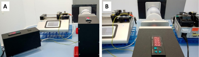
Electrospinning set up for (A) unimodal and (B) bimodal diameter distributions.
The meshes obtained from 8% PCL (random and aligned meshes), 15% PCL (aligned mesh), and co-electrospun 8% and 15% (aligned mesh) solutions were coated with a 10 nm layer of gold using a sputter coater (Turbomolecular pumped coater Q150T, Quorum Technologies, UK) and imaged under SEM (JSM-IT200 JEOL, Tokyo). The micrographs were acquired at an accelerating voltage ranging from 5 to 20 kV, working distance of 4–11 mm, probe current around 30, and magnifications values of 300 – 20 K.
The average fiber diameter and diameter distribution of meshes were determined using over 200 fibers per image (n = 3 images/group). Measurements were performed using ‘ImageJ’ software (NIH, USA) as described elsewhere [18]. Briefly, each image was segmented with five vertical lines of equal spacing, and fibers intersecting with the verticals were marked. The diameter of these fibers was measured by ‘ImageJ’, and fiber diameter distribution was calculated and reported in the form histogram.
Statistical analysis
A comparison of groups of scaffolds with different fiber diameters was performed using the Mann-Whitney U test due to the small sample size, and significance was achieved at p < 0.05.
Results
Collagen fibril diameter distribution of bovine ACL tissue
The results of the measurement is given in Fig. 4B1 and B2. For convenience these were reported together with the fiber diameter distribution of the bimodal scaffold formed from PCL with two different concentrations. Collagen fibrils in native bovine ACL exhibited a bimodal distribution with two peaks at 80 nm and 170 nm. The range of fibrils was between 40 nm and 300 nm. These findings were consistent with our previous report on the collagen fibril diameter distribution of bovine ACL [18].
Fabrication of fibers with diameters below 100 nm
With the formulations described in the Materials and Methods section, the composition of the PCL solution was optimized based on fiber diameter, surface properties (bead formation), spinnability (spinning or spraying), and flow stability. Some formulations formed regular uniform fibers, whereas others exhibited unstable flow, and favored electrospraying or formation of beads. As a result of different solutions screening with different process parameters (Supplementary Table S2), an 8% PCL solution was chosen for further scaffold fabrication. A 0.4 v/v% of pyridine was added to a 1 ml solution of acetone and formic acid in the ratio 1:1. Then, 0.08 g of PCL pellets (MW = 80 000 g/mol) was added to the solution and mixed by a magnetic stirrer for 2 hours at 40 °C. A summary of the runs for material and process parameters is tabulated below (Table 2).
Material and process parameters for fibers below 100 nm
Material and process parameters for fibers below 100 nm
DC: Dielectric constant (calculated based on composition as described in Du et al. [19]). TCD: Tip to collector distance.
From all electrospun solutions, three solvent systems, AA: FA (1:9), CF: DMF (2:8), and DCM: DMF (3:7), resulted in the diameter of the fiber close to 100 nm. Although the diameters of these fibers ranged from 45 to 145 nm, they exhibited beads in the form of spheres (Fig. 2A) or elongated spindles (Fig. 2B). The only solution formed bead-free ultra-thin nanofibers with a diameter of less than 100 nm was the solution of 8% PCL in the mixture of Ace: FA (1: 1) with the addition of 0.4% pyridine (#4 in Table 2, Fig. 2C).
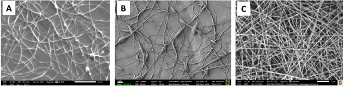
SEM images of (A) 12% PCL with DCM: DMF (3:7), (B) 13% PCL with CF: DMF (2:8), and (C) 8% PCL with Ace: FA (1:1). Scale bar 1 μm in A, B and C.
To produce the aligned meshes, an 8% solution of PCL was electrospun on a rotating drum. During initial trials, a flow instability leading to an accumulation of a tiny drop at the needle tip was noticed. Then the optimization of the process by the spinning of 8% PCL at different process parameters was attempted (Table 3), and the addition of 0.6% pyridine and spinning at 0.03 ml/h, 9 kV, 7 cm distance with 21 G needle formed fibers of sufficient quality(#10 in Table 3).
Fine-tuning of parameters for improved fiber quality (8% PCL, Ace: FA = 1:1)
TCD: Tip to collector distance.
Randomly oriented and aligned fibers from optimized 8% PCL solution were collected, imaged by SEM (Fig. 3A1, B1), and their diameter distributions were plotted (Fig. 3A2, B2).
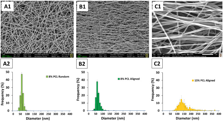
SEM images of meshes with 8% random PCL (A1), 8% aligned PCL (B1), and 15% aligned PCL (C1) together with their respective fiber diameter distributions (A2, B2, and C2). Error bars show SD with n = 3. Scale bar in SEM images = 1 μm.
Three images were used to calculate the average diameter and to determine the fiber diameter distribution. The plots for average diameter are shown in Fig. 3. Clearly, the random and aligned fibers produced with 8% PCL exhibited narrow distribution with a similar range between 30 nm and 100 nm (Fig. 3A2, B2). Also, their average values were found to be 55.99 ± 0.6 nm for the random and 60.60 ± 2.3 nm for the aligned fibers (p > 0.05).
The fibers with diameters greater than 100 nm were generated using higher PCL concentration. The runs performed to obtain quality fibers are given in Table 4.
Material and process parameters for fibers above 100 nm
Material and process parameters for fibers above 100 nm
As seen from the table, an increase in PCL concentration from 10% to 20% formed fibers with increasing diameters. Apparently, solutions greater than 15% PCL either resulted in fiber diameters beyond the diameter seen in native ACL tissue (with a peak of larger diameter of 170 nm) or were not of sufficient quality, and were not considered further. Run #4 was selected for further fabrication of the bimodal PCL scaffolds.
The 8 and 15% solutions were used for the fabrication of bimodal mesh by two-spinneret-electrospinning (Fig. 1B). The morphology of the co-electrospun scaffold is shown in the SEM micrograph (Fig. 4A1). As it is hypothesized, the two-spinneret electrospinning process generated fibers with a bimodal distribution. More specifically, as shown in the histogram (Fig. 4A2), there are two peaks in the chart, one at 80 nm and the other at 180 nm, with a range between 40 nm and 320 nm.
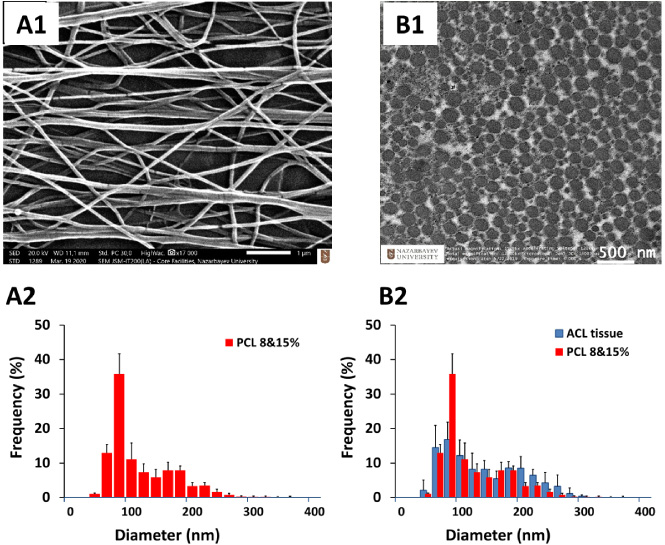
Comparison of bimodal aligned PCL mesh with bovine ACL tissue. (A1) SEM image of aligned PCL mesh formed by electrospinning 8&15% PCL solutions together and (A2) bimodal distribution of fiber diameter of this mesh. (B1) TEM image of the native bovine ACL and overlap of diameter distributions (B2). Bovine ACL tissue has two peaks at 80 nm and 170 nm, and bimodal PCL scaffold exhibits two peaks at 80 nm and 180 nm. Error bars in the charts show SD. Scale bars in A1 and B1 are 1 μm and 500 nm, respectively.
The fiber diameter distribution of the bimodal scaffold was compared with the distribution of collagen fibrils of bovine ACL. It is seen from Fig. 4B2 that the designed bimodal scaffold matches with the collagen fibril distribution of healthy ACL both qualitatively and quantitatively.
In this study, we hypothesized that the diameter distribution of native ACL tissue harvested from bovine can be replicated in a nanofiber structure. To test this hypothesis, we first determined the distribution of collagen fibrils in bovine ACL, and then investigated the conditions for the fabrication of fibers in the same diameter range. We, then, fabricated nanofiber mesh and compared the diameter distribution of electrospun meshes with that of the native tissue. Clearly, our data verified that our hypothesis was true. We were able to replicate the collagen fibril diameter distribution of bovine ACL tissue using nanofibers of PCL.
Amongst the parameters of materials, the solution concentration is the most influential one in controlling the diameter of fibers. The jets from the solutions of low polymer concentrations elongate and stretch at a higher extent to produce small diameter fibers. Also, the smaller quantity of polymer spun on the collector forms smaller diameter fiber as expected (conservation of mass). Production of nanofibers with a diameter of less than a hundred nanometer remains a challenge [20] because, at low concentrations, the quality of fibers is often compromised by the formation of beads in the form of droplets or spindles on fibers. Beads were formed on fibers from solutions with a concentration lower than 6% in our study, which was consistent with reported results for nylon [21] and PCL [22]. Uniform fibers were also produced at concentrations as low as 2% but for a different polymer, namely PAA [23]. At lower concentrations, bead formation can be avoided by adding process aids to reduce surface tension and improve stretching [24]. It is eventually the balance between the interaction of molecules in the solvent and air. For example, 5–10% PVP fiber morphology improved with the addition of salts, iron (III), and cobalt (III) nitrates, which decreased the beads content from 71% to 7% [25]. Also, many beads were observed in 50–103 nm PCL fibers from the solutions with less than 10% PCL concentration and few beads were found in 13% PCL solution [22]. Pyridine [21], silver nitrate [26], tetraethylammonium [27], hexadecyl trimethyl ammonium bromide (HTAB) and SDS [28] were also recommended as additives to improve fiber uniformity. When combined with the polymer and solutions, these additives enhance the free electrical charges, assist the ions to carry the current, increase solution conductivity, remove beads, and decrease fibers diameter.
In our study, we also observed beads at low concentrations and needed additives to eliminate the beads to improve the fiber uniformity. The 5% PCL generated fibers in Ace: FA (1:1) solution, but the fibers were beaded. The increase of the concentration up to 8% PCL decreased the number of beads, however, did not produce bead-free uniform fibers. Uniform fibers were generated only when pyridine was added to the solution. Pyridine is a weak base, which in reaction with weak formic acid generates an organic salt, thus enhancing the accumulation of the ions carrying the current and the electrical conductivity growth. The effect of pyridine was demonstrated when it was added to the solution of nylon-4.6 in formic acid [21]. An addition of 0.3% by weight pyridine increased the conductivity of an 8% solution by almost 26% and eliminated beads. When the concentration and, as a result, the viscosity are low, the fiber cannot keep its shape and results in electrospraying. On the other hand, when the polymer concentration increases, the surface tension of the solution increases too. This causes instability that forces a solution to take a smaller surface area per unit mass thus converting the jet into droplets [20].
The physical and chemical properties of the solvents are also critical for fiber quality. PCL is only partially soluble in both acetone and formic acid, hence our solution was heated up to 40 °C to increase solubility. The solvents that partially or fully dissolve PCL such as chloroform, DCM, and THF were found to produce either microfibers or beaded nanofibers [15]. It was previously shown that the relatively low values of the dielectric constant from 4.8 to 9.1 account for the large diameter [15]. The solutions with lower dielectric constants do not strongly uphold the electrostatic field and cause microfibers. The increase of the solution’s dielectric constant, by the addition of solvents with higher dielectric constants, contributes to the elevated electrical energy and the thinner fibers generation. The dielectric constants of certain solutions were calculated and it was observed that the solutions with the constants equal to 28 or above produced thinner fibers with fewer beads. The dielectric constant of Ace: FA (1:1) solution (∼39) is in the effective range, which is reported to be equal to 19 or higher [15]. Despite this claim in literature, uniform fibers were also formed in our study from solutions with even lower dielectric constants.
The 10% PCL solution in acetone alone demonstrated that low boiling point (b.p. ∼ 56 °C), that is to say high volatility of the solvent, hindered fiber formation. Acetone evaporated too fast, thus depriving the extruded jet an opportunity to stretch and reach the collector. This causes the jet to dry at the needle tip. In contrast, formic acid has a high boiling point (b.p. ∼ 100.8 °C) and low volatility, which is also not desirable as such solvents may not have time to evaporate, remain in the fibers and then produce thick and flat fibers. However mixing of acetone and formic acid resulted in an acceptable volatility.
Although the surface tension and viscosity of acetone and formic acid also vary significantly and were optimized by mixing, they are still governed by polymer concentration which is defined by molecular weight. According to Chaurey [27], a 15% PCL (MW = 80000 g/mol) solution in Ace: AA (1:1) formed non-uniform fibers with a mean diameter of about 388 nm and a wide diameter range from 200 to 1400 nm. Unlikely, electrospinning of the same solution of PCL but with MW = 45000 g/mol formed ∼ 88 nm fibers. For this reason, PCL with higher MW was recommended for the production of microfibers, whereas lower MW PCL is recommended for nanofibers. However, a current study showed that 80000 g/mol PCL is also suitable for the fabrication of ultra-thin nanofibers yet only when combined with an additive.
Considering the process parameters, flow rate seems to be the most important factor. Although the chosen rate of 0.03 ml/h is very low, it generated more uniform fibers without droplets which were present at 0.1 ml/h. As stated previously, a higher flow rate favors the generation of thicker fibers. Fiber diameter also increases due to increased tip-to-collector distance. However, a small increase in the tip-to-collector distance did not substantially affect the diameter of the fibers obtained from 8% PCL. These fibers had similar diameters when electrospun at 7 cm and 5 cm tip-to-collector distance.
To produce fibers with a diameter of nearly 200 nm, the concentration of PCL in Ace: FA (1: 1) was increased. As expected, more concentrated solutions formed larger fibers. For instance, the fibers with diameters around 125 nm were generated from 13% solution, around 150 nm from 15% solution, and around 1200 nm from 17% solution. Further increase in concentration led to the formation of thicker fibers, however, these were beyond the range of our interest. A similar effect was also reported by Bolgen [22] when an increase in PCL concentration from 13 to 15% resulted in a sharp increase of the diameter of the fibers from 100 nm to 250 nm, respectively.
In comparison with the separately electrospun 8% and 15% PCL fibers, the peak diameters of the bimodal distribution (8% and 15% PCL spun together) slightly increased from 60 nm to 80 nm and from 145 nm to 180 nm. However, this increase turned out to be beneficial when the diameter distribution of these fibers and the collagen fibrils from native ACL [18] were compared. Both qualitatively and quantitatively, the mesh with bimodal diameter distribution replicated the collagen distribution of the ACL tissue.
Overall, collagens of native ACL tissue exhibited a bimodal distribution with peaks at 80 nm and 170 nm, and we were able to replicate this distribution using nanofibers of PCL, which had two peaks at 80 nm and 180 nm. As a further study, it is recommended that grafts fabricated with bimodal distribution in the given range should be tested in vitro and in vivo.
Conclusion
The bimodal scaffold generated in this study is expected to serve as a graft for ligament regeneration because it closely mimics the structural properties of healthy ACL tissues. It was found in this study that i) the random and aligned fibers produced with 8% PCL exhibited narrow diameter distribution, all fibers possessing diameters less than 100 nm, with a range between 30 nm and 100 nm, and ii) the designed bimodal scaffold produced by simultaneous electrospinning of 8% and 15% PCL using AA:FA ratio of 1:1 matched with the collagen fibril distribution of healthy ACL both qualitatively and quantitatively. The findings of this study and forthcoming studies using these scaffolds will undoubtedly have a significant contribution to the efforts of the orthopedic research community to solve an important societal and economic healthcare problem related to ligament injuries. Future in vitro and in vivo studies using the graft proposed here should benefit from the biomimicry of these grafts.
Footnotes
Author contributions
Conflicts of interest
Funding
Data availability statement
Supplementary materials
 .
.