
Abstract
BACKGROUND:
The emergence of the global problem of multi-drug resistant bacteria (MDR) is closely related to the improper use of antibiotics, which gives birth to an urgent need for antimicrobial innovation in the medical and health field. Silver nanoparticles (AgNPs) show significant antibacterial potential because of their unique physical and chemical properties. By accurately regulating the morphology, size and surface properties of AgNPs, the antibacterial properties of AgNPs can be effectively enhanced and become a next generation antibacterial material with great development potential.
OBJECTIVE:
The detection of the inhibitory effect of AgNPs on MDR provides more possibilities for the research and development of new antimicrobial agents.
METHODS:
Promote the formation of AgNPs by redox reaction; determine the minimum inhibitory concentration (MIC) of AgNPs to bacteria by broth microdilution method; evaluate the killing efficacy of AgNPs against multi-drug-resistant bacteria by plate counting; evaluate the inhibitory effect of AgNPs on biofilm construction by crystal violet staining; study the drug resistance of bacteria by gradually increasing the concentration of AgNPs; and detect the toxicity of AgNPs to cells by CCK-8 method.
RESULTS:
AgNPs has a significant bactericidal effect on a variety of drug-resistant bacteria. After exposure to AgNPs solution for 12 hours, the number of E. coli decreased sharply, and S. aureus was basically eliminated after 16 hours. In particular, AgNPs showed stronger inhibition against Gram-negative bacteria. In addition, AgNPs can effectively hinder the formation of bacterial biofilm, and its inhibitory effect increases with the increase of AgNPs solution concentration. When AgNPs is used for a long time, the development of bacterial resistance to it is slow. From the point of view of safety, AgNPs has no harmful effects on organisms and has biosafety.
CONCLUSION:
AgNPs can inhibit MDR, and the bacteriostatic ability of Gram-negative bacteria is higher than that of Gram-positive bacteria. It can also inhibit the formation of bacterial biofilm, avoid drug resistance and reduce cytotoxicity.
Introduction
Multi-drug resistant bacteria (MDR) refer to bacteria that show resistance to multiple antibiotics [1,2]. These bacteria often possess multiple resistance genes or mechanisms, which weaken or even negate the efficacy of antibiotics (Fig. 1) [3]. Antibiotics are a type of drugs that can hinder or eliminate bacteria, treating infections by inhibiting the growth and replication of bacteria [4,5]. However, with the widespread use of antibiotics, the problem of MDR has become increasingly prominent [6]. Therefore, there is an urgent need to find new methods of bacteriostasis.
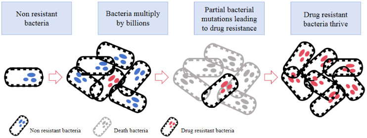
The process of bacterial resistance development.
Silver nanoparticles (AgNPs) have broad-spectrum antibacterial activity due to their unique physical and chemical properties, making them an effective strategy against MDR [7–9]. In recent years, a large amount of research has focused on the antibacterial mechanisms and performance optimization of AgNPs against MDR [10]. By adjusting the shape, size, and surface characteristics of AgNPs, their antibacterial efficacy can be further enhanced [11,12]. AgNPs enhance antibacterial effects due to their small size and larger surface area, which leads to a closer interaction with bacteria [13]. Additionally, AgNPs can effectively disrupt the biofilm formed by MDR, eliminating the protective function of bacterial biofilms and contributing to improved antibiotic treatment outcomes [14]. This characteristic is of great significance for the treatment of MDR infections.
The purpose of this study is to explore the effects of AgNPs on the bacteriostatic effect, drug resistance, biofilm formation and cytotoxicity of MDR, hoping to provide a basis for the study of MDR.
Materials
ATCC25922 E. coli, ATCC27853A P. aeruginosa, ATCC29213 S. aureus, ATCC10389 S. pyogenes were provided by the Biology Laboratory of Sichuan University. Multi-drug resistant E. coli (MDR. EC) and multi-drug resistant S. aureus (MDR.SA) were isolated from mice (Table 1 for details).
AgNO3, C76H52O46, NaCl, Na2CO3, HCl, C6H12O6, beef extract, peptone, and agar were all purchased from China National Pharmaceutical Group Chemical Reagent Co., Ltd.
The CCK-8 kit was purchased from Shanghai Yuchun Biotech Co., Ltd., and mouse umbilical vein endothelial cells (MUVEC) were purchased from Shanghai Jining Industrial Co., Ltd.
Susceptibility results of MDR
Susceptibility results of MDR
GH-5009C Constant temperature incubator (Beijing Yongguangming Medical Equipment Co., Ltd., China); SY-601 Constant temperature water bath pot (Tianjin Ouno Instrumentation Co., Ltd., China); D-8041 Multipurpose mechanical agitator (Tianjin Huaxing Technology Instrument Co., Ltd., China); 7DR-Z200 Bacteria turbidity meter (Hunan Changsha Tian Di Ren Biological Co., Ltd., China); Cary500 UV-visible spectrophotometer (Varian Medical Systems, USA).
Silver nanoparticles synthesis
Prepare 0.1 mol/L AgNO3 solution, 0.01 mol/L C76H52O46 solution, and 0.1 mol/L Na2CO3 solution in advance. Transfer the C76H52O46 solution into a three-necked flask and heat it moderately while stirring continuously. Then, slowly add an appropriate amount of Na2CO3 solution to the C76H52O46 solution. After 10 min of reaction, start adding AgNO3 solution dropwise until the color no longer changes. Continue heating and stirring for 30 min to obtain the AgNPs solution. Collect colloids or further centrifuge and collect powders for measurement.
Determination of minimum inhibitory concentration
Add 100 μL of LB liquid culture medium to each well of a 96-well plate. Following a serial dilution method, drop 100 μL of AgNPs solution into the first well, mix thoroughly, and transfer 100 μL to the second well. Repeat this procedure until the ninth well. Proceed to add 10 μL of diluted normal bacteria (E. coli, P. aeruginosa, S. aureus, S. pyogenes) and MDR into each well. Place the plate in a constant temperature incubator at 37 °C for 24 h, and observe and analyze the results.
Determination of sterilization efficiency
The appropriate concentration of AgNPs solution was selected, and three MDR.EC and three MDR.SA strains were measured separately. Meanwhile, a blank control group was set up to observe the number of bacteria on the plates at different time points (0, 2, 4, 6, 8, 12, 16 h).
Determination of inhibitory effect on biofilm formation
P. aeruginosa and S. aureus were pre-cultured and diluted. 100 μL of bacterial solution was taken into 96-well plate, and silver nano-solutions of different concentrations (0, 16, 32, 128 μg/mL) were added. The plate was incubated at
Silver nanoparticles induced bacterial resistance experiment
This experiment examined the resistance of E. coli to ampicillin and AgNPs, as well as the resistance of S. aureus to oxacillin and AgNPs. Firstly, the MIC of ampicillin, oxacillin, and AgNPs were determined. Then, the concentration was doubled, and both types of bacteria were cultured for 24 h before measuring the MIC again. This process was repeated for 30 d.
CCK-8 cytotoxicity experiment
MUVEC was used as the cellular model for the study, and WST-8 was utilized to measure cell proliferation and toxicity. In the mitochondria of cells, WST-8 is reduced by electron carriers to form highly water-soluble yellow formazan products. Cell quantity can be determined by measuring absorbance at 450 nm. The cell viability under different concentrations (0 μg/mL, 2 μg/mL, 4 μg/mL, 8 μg/mL, 16 μg/mL, 32 μg/mL, 64 μg/mL, 128 μg/mL) of AgNPs was measured, with a blank control set up. The experiment was repeated 3 times.
Cell survival rate = [(experimental - blank) / (control - blank)] × 100%
Experimental (contains cell culture medium, CCK-8, test substance)
Control (contains cell culture medium, CCK-8)
Blank (CCK-8)
Statistical analysis
Using SPSS 25.0 statistical software for data processing, the inhibitory rates of different strains were described using mean ± standard deviation (x ± s). An independent samples t-test was used to compare the direct inhibitory rates among different strains. All experiments were repeated three times.

TEM image of silver nanoparticles [34].
Characterization of AgNPs
After synthesizing the AgNPs solution (Fig. 2), a portion was taken to measure the absorption spectrum. The absorption spectrum of silver nanoparticles solution is measured as shown in Fig. 3A, the absorption peak is 400 nm, which accords with the absorption range of AgNPs 390–420 nm [15]. In addition, Fig. 3B shows the particle size range of 200 AgNPs, and the average particle size is 16.70 ± 3.8 nm. Therefore, it can be determined that the reaction produces AgNPs.

Analysis of AgNPs. (A) AgNPs UV vis spectrogram; (B) AgNPs diameter distribution.
First of all, four common strains were detected, among which E. coli and P. aeruginosa were Gram-negative bacteria, S. aureus and S. pyogenes were Gram-positive bacteria. According to the bacteriostatic results in Table 1, the MIC of AgNPs solution to E. coli and P. aeruginosa was 2 μg/ml and 4 μg/ml respectively. The MIC of S. aureus and S. pyogenes was 16 μg/ml (Table 2). Overall, AgNPs has a certain inhibitory effect on four kinds of bacteria, but the inhibitory effect on Gram-negative bacteria is better than that on Gram-positive bacteria.
Determination of MIC of AgNPs against on common bacteria
Determination of MIC of AgNPs against on common bacteria
Next, MIC detection was carried out for MDR E. coli and MDR S. aureus. As shown in Table 3. AgNPs also had inhibitory effect on these six MDR strains, and the MIC value of E. coli with multiple drug resistance was 4 μg/mL. The MIC of multidrug resistant S. aureus was 16 μg/mL.
Determination of MIC of AgNPs against on MDR
The number of colonies on the petri dish was observed at 0 h, 2 h, 4 h, 6 h, 8 h, 12 h and 16 h. As can be seen from the results of Fig. 4, when the AgNPs solution is not added, the number of both MDR increases with time, reaching the highest number on the petri dish at 6 h. After adding AgNPs solution, the amount of MDR decreased gradually. The bactericidal effect of AgNPs solution on E. coli was more significant, which was completely killed at 12 h (Fig. 4A), while S. aureus took 16 h to be completely eliminated (Fig. 4B). Therefore, with the passage of time, the antibacterial effect of AgNPs on MDR is getting better and better.
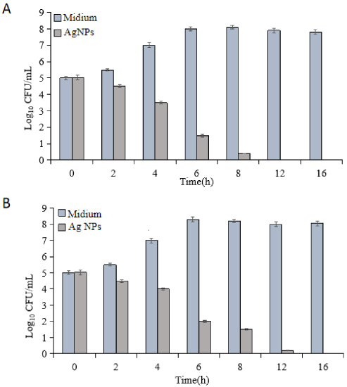
Bactericidal effects of AgNPs at different time points. (A) Bactericidal effects on MDR.EC; (B) Bactericidal effects on MDR.SA.
By using the crystal violet staining method, the effects of AgNPs at different concentrations (0, 16, 32, 64, 128 μg/mL) on the formation of biofilms by two types of MDR were observed. The absorbance at 595 nm was measured in the experiment, and the absorbance result at a concentration of 0 was set as 1. As shown in Fig. 5, the biofilm of E. coli decreased by 49% at 16 μg/mL, 64% at 32 μg/mL, 68% at 64 μg/mL and 70% at 128 μg/mL. S. aureus decreased by 24% at 16 μg/mL, 35% at 32 μg/mL, 34% at 64 μg/mL, and 39% at 128 μg/mL. Generally speaking, the higher the concentration of AgNPs solution is, the stronger the inhibitory effect is. Therefore, the inhibitory effect of AgNPs on the biofilm formation of E. coli is stronger than that of S. aureus.
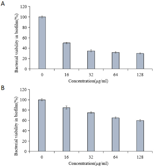
The impact of AgNPs on the formation of biofilms by MDR. (A) The impact on the formation of biofilms by E. coli; (B) The impact on the formation of biofilms by S. aureus.
Based on Table 2, it can be observed that E. coli has developed resistance to ampicillin, while S. aureus has become resistant to oxacillin. Therefore, ampicillin and oxacillin were chosen as controls to test the resistance of E. coli and S. aureus to AgNPs. The results are shown in Fig. 6.
The MIC of ampicillin against E. coli was doubled on day 30; however, the MIC of AgNPs solution increased 2-fold on day 11 and 4-fold on day 29 (Fig. 6A). The MIC of oxacillin against S. aureus increased 32-fold on the 30th day; however, the MIC of the AgNPs solution only tripled on the 15th day and remained stable until the 30th day (Fig. 6B). This shows that the application of AgNPs solution can prevent bacteria from developing drug resistance.
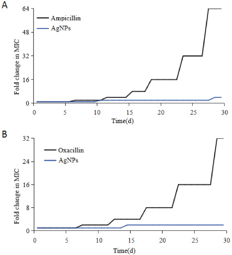
Folding changes of AgNPs and MIC of antibiotics. (A) Changes in resistance of E. coli (B) Changes in resistance of S. aureus.
The production of formazan was positively correlated with the degree of cell activity, that is, the more the number of formazan, the more stable the cell activity [16]. In this experiment, we took the cells without any treatment as the blank control, and the measured data was defined as 1, based on which the effect of AgNPs on cell activity was calculated. As shown in Fig. 7, the concentration of AgNPs did not significantly affect cell survival rates. When the concentration of AgNPs was at 0 μg/mL, 2 μg/mL, 4 μg/mL, 8 μg/mL, 16 μg/mL, 32 μg/mL, 64 μg/mL, and 128 μg/mL, the survival rates of MUVEC cells were 100%, 94.8%, 96.3%, 97.8%, 98.1%, 102.1%, 96.7%, and 93.8%, respectively, all of which were higher than 90%. This indicates that AgNPs have good safety.
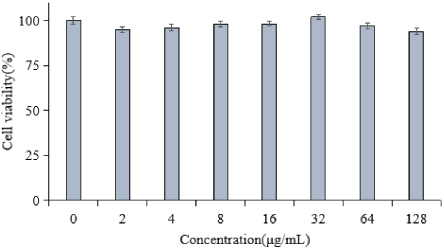
MUVEC cell survival rate.
With the emergence of MDR, the treatment of clinical bacterial infection becomes more and more difficult. Many patients with MDR are facing the situation that there is no drug available. Traditional antibiotics can no longer meet the needs of clinical treatment of drug-resistant bacterial infections. The exploration of new antibacterial mechanisms and the research and development of new antibacterial drugs and materials have become urgent [17–20]. AgNPs is a new type of antimicrobial agent, which has more powerful antibacterial activity and broad-spectrum antibacterial effect than traditional antimicrobial agents [21,22]. It is a new generation of antibacterial substance with great development prospect. AgNPs have very high bacteriostatic ability, and very low doses of AgNPs can produce strong bacteriostatic ability [23]. The strong bacteriostatic ability of AgNPs is closely related to its small particle size which leads to large surface area [24]. In this study, AgNPs were prepared by redox method. The absorption peak is at 400 nm, and the average particle size is 16.70 ± 3.8 nm, which is consistent with the general range of AgNPs. AgNPs destroy the cell membrane by binding to the negatively charged phosphate groups in the microbial cell membrane, and bind to the negatively charged sulfhydryl and carboxyl groups in the pathogen, resulting in the loss of the enzyme effect of some groups so as to achieve the effect of inhibiting bacteria [25,26]. The bacteriostatic effect of AgNPs is different in different bacteria. The results showed that AgNPs showed significant bacteriostatic effect on common strains and MDR. With the increase of the concentration of silver nano-solution, the bacteriostatic effect is also enhanced, especially on Gram-negative bacteria. The germicidal efficacy of AgNPs on MDR was also significant. E. coli MDR almost died in 12 hours, and S. aureus MDR died in 16 hours. According to research reports, AgNPs produce bacteriostatic effect by combining with peptidoglycan and plasma membrane components of bacterial structure [27]. The cell wall of Gram-positive bacteria has a thick (20∼80 nm) and dense peptidoglycan layer, up to 50 layers, accounting for 40%-95% of the cell wall components [28], while the cell wall of Gram-negative bacteria is thinner than that of Gram-positive bacteria (15∼20 nm) [29]. The results show that AgNPs can effectively inhibit the formation of bacterial biofilm, the higher the concentration of AgNPs, the stronger the inhibitory effect, and the inhibitory effect on E. coli is stronger than that on S. aureus. The results of this study are consistent with the current research. In addition, with the increasing resistance of MDR to antibiotics, there have been many cases in the market that are difficult to eradicate, and these bacteria have a very high resistance to traditional drugs [30]. In this experiment, E. coli and S. aureus were used continuously for one month to detect their drug resistance. It was found that the drug resistance of E. coli increased 4-fold after 29 days, and the drug resistance of S. aureus increased 2-fold after 15 days, and there was no further increase within 30 days. The mechanical damage of cell membrane caused by metal dissolved ions, surface protective agents or small size particles may lead to cytotoxicity of materials [31–33]. Through the CCK-8 cytotoxicity test, it is shown that no matter what the concentration of AgNPs is, the cell survival rate is more than 90%, which proves that AgNPs have good performance in biosafety.
Conclusion
This study confirmed that AgNPs have strong bacteriostatic activity, and the bacteriostatic ability of AgNPs against Gram-negative bacteria is higher than that of Gram-positive bacteria. AgNPs materials can be used to eliminate MDR, inhibit the formation of bacterial biofilm, avoid drug resistance and reduce cytotoxicity. It is helpful to reduce the dosage of antibiotics, reduce the occurrence of drug resistance of more bacteria, and improve the situation of a large number of pan-drug-resistant or even fully drug-resistant bacteria.
Footnotes
Conflict of interest
None to report.