
Abstract
BACKGROUND:
Polymeric electrospun mats have been used as scaffolds in tissue engineering for the development of novel materials due to its characteristics. The usage of synthetic materials has gone in decline due to environmental problems associated with their synthesis and waste disposal. Biomaterials such as biopolymers have been used recently due to good compatibility on biological applications and sustainability.
OBJECTIVE:
The purpose of this work is to obtain novel materials based on synthetic and natural polymers for applications on tissue engineering.
METHODS:
Aloe vera mucilage was obtained, chemically characterized, and used as an active compound contained in electrospun mats. Polymeric scaffolds were obtained in single, coaxial and tri-layer structures, characterized and evaluated in cell culture.
RESULTS:
Mucilage loaded electrospun fibers showed good compatibility due to formation of hydrogen bonds between polymers and biomolecules from its structure, evidenced by FTIR spectra and thermal properties. Cell viability test showed that most of the obtained mats result on viability higher than 75%, resulting in nontoxic materials, ready to be used on scaffolding applications.
CONCLUSION:
Mucilage containing fibers resulted on materials with potential use on scaffolding applications due to their mechanical performance and cell viability results.
Background
Biomaterials prepared by the electrospinning technique have shown great performance on biomedical applications because of their properties as large contact surface, high porosity, biocompatibility, and biodegradability [1–10]. Both synthetic and natural polymers have been successfully electrospun into non-woven mats with potential use in biomedical areas such as drug delivery, wound healing, and tissue engineering [11–20]. Several active compounds have been added to polymeric materials including drugs, natural extracts, and growth factors, to achieve a better performance as scaffold applications for cell growing, farming and differentiation. Nowadays, natural medicine is chosen above synthetic drugs because of their minimum side effects and the current trend of using ancient medicine as an alternative [21–23].
Synthetic polymers have been used on biomedical fields due to stability and low degradability when it is needed for long term applications. Polylactic acid (PLA) is a biocompatible polyester used in different biomedical applications, it has been approved by the Food and Drug Administration and is widely used in surgery and traumatology. PLA nanofibers have showed good properties as cell support for its biocompatibility and mechanical performance [24–29]. Polycaprolactone (PCL) is a biocompatible polyester usually used in medicine, especially on bone and soft tissue applications, and has been successfully electrospun into fibers for sustained release of active compounds [30–35]. Gelatin is a biopolymer from natural origin that presents high biodegradability, biocompatibility, and water absorption, and it is available commercially [36–38]. Gelatin has been widely used in various forms and has been successfully electrospun into membranes and tested on biomedical applications as drug delivery and wound dressing [39–43].
Aloe vera is a plant well known for its potential therapeutic action, numerous benefits have been attributed to it, including immunomodulation, healing of wounds and burns, hypoglycemic action, gastric protector, anticancer, antifungal, and anti-inflammatory action [44,45]. That is why it has been used in numerous commercial applications [46–52]. The part of the plant that presents these activities is the mucilage, which is a viscous substance like rubber that is found inside the leaves. In the case of Aloe, the mucilage is made up mostly of water and the rest by polysaccharides where glucomannans, xylose, ramose, galactose and arabinose are included in greater proportion, and to a lesser extent by minerals, lipids, and proteins. Plant-derived carbohydrates have exhibited diverse biological activities.
For this work, Aloe vera mucilage from Hermosillo, Sonora, Mexico was extracted and included into polymeric membranes based on gelatin electrospun fibers on different arrangements and characterized as biomaterials for scaffolding applications.
Materials and methods
Materials
PLA 4060D was supplied by Nature Works (Minnetonka, MN, USA). Poli(e-caprolactone) Mn = 80,000, Gelatin from porcine skin, gel strength 300 type A, Acetic Acid, glacial ACS reagent, and Epidermal growth factor from mouse were supplied by Sigma-Aldrich. Acetone and Chloroform were supplied by Fagalab (Sin, México). Aloe vera leaves (Aloe Barbadensis Milller) come from Hermosillo, Sonora, México (29°04 ′ 30 ′′ N 110°57 ′ 30 ′′ O).
Aloe vera Mucilage (Av)
Aloe vera (Aloe Barbadensis Milller) leaves with a healthy appearance were collected locally in Hermosillo, Sonora, México (29°04 ′ 30 ′′ N 110°57 ′ 30 ′′ O) from adult plants located in an urban area before spring season (January to March) to avoid floriation time. The obtention of Aloe vera mucilage powder was adapted from Medina-Torres et al. [53], as follows: The external part of the leaf was removed. Gel was semi-frozen and processed into a blender. The juice obtained from this process was filtered and centrifugated at 15000 rpm for 30 min to eliminate bigger solids. Supernatant was frozen and then freeze-dried for 4 days. The obtained material was minced resulting in aloe vera powder. Aloe vera mucilage powder was obtained as a yellowish material, and it was stored on a freezer (−4°C) prior to use.
Membranes preparation
PLA at 10%, PCL at 15% and Gelatin at 20% were dissolved with magnetic stirring for 24 h in acetone, chloroform, and acetic acid 90% (aqueous solution) respectively. Aloe vera mucilage (Av) was added into gelatin loaded membranes by adding the powder in the polymeric solution at 1% (w/v) and stirring until dissolution. Coaxial configuration fibers were prepared using PLA solution as the shell and Gelatin as the core.
Tri-layer membranes were obtained using PCL fibers as support. On a first step, PCL fibers were electrospun onto an aluminum collector; after evaporation of residual solvent, a second layer formed by gelatin-based fibers was placed above the first material, afterwards, a third layer was formed by fibers of a biocompatible polymer obtained from a PLA solution. For this material, the second layer was supplemented by bioactive compounds aloe vera mucilage, and epidermal growth factor (EGF) by dissolving them into the gelatin solution at 1% (w/v) for aloe vera powder and 0.2% (w/v) for EGF.
The electrospinning parameters for the different polymeric systems are shown in Table 1. Systems of Polylactic acid (PLA), Poli-(e-caprolactone) (PCL), Gelatin (Gel), Gelatin-Aloe Vera (Gel-AV) Coaxial (C), Coaxial-Aloe Vera (C-AV), tri-layer membranes (T), tri-layer aloe vera (T-AV), tri-layer Epidermal Growth factor (T-EGF), and Tri-layer Aloe vera and Epidermal growth factor (T-AV-EGF) are listed in Table 2.
Electrospinning parameters used in the preparation of polymeric fibers
Electrospinning parameters used in the preparation of polymeric fibers
Prepared polymeric systems
All the membranes were obtained by electrospinning technique, polymeric solutions were set into 6 mL syringes and placed in a KDS Scientific peristaltic pump (Holliston, MA, USA), high voltage was supplied using a Spellman CZE 1000R power supply (Hauppauge, NY, USA), flow rate and distance between the needle varied in each arrangement. An aluminum plate (10 × 10 cm) was used as collector. All membranes were stored at room temperature prior to use.
Membranes obtained from each system were morphology characterized by scanning electronic microscopy, using a JEOL 5410LV microscope (Tokyo, Japan). An electron beam of 15 kV intensity was used at high vacuum, the samples were coated in a gold film. Diameter distribution of fibers was measured using ImageJ software, at least 50 measures of each system were performed.
ATR-FTIR spectroscopy was executed to confirm the presence and interaction of the components in each system, a Frontier Perkin Elmer infrared spectrophotometer (Shellton, CT, USA) was used in a range from 4000 to 500 cm−1, 16 scans and a resolution of 1 cm−1 were set. Mats were measured as obtained with no additional treatment.
Thermogravimetric Analysis (TGA) was obtained using a Perkin Elmer Pyris 1 TGA apparatus (Shellton, CT, USA). Samples (2-4 mg) were weighed and heated from 25 to 800 °C in
Tensile mechanical properties were measured using a mechanical testing machine Electroforce 5110, TA Instruments (New Castle, DE, USA) with a 200N load cell and a cross-head rate of 1 mm s−1. Samples were cut in rectangles of 10 mm length, 5 mm width and approximately 0.2 mm thick. Young’s modulus, tensile strength and fracture strain were reported. At least three independent measurements were performed for each sample. Statistical analysis was performed by JMP program using ANOVA one-way to determine statistically significances by Tukey test.
In vitro cytotoxicity tests
Cell line L929 fibroblasts were grown in tissue culture treated dishes (D × H 60 × 15 mm, Sigma Aldrich) at 37 °C and 5% CO2, in Dulbecco’s Modified Eagle’s Medium (DMEM) with high glucose, sodium pyruvate and L-glutamine. Culture medium was supplemented with 10% of fetal bovine serum and 1% of penicillin-streptomycin solution. When a confluent growth was reached, cells were detached with 0.25% trypsin-EDTA solution for 3 min. Cell suspension was centrifuged (5 min, 1000 rpm), supernatant was separated, and cells were resuspended in fresh culture medium.
Cytotoxicity assay was performed using extracts from the materials: membranes were cut into 7 mm diameter discs that were sterilized using UV radiation (4 cycles, 15 min per side), and then placed in a 96 well plate, seed a total of 5 × 10³ cells per well and incubated. Viability test was carried out by triplicate at 24, 48 and 72 h of incubation using the resazurin method.
Measurements of cell viability was performed as follows: after 24, 48 and 72 h of incubation, the culture medium was removed and replaced by 100 μL of resazurin-medium solution and incubated at 37 °C for 6 h. Then, the absorbance was measured at 570 and 600 nm in a Synergy HTX multi-mode microplate reader (Biotek Instruments, San Diego, CA, USA).
Cell viability was obtained from the following equation:
Physicochemical characterization
Scanning electron microscope (SEM)
SEM images from electrospun fibers are shown in Fig. 1. PLA fibers present a smooth surface with ribbon morphology and a diameter of 424 nm, and PCL fibers display a round morphology with a diameter around 3.07 μm. Gelatin containing fibers show a round morphology, smooth surface, and no beads. Pure gelatin fibers exhibit a diameter of 492 nm, meanwhile, aloe vera loaded fibers show a diameter of 939 nm. In those fibers, the changes on the charge of the polymeric solution led to an increment on fibers diameter [54]. A prevalence of rounded fibers with slightly ribbon morphology appears in coaxial fibers. Load-free mats display diameters around 1072 nm and loaded fibers a diameter of 703 nm. Addition of Av decreases the diameter due to changes in the charge of the gelatin solution involved in the electrospinning process [46]. Tri-layer membranes were obtained as fiber mats as shown in Fig. 1, ribbon and round fibers were obtained, similarly to pure gelatin, PCL, or PLA materials. The mean diameter of unloaded tri-layer mats is 674 nm. Mean diameter values of 580 and 900 nm were observed for EGF and loaded Av-EGF, respectively. Addition of both compounds increase the diameter due to charges distribution in the gelatin solution. Tri-layer membranes transversal SEM images are shown in Fig. 2, it illustrates the nature of the mats composed of three kinds of fibers in different layers with no beads.

SEM images of prepared polymeric systems having smooth surfaces with a ribbon or rounded morphology 5000×.

SEM micrographs of Tri-layer membranes (T-Av-EGF). (a) SEM image of tri layer membranes 50×; (b) three kinds of fibers in different layers with gelatin (arrow) 200×.
Figure 3 shows the FTIR spectra for electrospun mats, pure gelatin fibers present peaks at 3287 and 2935 cm−1 for N–H and C–H stretching, respectively. Peaks at 1639, 1411 and 1085 cm−1 are attributed to amide I, II, and III coupling bonds [54]. PLA fibers show peaks at 1754 and 1452 cm−1 corresponding to C=O and C–H stretching, respectively. PCL fibers present peaks at 1721 cm−1 for C=O, 2940 cm−1 for C–H and 1161 cm−1 for C–O–C bond [55]. AV powder presents peaks at 3232 cm−1 due to O–H bonds, and 1578 and 1026 cm−1 for C-O bonds in the polysaccharide structure, a peak at 1715 cm−1 is also observed, related to C=O stretching of structures such as galactomannan [56,57].
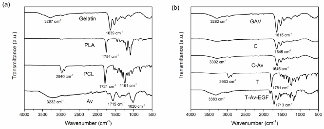
FTIR spectra of (a) Gelatin, PLA, PCL fibers and Aloe vera (Av) mucilage powder, and (b) Polymeric composite mats of Av-loaded gelatin fibers (G-Av), Coaxial fibers (C), Av-loaded coaxial fibers (C-Av), Tri-layer membranes (T), and Av-EGF loaded tri-layer membranes (T-Av-EGF).
Gelatin loaded fibers spectra show similar peaks as gelatin free mats with slightly slips in particular wavenumbers due to interactions of carbonyl bonds [39], C=O peak has an increase in wavenumber for these membranes from 1639 to 1645 cm−1 in G-Av and C-Av materials, meanwhile, T-Av-EGF membrane varies from 1781 cm−1 to 1713 cm−1 when active compounds were added, this is attributed to the interaction due to hydrogen bonds between the carbonyl group and hydroxyl groups in the active compounds of the loaded mats [11].
The thermogravimetric assay shows the thermal degradation of the prepared materials. Thermograms of the different materials are shown in Fig. 4. PLA and PCL fibers display a one-step decomposition at 350 and 410 °C, respectively, gelatin containing fibers show a two-step degradation curve, the first stage is related to water evaporation, around 100 °C, and the second stage at 334 °C, corresponding to the aminoacidic structure thermal degradation. Av exhibits a multi-step thermal decomposition due to the variety of compounds in the mucilage, such as proteins, polysaccharides, galactomannans, and minerals [51,58], a

TGA thermograms of (a) Gelatin, PLA, PCL fibers and Aloe vera (Av) mucilage powder, and (b) Polymeric composite mats of, Coaxial fibers (C), Av-loaded gelatin fibers (G-Av), Av-loaded coaxial fibers (C-Av), Tri-layer membranes (T), and Av-EGF loaded tri-layer membranes (T-Av-EGF).

DSC thermogram of Av powder, PLA fibers, Gelatin fibers and Av-loaded gelatin fibers.
Composite and gelatin loaded membranes display similar thermograms as their components, loaded gelatin fibers and coaxial fibers (loaded and unloaded) show similar decomposition behaviors starting with a stage of dehydration around 100 °C, followed by thermal decomposition of the organic structure, presenting the highest degradation rate at 328 °C for G-Av, 320 °C for C, and 327 °C for C-Av. On single gelatin fibers, the addition of aloe vera mucilage leads to a decrease on the temperature of degradation and
Different mechanical properties data of the polymeric mats are condensed in Table 3. Elastic modulus, tensile strength, and strain at break were calculated from stress-elongation curve for each test. Gelatin fibers display low mechanical properties as is commonly reported for biomaterials. On the other hand, PLA and PCL mats show enhanced performance due to their synthetic nature [59]. Single, coaxial, and tri-layer materials have similar mechanical performance but different mechanic properties. For Elastic modulus, unloaded mats exhibit greater values. Addition of Aloe vera mucilage decreases Young’s modulus and tensile strength as shown for single and tri-layer membranes, due to Av mucilage powder distributed around gelatin fibers, causing break points around the fibers inducing the break of the mats [60].
Mechanical properties of electrospun membranes. Means ± standard deviation
Mechanical properties of electrospun membranes. Means ± standard deviation
Values are shown as mean ± standard deviation of triplicate determinations. Means with different letters in the same column are significantly different (p < 0.05).
A good material performance and adequate values of strain at break are important on polymeric scaffolds when they are designed for topical applications [61]. Polymeric membranes are applied in tissue engineering for soft tissue and topical applications. Fragile materials lead to rupture during performance on topical uses. Natural polymers (G, G-Av,C and C-Av mats), present elongation at break around 50% for single systems and 33% for coaxial membranes. Meanwhile, synthetic based mats (PLA, PCL, T and T-Av-EGF) present values greater than 130%, even when natural polymers are present in the material.
Single and coaxial systems show no significant difference due to the similar performance of gelatin and PLA, but in the case of PCL and Tri-layer membranes, mechanical properties are enhanced due to the presence of polycaprolactone in the materials resulting in a resistant and rigid material that can perform in applications such as wound dressings.
Figure 6 shows the cell viability of L929 fibroblast in contact with polymeric extracts after 24, 48, and 72 h of incubation. Single PLA, Gel, Gel-Av, C-Av, T, and T-Av-EGF mats display cell viability greater than 75% (compared with positive control) and are considered nontoxic materials, the other materials present results below the accepted value [62–64]. Single and coaxial materials show an increment on cell proliferation when Aloe vera mucilage is added. Av mucilage is released from the gelatin matrix and mixed with culture media enhancing nutriments for the cultivated cells.

Cell viability of fibroblasts L929 at 24, 48, and 72 h of incubation in contact with polymeric extracts. PLA, Gelatin (Gel), Gel-Av (Gelatin-Aloe vera), PCL, C (Coaxial), C-Av (Coaxial-Aloe Vera), T (Tri-layer), T-Av (Tri-Layer- Aloe Vera), T-EGF (Tri-layer-Epidermal growth factor), T-Av-EGF(Tri-layer-Aloe Vera-Epidermal growth factor) and C+ (positive control).
Tri-layer membranes show good proliferation for 24 h and 48 h of cell culture and a decrease after 72 h. First stage of cell growth for these membranes is improved due to the dissolution of gelatin fibers into the culture media but it decreases along time. Tri-layer loaded mats (Av, and EGF) display lower cell viability than unloaded ones. Active compounds on polymeric systems show less efficacy due to its treatment, it is enhanced when both components are linked on the same system as shown for T-Av-EGF mat.
Polymeric membranes based on gelatin and Aloe vera (Aloe Barbadensis Milller) mucilage with potential use in tissue engineering were prepared by electrospinning technique. Fiber morphology and diameter were determined by SEM, and three different kinds of fibers were displayed on tri-layer membranes by transversal SEM images. The thermal characterization of aloe vera mucilage revealed that it contains phenolic compounds as galactomannan and polysaccharides, these molecules are responsible for biological performance of the materials. Chemical and thermal characterization revealed interaction among Aloe vera mucilage and gelatin by displacement of wavenumber on the signal of C=O group by FTIR, and modification of the temperature of thermal degradation in loaded fibers which lead us assume that hydrogen bonds occur, causing a good dispersion of the active compounds on the matrix.
Addition of active compounds on polymeric fibers results on decrease of elastic modulus and strain at break values, which is attributed to dispersion of particles among the matrix resulting on rupture points. Elongation at break was not affected by addition of mucilage on the membranes, and it results on acceptable values for tri-layer membranes with elongation values above 150%, this value is suitable for materials used in soft tissue engineering. Cell viability of fibroblast L929 is affected by the polymer in contact with the cells. Most of the membranes prepared in this work showed viability values higher than 75% after 72 h of cell culture. Addition of Aloe vera increased cell proliferation due to interaction of biomolecules from mucilage with cells.
The materials G, G-Av, C-Av, T and T-Av-EGF are suitable for applications on tissue engineering as dressings for wound healing, or scaffolds for cell growth and differentiation, due to the mechanical properties and biological performance. It is recommended that these materials should be tested as scaffolds on “in vivo” experiments for the use on tissue regeneration for diabetic foot treatment and burn dressing.
Footnotes
Acknowledgements
Damian Francisco Plascencia Martínez acknowledges CONAHCYT (Consejo Nacional de Humanidades, Ciencia y Tecnología, México) for the financial support provided for his graduate studies during this study. The authors thank Irela Santos Sauceda and Silvia Elena Burruel Ibarra for their contributions to the thermal and SEM characterizations.
Conflict of interest
The authors declare no competing financial interests.