
Abstract
Background
Polylactic acid (PLA) has been extensively used in tissue engineering. However, poor mechanical properties and low cell affinity have limited its pertinence in load bearing bone tissue regeneration (BTR) devices.
Objective
Augmenting PLA with β-Tricalcium Phosphate (β-TCP), a calcium phosphate-based ceramic, could potentially improve its mechanical properties and enhance its osteogenic potential.
Methods
Gels of PLA and β-TCP were prepared of different % w/w ratios through polymer dissolution in acetone, after which polymer-ceramic membranes were synthesized using the gel casting workflow and subjected to characterization.
Results
Gel-cast polymer-ceramic constructs were associated with significantly higher osteogenic capacity and calcium deposition in differentiated osteoblasts compared to pure polymer counterparts. Immunocytochemistry revealed cell spreading over the gel-cast membrane surfaces, characterized by trapezoidal morphology, distinct rounded nuclei, and well-aligned actin filaments. However, groups with higher ceramic loading expressed significantly higher levels of osteogenic markers relative to pure PLA membranes. Rule of mixtures and finite element models indicated an increase in theoretical mechanical strength with an increase in β-TCP concentration.
Conclusion
This study potentiates the use of PLA/β-TCP composites in load bearing BTR applications and the ability to be used as customized patient-specific shape memory membranes in guided bone regeneration.
Keywords
Introduction
Bone grafting is a widely utilized treatment modality, with over 4 million bone grafting procedures performed annually. 1 Bone grafts have the capacity to facilitate the regeneration of hard tissue and gradually restore both form and function. 2 Autografting is most commonly employed for bone tissue regeneration (BTR); however, it is accompanied by several disadvantages including restricted tissue accessibility, discomfort, and morbidity at the donor site.3–5 These limitations have driven the advancement of alternative options, such as synthetic grafts (alloplasts).
Research has been conducted on alloplasts to accurately reproduce the strength, bioactivity, shape, and load-bearing capacity of natural bone.6,7 Polymer-based grafts are often selected over many other types of alloplastic materials as they are readily accessible and inexpensive. 8 Crucially, they provide a biological setting that promotes the formation of new bone tissue as well as often being resorbable. Per a recent study, the Food and Drug Administration (FDA) has only authorized a small number of polymeric biomaterials for use in BTR applications. 1 Polylactic acid (PLA), an aliphatic polyester, is one such polymer notable for its biocompatibility and biodegradability. It undergoes hydrolytic degeneration by de-esterification, following the body’s natural processes eliminate the monomeric components.9,10 The production of lactic acid by hydrolytic breakdown occurs through a series of chain cleavage processes. The presence of catalytic carboxylic end groups facilitates the destruction of the molecular structure through bulk and surface erosion. 11 Consequently, soluble oligomers located near the surface are released, while those within the inner part of the structure stay trapped, the latter portion of which begins to break down into lactic acid.12,13 Nevertheless, the restricted applicability of load bearing BTR applications has been attributed to its weak mechanical qualities and low cell affinity.14,15
β-Tricalcium Phosphate (β-TCP), a calcium phosphate ceramic, has been widely used in BTR.16–20 It has been demonstrated to be effective in healing critically sized bony defects in preclinical models.16,21 Prior studies have indicated that the addition of osteoconductive bioceramics like β-TCP can increase the mechanical characteristics and osteogenic capabilities of pure PLA constructs such as membranes, and scaffolds.22–24 Considering these assertions, the incorporation of β-TCP into PLA has the potential to yield a polymer composite that exhibits enhanced mechanical robustness, bioactivity, and a higher capacity for use in BTR. Nevertheless, the processing conditions and the method of composite synthesis also have a significant impact in this regard. 25
Numerous synthesizing techniques have been used to fabricate PLA constructs including but not limited to injection molding, film extrusion, blow molding and thermoforming.25–27 Of note, solution processing or gel casting of PLA has found applications in the synthesis of resorbable membranes for biomedical use such as in Guided Bone Regeneration (GBR). 28 The gel casting approach serves to produce low-cost, high-quality, near-net-shape alternatives to other techniques in the synthesis of tissue engineering constructs. 29 Furthermore, it allows for the synthesis of high-ceramic particulate loading within the polymer phase 30 during the processing steps and has been shown to be capable of producing complex geometries with tortuous structures critical in determining the osteoconductive nature of the biomedical device. 31 The gel casting method involves several key steps including: (1) polymer dissolution, (2) polymer precipitation in a nonsolvent, (3) partial drying of the precipitate, (4) molding followed by heating/drying. Coombes et al. employed the gel casting approach to create mono-disperse and multi-polymer solutions using mild solvents that induced full dissolution and subsequent gelation of polymers. 32 This facilitated their relocation into a mold to produce dense, porous membranes. The presence of induced microporosities further enhanced the convoluted structure of membranes created using this approach. 32 As previously described by Kokabilta et al., the solution served as a fluid with low viscosity to carry the ceramic powder. 33 After pouring the polymer solution into a mold, the subsequent drying process, whether partial or complete, acted as means to permanently immobilize the ceramic particles in the appropriate gel-cast form. 33
Prior research has investigated gel-cast mixes like PCL/β-TCP and PLA/HA. Yet, there is limited information on the characterization of PLA/β-TCP generated using gel casting.34,35 There is hence a lack of a comprehensive comparison and study of the impact of ceramic loading on the mechanical and biological characteristics of PLA/β-TCP. The objective of this study was to analyze the physical and chemical characteristics, as well as the behavior of human osteo progenitor cells, in relation to the amount of ceramic contained in PLA/β-TCP composite membranes produced by gel casting. The postulated hypothesis was that the polymer-ceramic composites would result in increased biocompatibility and theoretical mechanical strength (computed through Rule of Mixtures models and Finite Element Analyses) when compared to their analogous, pure polymer counterparts.
Summary of the control and various experimental groups used in this study.

Materials and methods
Polymer gel synthesis and gel casting
A gel consisting of a single component was synthesized by dissolving Poly-D,L-Lactic Acid (Polysciences, Warrington, PA, USA − density = 1.25 g/cc, glass transition temperature (T g ) = 55 °C) in acetone at a concentration of 15% w/v. The solution was kept at a constant temperature of 46 °C while being continuously agitated by stirring at a speed of 60 rpm. As a result, full dissolution took place after approximately 60 min.
Furthermore, a gel consisting of two components was created by adding β-TCP (Sigma Aldrich, St. Louis, MO, USA) particles to the solution once the polymer had completely dissolved (summary of experimental groups provided in Table 1). However, prior to the addition, the β-TCP particles were calcined at 800 °C for 11 hours. Subsequently, the particles were milled in distilled water for 30 min using an attritor stirred ball mill (STD-01 Attritor, Union Process, Akron, OH, USA) in the presence of zirconia milling media (3 mm in diameter) at 120 rpm. After milling, the slurry was separated from the milling media by a sieve and dried in a low-temperature oven (∼37 °C) for 48 hours to yield a dry ceramic powder, as conducted in previous studies. 36 This process was necessitated owing to nano-porosities and non-equiaxial shapes of the commercially available particles, as described previously. 36 After the addition of β-TCP into the polymer solution, the stirring process was extended for an additional 15 min. The solutions were subsequently transferred to a borosilicate glass mold (as seen in Fig. 1a) and left undisturbed for about 24 hours to facilitate the evaporation of acetone. After the drying process, the membranes (Fig. 1b) had a thickness of approximately 1.5 mm at the bottom of the mold. The membranes were immersed in methanol for a duration of 30 min as a supplementary solvent treatment and dried at 25 °C for approximately one hour.

(a) Illustration of the gel-casting workflow (created with Biorender.com) and (b) pictograph of the gel cast membrane.
Material characterization
Gel permeation chromatography (GPC)
Recrystallization of PLA in acetone (C3H6O) may result in alterations to its molecular configuration. To examine these modifications, GPC was conducted using a liquid chromatograph that was equipped with an autosampler (Shimadzu LC-20AT/ SIL-20A HT Shimadzu Corporation, Kyoto, Japan). The molecular weight distributions of the polymer materials were analyzed using polystyrene standards in Tetrahydrofuran (THF). Prior to analysis, the concentrations of the solutions were adjusted to 3 mg/ml, and the flow rate was set at 1.0 ml/min.
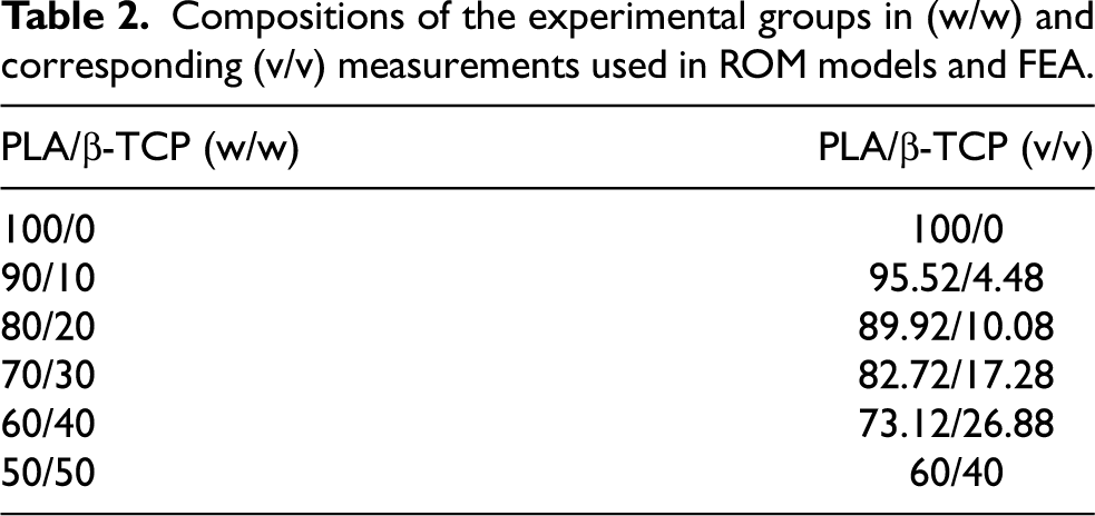
Compositions of the experimental groups in (w/w) and corresponding (v/v) measurements used in ROM models and FEA.
Scanning electron microscopy (SEM)
SEM images were obtained to gather topographical data of the gel-cast membranes. Images were obtained using a scanning electron microscope (Zeiss Gemini 300 FE-SEM, Oberkochen, Germany) at an acceleration voltage of 1 kV. SEM images of the β-TCP particles were obtained (Hitachi TM 4000 SEM, Tokyo, Japan), and subsequently, ImageJ (National Institutes of Health, Bethesda, MD, USA) was employed to carry out photoanalysis of β-TCP particles to generate the particle size distribution.
X-ray diffraction (XRD) and Fourier transform infrared spectroscopy (FTIR)
Gel-cast membranes were pulverized and analyzed using XRD (X’Pert Powder Panalytical device, Panalytical, Almelo, The Netherlands) to confirm the presence of β-TCP inside the PLA matrix. The spectra were obtained in the range of 20° to 80° with a step size of 0.013°. The voltage and current were adjusted to 45 kV and 40 mA, respectively. For FTIR, the pulverized samples were analyzed (Magna 550-IR Series, Waltham, MA, USA) within a wavelength range of 4000 to 400 cm−1. The absorbance peaks of the groups were measured using Potassium Bromide (KBr) as the reference background.
Differential scanning calorimetry (DSC)
In order to assess the alterations in the crystalline structure caused by the presence of β-TCP in PLA, DSC measurements were obtained (SDT Q600, TA instruments, New Castle, DE, USA). Three tests were conducted on each group, using 10 mg of sample per test. The temperature was increased at a constant rate of 10 °C/min, from 25 °C to 200 °C. The tests were conducted in an N2/O2 atmosphere. The acquired data was analyzed (TA Universal Analysis software program, TA instruments, New Castle, DE) to determine the melting point, T
m
; enthalpy of fusion at melting point, ΔH
f
(T
m
); and the enthalpy of fusion of the crystalline polymer, 
Rule of mixtures (ROM) model and finite element analysis (FEA)
The theoretical mechanical strength of the experimental groups was assessed using mathematical (ROM), models, namely, Voigt-Reuss, Hashin-Shtrikman, and Ravichandran. The methodology employed in this investigation adhered to the approach previously conducted by Hsieh et al. in assessing the elastic characteristics of ceramic-metal particle composites.
38
The utilization of ROM models was facilitated by converting the %w/w to %v/v measurements (ϕ) using the provided equation. The results of this conversion are presented in Table 2.
FEA was used to validate the results produced from the ROM models. FEA was conducted using a Unit Cell (UC), which was described in this work as a structure consisting of a regular arrangement of spherical β-TCP particles within a PLA cube (Fig. 2a). The particles were positioned so that their centers aligned with the vertices of the cube. The ceramic phase’s diameter was equivalent to the average particle size obtained from the particle size distribution (Fig. 2b). The unit cell size was adjusted based on the weight fraction of the β-TCP particles in each experimental group, using the equation provided:

(a) Illustration of the UC (red arrow is the polymer matrix, black arrow represents ceramic particle) produced for the FEA by applying a unit compressive load (represented by a solid blue arrow) and imposing a fixed boundary condition (shown by a translucent blue arrow), (b) Particle size distribution of β-TCP particles, where insert (b.1) shows a scanning electron micrograph of the particles.
The UC model was created using Onshape (PTC Inc., Boston, MA, USA). The model was then imported into Ansys Workbench 2021 FE package (Ansys Inc., Canonsburg, PA, USA). The static structural module was utilized, and the model was discretized utilizing higher order elements. Since compression data is free from many of the effects of debonding, a fixed boundary condition was applied on one of the faces of the system while the opposite face was subjected to a unit compressive load after which maximum principal stress theory was used to determine Young’s moduli (E).
In vitro experimentation
Cell culture
Bone fragments were acquired from a patient undergoing surgical intervention for the treatment of prognathism in accordance with the Institutional Review Board of New York University (#S18-01579). Briefly, bone samples were retrieved immediately after harvesting from the operating theatre in sterile 50 ml falcon tubes containing 15 ml of Liebovitz (L-15) medium. Specimens were then handled with sterile forceps and washed thoroughly in Phosphate Buffer Saline (PBS) and were then further cut into smaller pieces (roughly 1 × 1 mm in size), plated onto a 24 well plate, and completely immersed in expansion media. Expansion media comprised of Dulbecco’s Modified Eagle Media (DMEM) with Glutamax, along with 1% antibiotic/antimycotic and 10% ES-fetal bovine serum (FBS) (Thermo Fisher Scientific, Waltham, MA, USA). The cells (human osteoprogenitors (hOP)) were routinely examined, with the medium being refreshed every 48 hours. After reaching a confluence of ∼70%, the medium was removed, and the cells were extensively washed with Phosphate Buffer Saline (PBS). PBS was aspirated, and cells were separated from the bottom of the well plate using 12 ml of Trypsin (Thermo Fisher Scientific, Waltham, MA, USA). The plates were subjected to incubation at a temperature of 37 °C for a duration of 5 min. Subsequently, 12 ml of DMEM was introduced to counteract the effect of Trypsin. The cells were examined under an optical microscope to confirm their separation from the growth plate and transferred to a 15 ml conical centrifuge tube. The cells were centrifuged for 5 min at 1000 rpm and the medium discarded. The pellet at the bottom of the tube was resuspended in 1000 μl of new expansion medium. A volume of 20 μl of the cell suspension was combined with Trypan Blue (Sigma Aldrich, St. Louis, MO, USA) at a concentration of 1:10. The mixture was then placed onto a cell-counter chamber slide (Countess II FL Automated Cell Counter, Thermo Fisher Scientific, Waltham, MA, USA). All in vitro cell culture experiments were performed using the tissue culture plate as the negative control (Ctrl), and stock PLA pellets (cPLA) as the positive control group.
Presto Blue
Presto Blue (Thermo Fisher Scientific, Waltham, MA, USA) was used to assess the viability of cells seeded onto the different groups used in this study. 39 To conduct the experiments, ∼50,000 cells were seeded onto the samples (n = 3/group). The experiments were performed at time points of 24, 48, 96, 192, and 384 hours after cell seeding. A 10% solution of Presto Blue was synthesized using DMEM. During the experiment, the expansion medium in the well plate was substituted with the Presto Blue solution. The plate was incubated at a temperature of 37 °C for a duration of 15 min. The solution in each well was transferred to a new plate and fluorescence quantified (FilterMax F3/F5 Multi-mode Microplate Reader, Molecular Devices, San Jose, CA). In brief, the fluorescence experiment was performed following a 5-second orbital agitation. A 535 nm excitation wavelength and a 595 nm emission wavelength were employed. The control group consisted of cells that were directly seeded into well plates. The fluorescence values of all experimental groups at each time point were normalized by dividing them by the fluorescence value obtained for the negative control group at the 24-hour time point. This normalization produced a time-series effect, represented by the relative fluorescence values.
Alizarin Red
Alizarin Red assay (Thermo Fisher Scientific, Waltham, MA, USA), was performed to evaluate the differentiation of hOP into fully developed osteoblasts and the subsequent process of calcification on the surface of the gel-cast membranes. Three samples per group were first seeded with ∼50,000 cells each and cultured for 168 hours in DMEM. A 4% solution of paraformaldehyde (PFA) was prepared by adding phosphate buffered saline (PBS) that made up 80% of the required volume of PFA. The PBS was heated to a temperature of 60 °C, and the appropriate amount of PFA powder was added to the solution. Sodium hydroxide (NaOH) was gradually added until the solution became transparent. The solution was cooled to ambient temperature, and the total volume was adjusted by adding PBS. The solution’s pH was modified to 6.9 using hydrochloric acid (HCl). At 168 hours, the medium in the wells was aspirated and the cells were treated with a 4% PFA solution for 10 min. The cells were then washed twice with PBS. The PBS solution was disposed, and the samples were stained with a filtered solution of 1% Alizarin Red for a duration of 5 min. The wells were treated with a solution of 10% acetic acid and allowed to incubate at room temperature for 30 min. The contents of the well plates were collected and transferred to 1.5 ml centrifuge tubes. The tubes were subjected to thermal treatment at a temperature of 85 °C for a duration of 10 min, after which they centrifuged at 1000 rpm for 5 min. The contents of the tubes were transferred to a new tissue culture plate, and the intensity of light absorption (IU/ml) was determined at a wavelength of 405 nm (FilterMax F3/F5, Molecular Devices, San Jose, CA, USA).
Immunocytochemistry
The purpose of immunocytochemistry was to assess the morphology of hOP cells, to visualize cell spreading and cellular affinity on the various groups 24- and 48-hours post-seeding. Each sample per group was initially seeded with ∼50,000 cells each. Cells were allowed to proliferate in expansion medium. Before imaging, the medium was extracted from the wells and the cells were fixed in a 4% PFA solution for 10 min after which cells were rinsed two times with PBS. The blocking buffer was prepared by combining 3% Bovine Serum Albumin (BSA) and 10% Fetal Bovine Serum (FBS), followed by the addition of 0.2% Triton-X (Sigma Aldrich, St. Louis, MO, USA). The samples were immersed in a blocking buffer for a duration of 60 min and washed three times in PBS for a duration of 5 min each. The cells were stained with Alexa Fluor 488 Phalloidin (for staining actin filaments) (Thermo Fisher Scientific, Waltham, MA, USA) and Hoechst 33258 (for staining nuclei) (Thermo Fisher Scientific, Waltham, MA, USA) after which fluorescence images were obtained on an inverted microscope (Leica DM IL LED, Leica Microsystems Inc., Wetzlar, Germany) and merged using an image processing software (ImageJ, U.S. National Institutes of Health, Bethesda, Maryland, USA).
Real time-polymerase chain reaction
A real-time polymerase chain reaction (PCR) was conducted to examine gene expression 168 hours after cell seeding. A total of around ∼50,000 cells were seeded upon the samples, (n = 6/group). Before conducting the experiment, the expansion media was removed from the wells and 500 μl of TRIzol (Invitrogen, Thermo Fisher Scientific, Waltham, MA, USA) was introduced into each well. The contents of the wells were transferred to 2 ml microcentrifuge tubes. Next, 0.1 ml of chloroform was added to each tube, and the tubes were subjected to centrifugation for 15 min at a temperature of 4 °C with a force of 13,000g. The liquid phase was separated, and 1 ml of isopropanol was introduced. The combination was subjected to another round of centrifugation using the same parameters, following which the liquid portion above the sediment was discarded. The RNA pellet at the bottom of the tubes was rinsed three times with 70% ethanol diluted in ultrapure water and left to air dry for 10 min. The pellets were suspended again in 100 μl of diethylpryrocarbonate (DEPC) water. The RNA quality was measured using a spectrophotometer (NanoDrop One/One C , Thermo Fisher Scientific, Waltham, MA, USA). Subsequently, cDNA synthesis was performed using the QuantiTect Reverse Transcriptase kit (Qiagen, N.V., Hilden, Germany). The PCR experiment was conducted using a 10 μl solution containing 5 μl of SYBR Green Master Mix (Thermo Fisher Scientific, Waltham, MA, USA), 1 μl of specific forward and 1 μl of reverse primers, 1 μl of cDNA, and 2 μl of nuclease-free water. Experiments followed the protocol: denaturation (95 °C, 15 s), annealing (60 °C, 30 s), and extension (72 °C, 30 s)), and were conducted with a Real-Time PCR system (QuantStudio 3, Thermo Fisher Scientific, Waltham, MA, USA). Runx2 (Forward primer: CCGTCCATCCACTCTACCAC; Reverse primer: ATGAAATGCTTGGGAACTGC) and Col1α1 (Forward primer: GGCTGAGTAGGGTACACGCAGG; Reverse primer: AACCAAGGCTGCAACCTGGA) gene expressions were obtained using the ΔΔCT method, with GAPDH (Forward primer: GACTCATGACCACAGTCCATGC; Reverse primer: AGAGGCAGGGATGATGTTCTG) and 18S (Forward primer: TGCCAGAGTCTCGTTCGTTATCG; Reverse primer: CGGACAGGATTGACAGATTGATAGC) serving as the housekeeping genes.

Elution characteristics of the cPLA and ePLA groups using GPC.
Statistical analysis
Outcome variables from the experiments were assessed for normality using the Shapiro-Wilk test. Data was analyzed using one-way analyses of variance (ANOVA) tests on IBM SPSS (v29, Armonk, NY, USA). Data was presented as mean values with corresponding 95% confidence intervals (mean ± 95% CI). p ≤ 0.05 was considered statistically significant.
Results
Material characterization
Gel permeation chromatography (GPC)
The molecular weight of the PLA phase in the gel cast PLA/β-TCP composites was presumed to be comparable to that of ePLA, as no grafting operation was performed. Therefore, GPC was conducted on both ePLA and cPLA membranes to examine the alteration in molecular weight distribution resulting from the gel casting procedure. Figure 3 illustrates the molecular weight distribution of PLA prior to (cPLA) and after (ePLA) the gel casting procedure. The ePLA group exhibited greater number-average molecular weight (M n ), weight-average molecular weight (M w ), and Poly Dispersity Index (PDI) values (92920, 167691, and 1.80, respectively) compared to the stock cPLA group (91051, 162316, PDI 1.78, respectively).
Scanning electron microscopy (SEM)
The SEM micrographs (Fig. 4) revealed that all gel cast groups had a rougher surface structure, in contrast to the smooth and uniform surface of the stock PLA pellets (cPLA) (Fig. 4b). In addition, groups with greater proportions of ceramic particles (60/40 and 50/50 ePLATCP groups) (Fig. 4g and 4h) exhibited a distribution of ceramic particles and their clusters.

Scanning electron micrographs of (a) TCP (low magnification overview of Fig. 2b.1) (b) cPLA, (c) ePLA, (d) 90/10 ePLATCP, (e) 80/20 ePLATCP, (f) 70/30 ePLATCP, (g) 60/40 ePLATCP, and (h) 50/50 ePLATCP constructs.
Differential scanning calorimetry (DSC), X-Ray diffraction (XRD) and Fourier transform infrared (FT-IR) spectroscopy
Although a trend was observed, suggesting a rising mean crystallinity with an increase in ceramic loading, differences were not significant (p > 0.05) (Fig. 5a). XRD spectra (Fig. 5b) demonstrated the presence of distinct, intense diffraction peaks occurring at angles of 28°, 30°, and 35°, which corresponded to the (2 1 4), (0 2 10), and (2 2 0) miller indices of rhombohedral β-TCP. The obtained results were consistent with the data reported in another investigation.
40
The aforementioned peaks were not present in the cPLA and ePLA groups, as anticipated, since there were no ceramic particle reinforcements within these two groups. The phosphate (PO

(a) % Crystallinity of the various groups used in this study presented as means and corresponding 95% confidence interval values. p < 0.05 is considered statistically significant, (b) XRD and (c) FTIR spectra, showing some of the most prominent peaks.
In vitro experimentation
Presto Blue assays
Cellular viability exhibited a progressive increase over time in all groups and at all examined time periods (Fig. 6a). The incorporation of β-TCP into the polymer matrix significantly enhanced cellular survival compared to the plastic control (p < 0.05). Additionally, a notable rise in fluorescence at all time intervals was observed between groups with high ceramic concentrations (60/40 ePLATCP and 50/50 ePLATCP) compared to stock PLA pellets (cPLA) and gel-cast (ePLA) groups (p < 0.05).

(a) Fluorescence measurements obtained through Presto Blue cellular viability assays (raw data from authors' own work 44 ), p < 0.05 is statistically significant with same letters above error bars representing statistically homogenous groups and (b) Alizarin Red (absorbance (IU/ml)) quantification. Data presented as means and corresponding 95% confidence intervals.
Alizarin Red assay
The Alizarin Red staining technique was used to assess calcium deposition in developed osteoblasts and to detect calcium in osteocytes that emerged following differentiation. The quantification of Alizarin (Fig. 6b) revealed that the presence of β-TCP in the membranes enhanced matrix mineralization compared to the negative control (empty wells) (p ≤ 0.003). The absorbance values (IU/ml) in the composite membranes showed a considerable rise 168 hours after cell seeding. The control group (empty wells) depicted low levels of calcification and was statistically similar to the pure polymer groups cPLA and ePLA (p = 0.150, p = 0.531, respectively).
Immunohistochemistry
Figure 7 displays the fluorescence micrographs of osteoprogenitor cells seeded onto the gel-cast membranes after 24- and 48-hours. The presence of selective green labeling of F-actin (Phalloidin) and blue nuclear counter (Hoechst) in all groups indicates that the materials utilized in this investigation effectively facilitated cell attachment. Cell spreading was found on all surfaces, exhibiting their characteristic trapezoidal shape and distinct rounded nuclei, indicating normal cellular development. The actin filaments were aligned over all surfaces (including the empty wells of the control group), indicating excellent biocompatibility during the initial phases following cell seeding. Of note, a greater presence of cells was observed upon the composite scaffolds relative to the empty wells and positive control group (cPLA), characterized by a greater cell (nuclei) density, qualitatively.

Fluorescence micrographs of the samples taken after 24- and 48-hours following cell seeding, with green and blue image channels indicating Phalloidin (actin filaments) and Hoechst (nuclei) staining respectively. White scale bars (bottom right of the pictographs) are 1000 μm in length.
Real time polymerase chain reaction
Effect of different amounts of β-TCP on runt-related transcription factor 2 (Runx2) gene expression in osteoprogenitor cells was evaluated after 168 hours (Fig. 8a). No significant difference in Runx2 gene expression was observed between the experimental composite groups and the control groups (pure polymer groups and empty wells) (p > 0.05). Col1α1 gene expression was also evaluated at 168 hours following cell seeding (Fig. 8b). No significant difference was detected between pure polymer groups (cPLA and ePLA) and negative control (empty wells) (p > 0.05). Experimental composite groups with low ceramic loading (90/10ePLATCP and 80/20ePLATCP) were statistically similar to control groups and the 50/50 ePLATCP group (highest ceramic loading) (p > 0.05). The 60/40 ePLATCP group resulted in the highest mean Col1α1 gene expression, statistically greater than cPLA and ePLA groups (p = 0.002 and p = 0.006, respectively).

Expression of (a) Runx2 and (b) Col1α1 (COL). Values are presented as means and corresponding 95% confidence intervals. p < 0.05 was considered statistically significant, with same letters atop the error bars indicating statistically homogenous groups.
Rule of mixtures (ROM) models and finite element analyses (FEA)
The results acquired by ROM are summarized in Table 3. Figure 9a demonstrates that the strength of the composite material increased when the concentration of β-TCP particles in the PLA matrix was raised. Lower concentrations resulted in narrower boundaries irrespective of the model utilized. In contrast, when there were larger concentrations of ceramic particle reinforcements, much broader limits were observed. Therefore, theoretical models often require verification through numerical analyses, such as FEA. Finite element simulations were in good agreement with the ROM model (Fig. 9a). The maximum principal stress theory was used to estimate the strength of the composite systems. Young’s moduli predicted by FEA were in agreement with the bounds previously established by the ROM models. Ceramic particle concentration was proportional to the predicted mechanical strength. Contrary to the ROM model, stress distribution was observed within the individual phases (Fig. 9b). To further elaborate, under the application of load, there was a formation of stress concentration regions within the ceramic particles (Fig. 9c). This phenomenon occurred in proximity to the interface between the 2 phases and could be attributed to the low ductility and low Poisson’s ratio of the reinforcing phase (ν = 0.22).

(a) Young’s moduli of the different groups in relation to the ceramic content. The Voight-Reuss, Hashin-Shtrikman, and Ravichandran bounds are displayed for the purpose of comparing them with the data acquired using FEA, (b) isometric representations of FEA simulations illustrating the compressive force on the unit cell models, with respect to the concentration of ceramic particles, (c) finite element depiction of the stress distribution within the polymer and ceramic phases in a cross-sectional view. Note the red probe depicting the highest level of primary stress near the interface between the ceramic and polymer phases.
A summary of the results obtained from comparing the results of FEA and ROM analysis as a function of the percentage of ceramic loading. Eβ-TCP = 162 GPa, EPLA = 3.986 GPa, νβ-TCP = 0.22, νPLA = 0.3.

Discussion
Previous studies have examined gel-cast mixtures such as PCL/β-TCP and PLA/HA. However, the available data about the characteristics of PLA/β-TCP produced using gel casting is currently limited. Consequently, there exists a dearth of a thorough examination and analysis of the influence of ceramic loading on the mechanical and biological properties of PLA/β-TCP. The primary aim of this research was to examine the physical and chemical properties, as well as the characteristics of hOP cells, in correlation with the quantity of ceramic present in PLA/β-TCP composite membranes manufactured using gel casting.
In this study, alterations in the distribution of molecular weights may have occurred throughout the process of polymer chain development. Coombes et al. previously elucidated the gel casting process as including the creation of lamellae from multilayer crystals. 32 The utilization of gel casting may have resulted in the creation of intersecting lamellae and microporosities. The alterations in microgeometry of gel cast samples may also be a key factor contributing to the variations in molecular weight distributions of ePLA membranes compared to cPLA pellets – which present a smoother surface texture. Specifically, within the ePLA group, the development of crystalline outer layers that appear as wafer-like structures may have caused an elevation in molecular weight, thus leading to an overall rise in PDI. Moreover, the elongated polymer chains that are created during gel casting as a result of flow effects during the composite membrane synthesis might potentially account for the rise in both M w and M n .
SEM revealed a varied surface morphology as a result of β-TCP inclusion within the polymer matrix, relative to pure PLA. Notably, agglomeration was observed in groups with a higher particle concentration (60/40 and 50/50 ePLATCP groups). Ko et al. previously described this phenomenon in a PLA/HA system employing chloroform as a solvent for dissolution. Chloroform, being a solvent with relatively low polarity for polymer dissolution, leads to considerable agglomeration even at reduced ceramic concentrations (less than 30% w/w). 45 In contrast, the presence of particle agglomeration that was noted in our investigation, predominantly in groups with high ceramic concentrations, maybe attributable to the increased particle packing fraction. Pertaining to the crystallinity measurements obtained through DSC, increasing the β-TCP particle concentration, the concentration and crystallinity of PLA in the acetone solvent could have decreased. As the total weight of solids dissolved in the solvent remained consistent across all samples, augmenting the concentration of β-TCP may have resulted in an overall reduction in the crystallinity/concentration of the PLA matrix. As a result, the crystallinity of the polymer-ceramic system was statistically similar. ShiraliPour et al. made a similar observation when analyzing PCL/β-TCP structures, where an increase in ceramic loading reduced the PCL concentration within the composite causing an analogous effect. 46
In this study, the pure polymer groups (ePLA and cPLA) exhibited slight variations in their spectra, but nonetheless were characteristic of crystalline PLA - a pseudo orthorhombic unit cell. 47 The spectrum of cPLA exhibited a solitary, broad, and narrow peak, whereas ePLA had two separate and well-defined peaks at 17° and 20° degrees. All other composite groups produced by the gel-casting process exhibited these two distinct peaks, potentially because of recrystallization occurring during gel casting and solvent evaporation. The spectra of the composite groups exhibited a combination of the polymer and ceramic phases. The slight deviations in the 2θ values could be ascribed to the zero error of the XRD instrument. 42 On the other hand, similar infrared (IR) bands were detected for all composite groups. The resemblance can be elucidated by the polymerization mechanism that takes place in aliphatic polyesters. During the progression of polymerization, the average molecular weight steadily rises. Over time, this might have completely used all the polymer’s functional groups. In addition, this could have led to the near-complete elimination of monomers and other side chains from the system, resulting in the production of identical IR spectra.
Among the various physicochemical properties, solubility and ion release can potentially influence cellular behavior. In particular, dissolution of β-TCP can change the extracellular ion concentration, thus impacting cell viability and differentiation. The cell viability studies demonstrated that all groups exhibited biocompatibility, with no detrimental impact of the materials on cellular growth. However, the quantitative differences in cellular proliferation could be attributed to variation in phosphate and calcium ions between groups.48,49
Calcium ions released from the composite material surfaces as a result of dissolution, has been shown to modulate osteoblastic differentiation through the cyclic adenosine monophosphate-response element binding protein (CREB) or the extracellular signal-regulated kinase (ERK) pathway.50,51 While this is indicative of the phenomenon that dissolved calcium from these composite surfaces can promote osteoblast differentiation, phosphate ions have been shown in previous studies to play a secondary role in osteoblastic differentiation. 51 For example, a previous study revealed that calcium ions are required for phosphate ions to activate the ERK dependent pathway through extracellular calcium phosphate precipitate formation.51,52 The effects of calcium and phosphate ions on cell proliferation and differentiation therefore appear to strongly depend on their extracellular concentrations. The greater matrix mineralization seen in composite membranes may be due to this increased release of Ca2+. Of note, greater calcium concentrations were shown to inhibit cell differentiation, yet promote matrix mineralization, similar to the findings of the current study.51,53
Aside from variations in composition, the surface’s form is also a crucial factor that impacts cellular activity. The plastic control and cPLA groups offered a two-dimensional surface for cell proliferation, whereas the other groups rendered a more three-dimensional environment. The lower cell viability seen in the control group can be attributed to the lower surface area available for cell attachment in 2D cultures, as demonstrated in prior research. 54 Furthermore, both the negative control and the cPLA groups exhibited a more even surface texture. The gel casting method, in contrast, created a rougher surface, which could have helped increase the accessible surface area for cells to adhere onto. Previous studies have also shown that the increase in specific surface area of a scaffold results in more protein adsorption sites that are necessary to stimulate osteogenic-related function, as mentioned above. 55
On a separate note, Runx2 is a transcription factor that is strongly linked to the osteoblast phenotype. The upstream promoter P1 regulates the production of type II Runx2 mRNA, which is reported to be the predominant form of Runx2 protein in osteoblasts.56,57 Therefore, Runx2 has been regarded as a key transcription factor in the process of osteoblast development. 57 Another crucial factor in bone development is the manifestation of Col1α1, which contributes significantly to the overall organic extracellular matrix in fully developed bone. The genes are regulated by a sequential set of cascade events throughout the differentiation and proliferation of osteoprogenitor cells. Runx2, a positive regulator, upregulates the expression of Col1α1. It induces the expression of major bone matrix protein genes in osteoprogenitor cells, allowing them to acquire the osteoblastic phenotype at an immature stage. 58 Literature suggests that in mature osteoblasts (in this case, at 168 hours), a low level of Runx2 expression might be required for the maintenance of the expression of Col1α1. The similarity in the expression of Runx2 gene among all the groups in this study might be a result of this feedback regulation - due to the increased expression of Col1α1. 59 This phenomenon was especially prevalent in the high ceramic loading group 60/40 ePLATCP at 168 hours. Nonetheless, all groups evidenced the expression of the most significant osteogenic markers indicating their ability to support osteoprogenitor adhesion, proliferation, and differentiation into mature osteoblasts.
On the other hand, at high concentration of β-TCP (50/50 ePLATCP), an increase in calcium and phosphate content may have caused an increase in steric hindrance leading to a reduction in the available adhesion sites, thereby affecting osteogenesis. To elaborate, Sen et al. described this phenomenon to be a result of the increase in steric hindrance and a greater stiffness of the material causing a possible decrease in the differentiation potential. 60 As such, while the addition of a higher quantity of ceramic particles into the polymer matrix can reduce the overall rate of dissolution of the composite construct, optimal particle concentrations can elicit a more favorable osteogenic response. 61 However, this phenomenon remains to be evaluated in future studies.
ROM models were utilized to ascertain the upper and lower limits of the Young’s modulus of the composite system, based on the concentration of reinforcement. The choice of the ROM used in the analysis of a material system is of paramount importance. To select a particular model for analysis, it is vital to compare the bounds presented by various models and to consider their assumptions. The Voigt–Reuss model assumed a laminated system, fundamentally different from the microstructure of particulate composites.62,63 As a result, neither the iso-stress nor the iso-strain conditions (assumptions of the Voigt–Reuss model) have been considered realistic in previous studies. 38 Furthermore, there appeared to be a linear relationship between mechanical strength of the composite system and the ceramic particle phase composition. The Hashin–Shtrikman and Ravichandran models, on the other hand, treated the system as one comprised of a continuous matrix and isolated particles.38,64,65
The Hashin-Shtrikman bounds have been proven to be valid for a wide array of composite materials. 64 However, a major limitation lies in the fact that the system assumes small differences in elastic constants between constituent phases. From Fig. 9a, it is evident that the H-S model presents the widest bounds of mechanical strength and as a result, does not provide an accurate estimate. Ravichandran et al. hence proposed a more robust model that could better predict properties of particulate based composites with vastly different elastic constants. 65 This model has demonstrated a high level of accuracy in estimating the strength of a two-phase system characterized by significantly differing elastic constants (>1 × 10² GPa) while also presenting the tightest bounds, compared to other ROM models applied to the same material system. While the Ravichandran model presented its fair share of advantages over other ROM models, it was not without drawbacks. A primary shortcoming of this method was the assumption that the stresses and strains within the matrix and reinforcement phases were uniform irrespective of location within that phase. Furthermore, the mathematical models served only as an approximation. On the other hand, FEA detailed microscopic patterns of stresses and strains. Nonetheless, an increase in ceramic concentration from 0% w/w to 50% w/w led to a ∼3-fold increase in theoretical mechanical strength (from 3.96 GPa to 11.55 GPa, respectively). As such, compression moduli of experimental groups with ceramic loading >30% w/w closely resembled that of cortical bone under compression (between 8.69 GPa and 14.1 GPa). 66
The UC of the material was employed to ascertain the effective macroscopic properties. Micromechanical models assume a periodic and homogenous arrangement of particles, allowing for the isolation of the UC. Uniform loading of a homogenous material results in a uniform stress and strain state. 67 This differs in a composite material comprised of reinforcement particles and a matrix with varying mechanical properties. However, materials at micrometer length scales are characteristically heterogeneous owing to dislocations, particle agglomerates, voids, and cracks. The uncertainty and randomness of such heterogeneities renders it difficult to account for in a UC model. Nevertheless, all the UCs display identical stress and strain fields. 67 Therefore, from a macroscopic perspective, the stress and strain fields will remain periodic in nature. Most importantly, the establishment of a UC for FEA is based on the assumption that the size of the heterogeneity is smaller than the size of the UC. 68 Lastly, the results obtained from FEA fall within the bounds of the well-established ROM models, thereby validating all aforementioned assumptions and as such, agglomeration of particles was not considered in this UC.
Novel use cases of PLA/β-TCP membranes for guided bone regeneration (GBR)
Significant periodontal disease, periapical pathology, dental fractures, or carious lesions can lead to tooth loss and eventually cause atrophy of the surrounding bony structures.69–72 Treating this absence of bone involves ridge augmentation by GBR. In order to guarantee the exclusion of soft tissue and permit compartmentalized hard tissue repair, the GBR treatment modality utilizes a membrane. 73 Nevertheless, the ability of the membrane to withstand the physiological loading at the defect site is limited by their physicomechanical properties owing to the pressure that is applied by the soft tissue covering the defect, masticatory forces, etc. 74 This may result in a decrease in the amount of space that is accessible within the membrane’s boundaries or the membrane becoming detached from the defect region. Titanium meshes have been used to solve this issue in the past; aiding in maintaining the space throughout the healing process and in restoring severely inadequate alveolar ridges.75,76 For similar GBR operations, titanium (Ti) and Ti reinforced Polytetrafluoroethylene (PTFE) membranes have also been utilized. These membranes have proven to be able to preserve a sizable, safe region for blood clot stabilization without the need for bone grafts to be added to the defect area. 77 Moreover, they offer increased mechanical rigidity to stop periosteal pressure from causing the tented defect location to collapse.76,77
Although these non-resorbable, reinforced membranes have several benefits, wound dehiscence has been identified as the main cause of their in vivo failure. 78 More significantly, non-resorbable membranes nearly always necessitate a second procedure of hardware removal, which raises surgical costs, increases the possibility of prolonged hospital stays, increases discomfort, and increases the risk of harm to newly regenerated bone. 79 Conversely, polymer ceramic composites have found application as shape memory structures – able to be trained to retain a transient shape via thermomechanical fixing and then returned to their initial shape via thermal actuation. In polymers such as PLA, the modulus changes drastically at glass transition temperature (T g ). As a result, strains can be induced easily into the polymer (Fig. 10a – point A to point B). In addition, an induced strain has been described to be ‘frozen in’ if it is maintained constant (Fig. 10b and 10c) while temperature is decreased below T g (Fig. 10a – point B to point C) and to be ‘recovered’ if it is once again heated above T g (Fig. 10a – point D to point A). 80

(a) A representative thermomechanical cycling curve showing SME. The 60/40 ePLATCP membrane is depicted pictorially during (b) shape memorization step, where a needle driver is used to deform the membrane, (c) trained/temporary shape, and (d) shape recovery step. Note the time-dependent actuation in water at ∼60 °C. (e) An illustration of a shape memory membrane applied to a mandibular defect for GBR; the black arrow indicates the defect space, and the green arrows indicate a close fit along the region of interest (image generated on Biorender.com).
Two distinct system domains are crucial to SME during these heating and cooling periods. The amorphous or switching parts, like the lengthy polymeric chains, and the crystalline domain or fixed parts, like the crystallized PLA chains. 81 While shape-switching portions store elastic energy to facilitate SME, shape-fixing parts, also known as net points, are in charge of preserving dimensional stability during deformation and recovery. Because of their intricate and customizable mechanical features, shape memory could allow for the training of a construct or membrane to maintain a specific form while also guaranteeing a tight fit inside the defect area, preventing collapse into the defect site. The tight fit that shape memory can provide would, above all, eliminate the need for extra tenting screws or tacks to limit motion that may arise from physiological loading after GBR surgical procedures. The PLA/β-TCP membrane shape memory effect, as demonstrated in the pilot experiments (Fig. 10b–d), may be used to quickly enable partial or complete actuation by immersing in water at a temperature of 60 °C (Fig. 10d), providing a surgeon with leverage to further tailor the membrane shape based on requirements during implantation. Additionally, shape memory GBR membranes may promote faster healing while also restoring anatomically accurate ridge contour (Fig. 10e). However, literature pertaining to this treatment modality is lacking and needs to be validated through rigorous translational experimentation in forthcoming studies.
Conclusions
The gel-cast polymer-ceramic membranes demonstrated greater biocompatibility compared to the pure polymer equivalents. All groups exhibited the expression of the most notable early osteogenic markers. Nevertheless, the group treated with a 60/40 ePLATCP mixture had notably elevated levels of Col1α1. The groups with high ceramic loading (50/50 and 60/40 ePLATCP) showed greater calcium deposition in differentiated osteoblasts. While the crystallinity percentages of the composite groups and the pure polymer membranes were statistically identical, the theoretical mechanical strength of the composite groups increased as the concentration of β-TCP was increased. The presence of β-TCP inclusions within polymer matrix has been reported to increase hydrophilicity, however this remains to be explored in follow up studies with elaborate surface characterization with contact angle and surface energy measurements. On the other hand, the ROM and FEA reported in this study elucidate the effect of particulate reinforcements on the Young’s moduli of the composite systems. However, these results warrant verification with bench top mechanical testing. The materials developed in this study also show potential to be used as shape memory membranes in GBR necessitating follow up studies to characterize their thermomechanical behavior followed by pre-clinical translation.
Footnotes
Acknowledgements
The authors would like to thank Dr. Marcel Rodrigues Ferreira for assisting with the in vitro experimentation. The Zeiss Gemini 300 FE-SEM was provided courtesy of the National Institutes of Health S10 Shared Instrumentation Program (1A10OD026989-01).
Ethics statement
The study was conducted in accordance with the Institutional Review Board of New York University (#S18-01579).
Conflicts of interest
The authors declare no conflict of interest.
Author contributions
V.V.N.: Conceptualization, Formal analysis, Software, Methodology, Investigation, Writing: original draft. E.T.P.B.: Supervision, Writing: review and editing. V.S.: Methodology, Supervision, Writing: review and editing. R.K.B.: Methodology, Supervision, Writing: review and editing. N.G.: Methodology, Supervision, Writing: review and editing. P.G.C.: Conceptualization, Formal analysis, Methodology, Investigation, Project administration, Resources, Supervision, and Writing: review and editing. L.W.: Conceptualization, Formal analysis, Methodology, Investigation, Project administration, Resources, Supervision, and Writing: review and editing. All authors have read and agreed to the published version of the manuscript.