



Abstract
BACKGROUND:
IVD degeneration is a widespread problem all over the world, which a variety of inflammatory cytokines have been implicated in, while Sphingosine 1-phosphate (S1P) is an important lipid mediator that may play a role in IVD degeneration.
OBJECTIVE:
To study the expression and role of S1PRs in the intervertebal disc (IVD) degeneration to enhance understanding of disc degeneration.
METHODS:
Degenerated and normal IVD were harvested from patients through surgery. Expression of S1P receptor subtypes was evaluated using real-time PCR, immunohistochemistry, and western blotting. The effect of S1PR on inflammation induced by interleukin-1
RESULTS:
The nucleus pulposus mainly expressed the S1PR1/2/3, and the expression decreased in the severe degenerated nucleus pulposus cells. The ligand, S1P, inhibited the up-regulation of matrix metallopeptidase-3 (MMP-3) and ADAM metallopeptidase with thrombospondin type 1 motif 4 (ADAMTS4) induced by IL-1
CONCLUSIONS:
The results show that an the expression of S1PRs in degenerative discs is down-regulated as degeneration, and S1P can inhibit the inflammation response induced by IL-1
Keywords
Introduction
Chronic low back pain (LBP) is a widespread problem all over the world, which affects about 50 to 80% of adults during their lifetime [1]. Numerous studies have shown that intervertebral (IVD) degeneration is one of the major underlying factors in chronic LBP [2, 3]. Various changes occur with the degeneration, such as fissures in the annulus fibrosus, and dehydration in the nucleus pulposus (NP). Alterations in the extracellular matrix and expression profile of the NP cells, including decreases in proteoglycan and type II collagen synthesis, increases in type I collagen synthesis and aggrecan fragment accumulation, is the major pathogenic characterization of IVD degeneration [4].
Furthermore, a variety of inflammatory cytokines have been implicated in IVD degeneration [5, 6, 7]. Previous studies have demonstrated that IL-1
Sphingosine 1-phosphate (S1P) is an important lipid mediator, formed by phosphorylation of sphingosine and catalyzed by sphingosine kinase. It has shown to be implicated in many biological processes, including cell migration, differentiation, inflammation and angiogenesis [10, 11, 12]. S1P exerts its various functions by binding to specific G protein-coupled receptors, and 5 functionally different isoforms (termed S1PR 1-5) have been identified. Strikingly, previous research have shown that in human chondrocytes, S1P can inhibit the catabolic response induced via activation of the S1P receptor [13, 14], thus the activation of the receptor may be a therapeutic target for OA. In light of similar biological property of nucleus pulposus cells, S1P and its receptors reflect a certain application prospect in the treatment and control of intervertebral degeneration. To the best of our knowledge, the expression of S1P receptors or the effect of S1P in IVD degeneration have not been explored.
In the current study, we investigated the expression of S1P receptors in nucleus pulposus, and compared the expression in NP cells with different degrees of degeneration in order to verify the relation between the S1PRs and IVD degeneration. Finally, we preliminary explored the effects of S1P on IL-1
Materials and methods
Patients and samples
Thirty-four patients with lumbar degeneration (lumbar herniation or spondylolisthesis) and 5 patients with vertebral fracture were selected for this study. Study protocols were approved by the Ethics Committee of our institution, and the patients provided informed consent forms. Patients with an infection, tumor, immunological and endocrine disease were excluded from the current study.
The patients with degeneration were 51.17
Histological grading of IVD tissues
To histologically assess the grade of degenerated IVD, samples obtained from the surgical operation were fixed with formalin and embedded with paraffin. Five microns sections of the tissue were acquired and processed into HE staining and Saf-O staining. The degree of degeneration was graded according to the previously published histological scoring system [15]. Two readers independently graded each of the lumbar IVDs. Consensus was reached when the initial reading of the two investigators differed.
Immunohistochemistry
Paraffin sections (5
Antigen retrieval was performed by heating the sections in 10 mmol/L citrate buffer (pH 6.0) up to 95
HE and Saf-O stain of the intervertebral disc: normal NP shows abundant extracellular matrix and stained red evenly, small-size colonies in mild degenerated NP and less stained red, severe degenerated NP shows huge stained and stained red mainly in the cell colonies, bar 100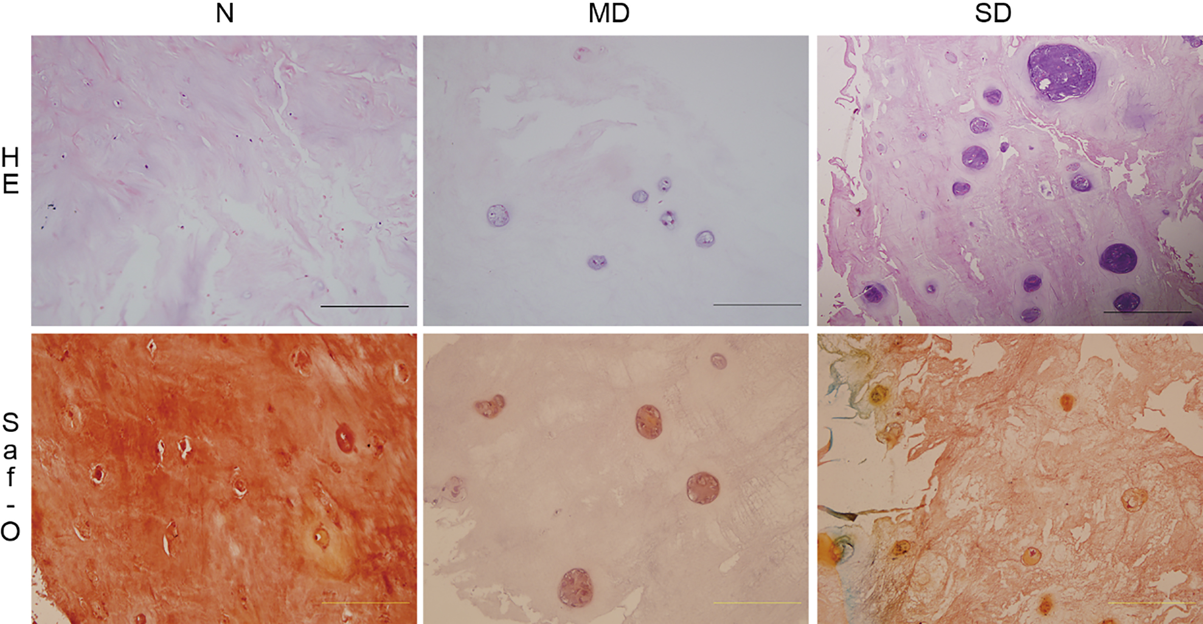
The expression of S1PR in NP was shown by immunohistochemistry. S1PR1, S1PR2 and S1PR3 are all positively expressed in NP tissues, and it is down-regulated with degeneration, bar 100 
The IVD tissues were harvested from three patients through surgery and transported to the laboratory within 30 min. NP tissue were carefully picked and digested in a 0.5% type II collagenase (Sigma-Aldrich, Saint Louis, MO, USA) solution in serum-free medium. The digested tissue/cell suspension was passed through a 100
Cell treatment
To explore the role of S1PRs in human NP cells, the cells were stimulated with ligand, Sphingosine 1-phosphate (S1P), and the effect was assessed by subsequent real-time PCR. Specific information is as follows: firstly, S1P (Sigma-Aldrich) was dissolved in methanol, evaporated, and then resuspended in 0.4% fatty acid-free bovine serum albumin. Recombinant human IL-1
Real-time PCR
Total RNA was extracted from the NP cells by Trizol (Invitrogen, Carlsbad, CA, USA) according to the manufacturer’s instructions. The mRNA was analyzed with real-time polymerase chain reaction using a real-time polymerase chain reaction system (Geneline9640, Bioer, Hangzhou, China). The cDNA was then reverse transcribed (model R0037A, TaKaRa, Dalian, China) according to the manufacturer’s instructions. Real-time polymerase chain reaction reactions were done in triplicate in 96-well plates in a final volume of 20
Western blotting
The total protein was extracted using RIPA supplemented with 1% protease inhibitor cocktail (both from Sigma-Aldrich). Four independent experiments using chondrocytes derived from different patients were performed. The protein concentration was measured with BCA method (Bio-Rad). Proteins were separated by SPS-PAGE electrophoresis using a 10% polyacrylamide gel, and then transferred to a nitrocellulose membrane (Bio-Rad). After blocking in 5% skim milk-Tris buffered saline, the membranes were incubated with primary antibodies overnight. Thereafter, the membranes were rinsed in blocking solution and incubated for 1 hour with a secondary antibody conjugated to horseradish peroxidase. Bands were visualized using an acridan-based substrate detection system (ECL Plus, Amersham, UK) and protein levels were relatively quantified by densitometry using ImageJ.
Statistical analysis
Histological staining and western blot data are described qualitatively. The data of Real-PCR is shown as means
The scores of histological grading of the IVD tissues harvested from patients through surgery
The scores of histological grading of the IVD tissues harvested from patients through surgery
Expression of 3 types of S1PRs (S1PR1/2/3) in NP cells with different degrees of degeneration were measured by real-time PCR and western blotting. The typical grayscale of western blot from some samples was also shown (B). They all decrease in the severe degenerated cells. The relative expression to the severe degenerated cells is shown as mean 
Expression of MMP-3 and ADAMTS4 in NP cells treated with IL-1
Evaluation of the degeneration of IVD samples
In order to evaluate the grade of degeneration in NP cells, histological grading was proceeded by HE and Saf-O staining. The scores of histological grading of the IVD tissues harvested from patients through surgery is shown in Table 1. The representative HE and SAF-O staining of NP tissue with different levels of degeneration is shown in Fig. 1. Overall, with the progress of the degeneration, the NP cells get together into colonies and the extracellular matrix loose.
Primer sequences for real-time PCR
Primer sequences for real-time PCR
According to the grading system [15], which generates a score between 0 and 12, we defined that a grade of 0 to 4 represents histologically normal degeneration (N), grades 5 to 8 indicate mild degeneration (MD), and grades 9 to 12 severe degeneration (SD). Then all the samples were divided into three groups, the N group included 5 samples, the MD group included 12 samples, and the SD group included 22 samples.
To investigate the distribution of S1P receptors in IVD tissue, we performed immunohistochemical staining. S1PR1/2/3 was detected on the IVD sections (Fig. 2). Both cytoplasmic and cytomembrane staining of S1PR1/2/3 immunoreactivity were observed in the nucleus pulposus cells. Then we correlated the immune-expression with degree of degeneration, it seems down-regulated during disease progression.
Expression of S1PRs in NP cells with different levels of degeneration
To confirm the trend shown in immunohistochem, we used real-time PCR to compare the gene expression of S1PR1/2/3 in human NP cells with different levels of degeneration (Fig. 3). The results confirmed that human degenerative NP cells demonstrated a significant decrease in S1PR1/2 expression at mRNA level in severe degenerated NP cells, S1PR3 was also decreased but not significantly. Western blotting shows that the decrease tendency is the same in the protein level, which the expression of S1PR1/2/3 decreases in severe degenerated NP cells.
The effect of the ligand (S1P) in NP cells
Previous studies have confirmed that in human chondrocytes S1P can reduce the induction of catabolic genes in the presence of IL-1
Discussion
The results of this study demonstrate for the first time that S1PR1/2/3 are expressed in the human nucleus pulposus, and it is down-regulated with the intervertebral disc degeneration. The activation of the receptor (stimulated by the ligand, S1P) inhibits the up-regulation of MMMP-3 and ADAMTS4 induced by IL-1
Even though we do not know whether there is S1P in IVD tissue, the presence in synovial fluid, blood plasma and cerebrospinal fluid has been confirmed [16, 17]. With the development of IVD degeneration, blood vessel infiltrated the nucleus pulposus tissue [18], the plasma component may affect the metabolism of the NP cells. Furthermore, immune cells, such as macrophages, neutrophils and T cells, can be recruited to degenerated IVD [19], thus as an important lipid mediator, S1P probably participates in the inflammatory response.
NP cells showed a different pattern of gene expression with degeneration [20, 21] and these variations may lead to distinct cells function, and interact with the degeneration. Here we compared the expression of S1PR1/2/3 in NP cells with different levels of degeneration, and found that the expression of S1PR1/2/3 all decrease with the development of degeneration. The change of S1PR2 is in agreement with a previous study [13], while S1PR1 and S1PR3 is not. The disagreement may be due to the difference between NP cells and chondrocytes, or the different experimental method. This result may be helpful to assess the risk of recurrence after surgery.
In addition to the change of the expression of S1PRs, we confirmed the anti-inflammatory effect of S1P. This is also in line with previous studies in chondrocytes [13, 14]. Together with the decrease of S1PRs, this result may explain the accumulation of matrix degrading enzymes, such as MMP-3 and ADAMTS4 in IVD degeneration. Here we test the effect in NP cells from IVD with mild degeneration, which express S1PRs representatively. As the angiogenesis and tissue defect along with IVD degeneration, the level of S1P in IVD may increase; while the S1PRs is down-regulated, the effect of S1P/S1PR in severely degenerated IVD is doubtful, which needs more research.
Other effects of S1P in chondrocyte have also been reported [11, 22]. Notably, Masuko’s study drew the conclusion that S1P attenuates proteoglycan aggrecan expression via production of prostaglandin E2 from human articular chondrocytes and may thus promote cartilage degradation. Furthermore, S1P is also involved in spinal nociceptive processing [17]. Taken this into account, the down-regulation of S1PRs may be advantageous for the nucleus pulposus cells to adapt to the deterioration of internal environment caused by degeneration.
The biological action of S1P is largely ascribed to specific S1PRs that may evoke distinct biological responses. The key receptor of the anti-inflammatory effect in NP cells has not been studied. This has been controversial even in chondrocytes. Moon’s study shows that S1PR1 is involved in the chondrocyte anti-inflammatory effect of S1P [14]; while Stradner’s study drew the conclusion that S1P reduces the induction of catabolic genes in the presence of IL-1
In the current study, we investigated the role of S1P and its receptors in intervertebral degeneration. We drew the conclusion that human nucleus pulposus cells mainly express S1PR1/2/3, and the expression of decreased when severe degeneration appears. S1P can inhibit the inflammation response induced by IL-1
Footnotes
Acknowledgments
We would like to thank Professor Yue Zhou (Department of Orthopedics, Xinqiao Hospital, The Third Military Medical University, China) for support of the study.
Conflict of interest
None of the authors have any conflicts of interest related to this manuscript.