



Abstract
BACKGROUND:
Cervical radiculopathy is characterized by pain, numbness, tingling, and weakness, mostly in an affected extremity, reflecting compression of a nerve in the neck is compressed or irritated where it emerges from the spinal cord. Diagnosis requires a detailed anamnesis, physical examination, and imaging. Physical therapy, exercise, medical therapy, and injections are the preferred treatments, but injections into the cervical region are only indicated if conservative treatment is ineffective.
OBJECTIVE:
This study explored the utility of selective cervical nerve root blocks (SNRBs) performed at various levels under ultrasound guidance (USG).
METHODS:
We evaluated patients diagnosed with cervical radiculopathy via physical examination and magnetic resonance imaging from November 2019 to March 2020. We included those who did not respond to conservative treatment and therefore received SNRBs at various levels. Sixty-three patients were evaluated over 6 months in terms of pain, functional status, and complications.
RESULTS:
We retrospectively evaluated patients with cervical herniated discs who received SNRBs at various levels between C4–7 under USG. Pain and functional status improved in month 1 and was maintained until at least month 6.
CONCLUSION:
SNRB injections performed under USG effectively treated pain and poor functional status in selected patients. The procedure is safe (especially) for patients who do not respond to conservative treatment.
Introduction
Cervical radiculopathy is characterized by pain, numbness, tingling, and weakness (mostly in an extremity) reflecting compression of a nerve [1, 2, 3, 4]. Diagnosis requires a detailed anamnesis, physical examination, and imaging. Magnetic resonance imaging (MRI) alone should not be used to plan treatment or evaluate the response. A patient may exhibit multi-level disc herniations, but the complaint may be related to a single region. Physical therapy, exercise, medical treatment, and injections are initially preferred; however, surgery is possible if conservative treatment is ineffective [5].
Cervical interlaminar epidural steroid injections (CIESIs) have been used for many years to treat radicular pain refractory to conservative treatment [6]. Symptoms subside when the mechanical pressure created by the hernia is reduced, and the inflammatory process that may occur during or after release of the pressure is inhibited [7].
CIESIs are usually performed fluoroscopically, with the needle targeted at the required anatomical region [8, 9]. The reported complications included spinal cord injury and infection, epidural hematoma, and abscess [8]. In addition, the patient and the physician face radiation risk from fluoroscopy. Alternatively, cervical selective nerve root blocks (SNRBs) have been performed under ultrasound (US) guidance in patients with cervical radiculopathy in recent years [10, 11]. Doppler US reveals soft tissue structures, i.e., vital structures of the cervical region and the needle targets [11]. Yamauchi et al. found that symptom improvement was evident at both 1 day and 1 month after US-guided injection [12]. In comparative studies, US-guided injections were safer than fluoroscopically-guided injections; fluoroscopy was found to be more difficult than US and associated with a radiation risk.
In this work we report 63 cervical SNRBs performed from November 2019 to March 2020. We injected dexamethasone under US guidance into patients with root compressions caused by herniated cervical discs and followed up with these patients for 6 months. The objective of the study is to explore the efficacy of SNRBs performed at various levels under ultrasound guidance (USG).
Methods
This retrospective study was approved by the ethics committee of Kanuni Sultan Suleyman Training and Research Hospital, Istanbul, Turkey. Ethical approval was obtained from the Sisli Etfal Trainig and Research Hospital Clinical Research Ethics Committee (approval no. 1042).
Participants and interventions
All patients provided written informed consent before their injections. We evaluated the records of patients who visited from November 2019 to March 2020 and were diagnosed with cervical radiculopathy via physical examination and MRI; we performed SNRBs at various levels because the patients did not respond to conservative treatment (medical therapy involving nonsteroidal anti-inflammatory drugs or opioids, or exercise and physical therapy performed for at least 4 weeks).
Inclusion and exclusion criteria
A total of 78 patients met the initial inclusion criteria of the study, but some were not evaluated. Six did not attend their 1-month check-ups (all were invited to attend our outpatient clinic at 1 month regardless of the injection site and drug, and all were required to log daily life activity/functional status using a visual analog scale [VAS] and monitor their injection status). Four patients could not be reached by phone, and two underwent surgery. A further three patients did not attend their regular check-ups. We thus finally enrolled 63 patients (Fig. 1). The exclusion criteria applied before injection were any psychiatric disorder or neurological deficit, cervical myelopathy, and/or a history of cervical spine surgery.
Flowchart of the study.
All patients received check-ups in first, second and sixth months and pain level was evaluated using a VAS and functional status using the Neck Disability Index (NDI). The NDI is designed to measure the rate of neck disability and was prepared in the form of a questionnaire consisting of 10 items. The index includes items related to activities of daily living such as pain and self-care, lifting weights, reading, headaches, concentration, work status, driving, sleep and rest [14].
The VAS is a subjective measure used for acute and chronic pain. It converts some values that cannot be measured numerically into numerical data. Scores are handwritten on a 10 cm line representing the continuum between “no pain” and “severe pain”. The test is safe and easy to apply, and is accepted all over the world [13].
Injection method
Before receiving the injection, all patients were given detailed information on the procedure and the expected benefits and risks and signed consent forms. All procedures were performed by one physician (B. Dernek) with more than 2 years of relevant experience. The US-guided SNRB device was fitted with a linear 6–10 MHz probe. Injections were administered with the patients sitting. The injection location in the neck was wiped three times with povidone iodine, and the neck was flexed by approximately 20
Marking the anatomic areas.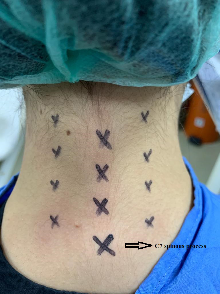
After the C5 level was determined, the probe was shifted up (in the same plane) by approximately 1 cm to detect C4. To identify C6, the probe was shifted 1 cm below C5 (in the same plane); to detect C7, the probe was moved approximately 1 cm below C6 (in the same plane). The injection target point was approximately 1 cm superior to the nerve root. A 5-mL 22-gauge syringe and a 30-mm needle was used to inject 2 mL of lidocaine and 1 mL of dexamethasone solution, as well as 4 mg of NaCl (2 mL of normal saline). Each injection commenced in the plane position under US guidance; the location was marked and sterilized. The needle was inserted approximately 0.5–1 cm superior to where the nerve root was located. The mixture was injected after bleeding control/aspiration. After the procedure was completed, the injection site was cleaned with alcohol and covered. The patients were kept in the observation room for around 1 h in case complications developed.
The NDI questionnaire and the VAS were routinely used to evaluate all patients prior to injection. NDI scores
We retrospectively evaluated patients with cervical herniated discs who received US-guided SNRBs at various levels between C4–7. Patients were followed-up for 6 months.
Demographic characteristics
Demographic characteristics
We calculated averages, standard deviations and medians, recorded the lowest and highest frequencies, and derived ratios. The Wilcoxon test was used to analyze dependent quantitative data and the McNemar test was used to analyze qualitative dependent data. SPSS ver. 26.0 software (IBM Corp., Armonk, NY, USA) was used.
Results
Fifty-two of the 63 patients were female (82.5%) and 11 (17.5%) were male. Thirty-eight were housewives (60.3%) and 36 had various comorbid diseases, the most common of which was type 2 diabetes mellitus (12 [17.5%] patients). Bilateral procedures were performed in 23 (36.5%) cases, and right and left unilateral procedures in 18 (28.6%) and 22 (34.9%) cases, respectively. The most common cervical discopathy was at level C5–6 (
The VAS scores in months 1, 3, and 6 were significantly lower than at baseline (all
Results of the visual analog scale (VAS) and Neck Disability Index (NDI)
Results of the visual analog scale (VAS) and Neck Disability Index (NDI)
Results of the Neck Disability Index (NDI)
We recorded significant recovery (especially in month 1) in terms of both pain and functional status. Patients were followed-up for 6 months. Recovery continued over that time but was less marked between months 3 and 6 than between months 1 and 3. However, the difference was not significant. Recovery commenced in month 1 and continued through month 3 to month 6.
This study explored the utility of SNRBs performed at various levels under USG. SNRBs can relieve cervical radiculopathy symptoms including head and neck pain, numbness, and tingling [15]. The procedure must be performed under guidance, as various complications (primarily intravascular) have been reported. We encountered no serious complications. Three patients developed hypotension that persisted for 15–20 min. In particularly difficult cases, it may be useful to inject under MRI, computed tomography, or fluoroscopic guidance [16, 17]. However, US is safer, as fluoroscopy and computed tomography are both more difficult and associated with radiation exposure [15]. The results of the study recovery persisted for 6 months. A significant improvement was observed in pain and functional results in the first 3 months. Recent anatomical and clinical studies have shown that US-guided SNRB injections are effective and safe, with the pain controlled for at least 1 month [15]. US is obviously not associated with an allergic reaction to contrast material or radiation exposure [15]. SNRBs avoid the need for intravascular or intraneural injection [15]. The most important difference between US- and fluoroscopy-guided SNRBs is that the injection is applied to the region around the nerve root [15].
The SNRB complication rate ranges from 1.64% to 19.4% [18, 19]. Serious complications include intravascular injection, vertebral artery dissection, and brain/spinal cord infection caused principally by intravascular injection of steroid or lidocaine [20, 21]. Four patients exhibited mild hypotensive symptoms (dizziness, blurred vision) immediately after injection that persisted for approximately 10 min. Such mild attacks are associated with pain or vasovagal stimulation.
US revealed vertebral artery pulsation in almost all cases. Prior to injection, we used Doppler US to check whether any vascular structure lay along the injection line. Yamauchi et al. found that US imaging of transverse processes, nerve roots, and surrounding vessels (especially around C3–7) increased US reliability [15]. The hydrostatic pressure and osmotic effects of fluid injected close to the nerve root allows the solution to spread toward the nerve fiber [15]. Narouze et al. found that US guidance placed the needle within 5 mm of the target area [22]. Our level of precision was approximately 5–10 mm.
In a retrospective study, Pak et al. evaluated the results of cervical SNRB injections given under US and fluoroscopy guidance at 1, 3, and 6 months; healing was similar in both groups in terms of both pain and functional status. Since similar results were obtained in this study, we believe that US-guided SNRB is effective, safe, and has a shorter procedure time in comparison to fluoroscopy-guided methods. The cited authors injected 10 mg of dexamethasone; we injected approximately 2 mg but observed significant recovery. We thus recommend a dose of 2 mg [6]. US does not reveal micron-sized vascular structures. We think that our study is superior to the study of Park et al., since we obtained the same result with lower doses in our study. In support of our findings, Tiso et al. [23] recommended the use of particle-free or small-particle steroids such as dexamethasone and betamethasone. We also favor the use of particle-free steroids such as dexamethasone when employing US guidance.
The limitations of the study are the relatively short follow-up (6 months), lack of recording the use of painkillers before and after the procedures and the lack of pathophysiological data.
Conclusions
US-guided SNRB injection effectively relieved pain and improved the functional status of selected patients. It is safe, especially for patients who do not respond to conservative treatment. The injection of particle-free steroids is recommended to minimize complications.
Footnotes
Conflict of interest
None to report.