
Abstract
Studying bladder cancer molecular biology revealed the presence of genetic alterations. So, detection of molecular biomarkers that help in monitoring the disease, evaluating the prognosis of the patients, and their response to therapy is needed. In this study, we investigated the expression and the prognostic significance of SATB-1 and ERBB2 mRNA and protein by quantitative RT-PCR and immunohistochemical analysis in urothelial bladder cancer cases and the surrounding normal bladder tissue. The correlations between the expression of both markers and the clinicopathological parameters were performed with further analysis of the correlation between the expression of SATB-1 and ERBB2. Compared to control, the expression of SATB-1 and ERBB2 mRNA and protein in cancer tissues were significantly up-regulated (
Introduction
Urinary bladder carcinoma (BC) is the third most common tumor in men and the eleventh most common tumor in women [1]. In Egypt, it constitutes 30% of all cancer cases with an incidence rate of 13.5/100,000 individuals according to the National Cancer Institute [2]. Urothelial cell carcinoma represents more than 90% of bladder cancer [3].
Bladder cancer patients suffer from the poor unpredictable clinical outcome as more than 50% show relapse within the first year despite of being diagnosed at an early stage. Also, cases with muscle invasion have a bad prognosis with a 15–68% 5-year survival rate. This indicates the importance of knowing the oncogenic pathway and the factors affecting bladder cancer behavior [4].
The management of BC depends on the patients’ clinicopathological parameters as TNM staging and grading of the tumor, as predictors of good/poor prognosis. However, many variations occur within the same grade/stage as a result of bladder cancer cell heterogeneity. So, the detection of new biomarkers can help early detection of the disease, prediction of high-risk patients, and help proper management [5].
Special AT-rich sequence binding protein-1 (SATB-1) is a nuclear matrix attachment region (MAR) DNA-binding protein. It acts as a transcription factor and affects chromatin organization. Therefore, it can regulate the expression of several genes associated with the occurrence of cancer, and the ERBB2 gene is one of them. Altered SATB-1 expression was associated with the occurrence, invasion, and metastasis of many malignant tumors [6, 7].
Liu et al. found that SATB-1 expression correlated with poorly differentiated breast cancer and indicated an unfavorable prognosis and lower survival rate than those with negative expression. SATB-1 might inhibit hormone receptor expression in cooperating with Her2 and promotes tumor progression or estrogen therapy resistance in breast cancer [8]. Han et al. showed that SATB-1 promotes tumor growth and metastasis in breast cancer. Moreover, MAPK signaling was activated and PI3K/mTOR signaling was suppressed by SATB-1 in breast cancer cells [9].
ERBB2 gene encodes a member of the epidermal growth factor receptor family of receptor tyrosine kinases. The product of the human ERBB2 gene (Her2) is a 185-kilodalton glycoprotein with protein-tyrosine kinase activity, which is a human version of the rat neu proto-oncogene. However, it is still an orphan receptor to which no specific ligand has been identified. Her2 activates the Phosphatidylinositol-3 kinase-AKT-NF-
Her2 is considered as one of the best-known therapeutic targets in oncology. Patients with ERBB2-amplified or -overexpressing breast cancer benefit from ERBB2-targeted therapy, including anti-ERBB2 antibodies (such as trastuzumab and pertuzumab) and small-molecule tyrosine kinase inhibitors (such as lapatinib and neratinib) [11]. The 2007 ASCO guidelines mandate that Her2 should be evaluated in every invasive breast cancer, either at the time of diagnosis or recurrence to guide therapy [12]. Her2-positive breast cancer patients respond to monoclonal antibody therapy with trastuzumab or tyrosine kinase inhibitors at least at early stages. However, the prognosis of such patients is poor since
Her2-positive breast cancers are more invasive than estrogen receptor positive ones. However, in Her2-positive and triple-negative breast cancer, the pathological complete response is high (60% or more), and correlated with long-term outcome [13].
The human ERBB2 gene is amplified in
Overexpression of the ERBB2 proto-oncogene is associated with amplification of the gene in breast cancer but increased activity of the promoter also plays a significant role. Some studies reported an apparent lack of a marked association between Her2 protein expression and gene amplification in bladder cancer, as well [14]. Members of two transcription factor families (AP-2 and Ets) show increased binding to the promoter in over-expressing cells [15].
This study was done to detect SATB-1 and ERBB2 (mRNA and protein) expression in bladder urothelial cell carcinoma, and to evaluate the relations between their expression and clinicopathological variables and prognosis. The correlations between the expression of SATB-1 and ERBB2 were analyzed.
Patients and methods
This work was carried out in the Medical Biochemistry and Molecular Biology Department with the participation of the Pathology and Public health and Community Departments, Faculty of Medicine, Zagazig University, and the Urology Department, Al-Ahrar Teaching Hospital, Zagazig, Egypt. It included 50 samples from patients with primary bladder urothelial cell carcinoma obtained by transurethral resection of bladder tumor (TURBT) or radical cystectomy from January 2020 to June 2020. Informed consent was obtained from participants. Adjacent normal urothelial specimens were enrolled in the study as control. The clinical data of the patients were obtained from their medical records of Al-Ahrar Teaching Hospital, Zagazig, Egypt. Grading of tumors was evaluated according to the World Health Organization’s (WHO) classification of tumors of the Urinary System and Male Genital Organs [16]. Pathological staging was evaluated according to the TNM system of tumors. All the cases were not subjected to any preoperative chemotherapy or radiotherapy. The study complied with the guidelines of the local ethics committee.
Immunohistochemistry (IHC)
Streptavidin-biotin-peroxidase complex (ABC) technique was used, 5 um sections of the formalin-fixed, paraffin-embedded tissues were cut, deparaffinized in xylene, rehydrated through a graded series of ethanol and washed in running water. Endogenous peroxidase activity was blocked by incubation with 0.3% H
Pathological analysis of immunohistochemistry
For SATB-1, nuclear staining was defined as immunoreactions. According to the intensity and the percentage of stained cells, the score was calculated. The predominant intensity was evaluated as follows: no nuclear staining (0), weak nuclear staining (1), moderate nuclear staining (2), and strong staining (3). The percentage of stained cells was evaluated as follows: less than 5% (0); 5 to 25 % (1); 25 to 50% (2); more than 50% (3). The final score was calculated by adding the two above scores. Scores of 0–2 were defined as low expression scores, and 3–6 were defined as high scores [17].
For Her2 immunostaining, scoring was based on the American Society of Clinical Oncology/College of American Pathologist guidelines 0: no staining, 1+: incomplete staining of the cell membrane, 2+: complete but weak or moderate membranous staining in
RNA extraction and quantitative RT-PCR
Bladder tissues were homogenized and total cellular RNA was extracted from tissue homogenate using IQeasy plus CTB RNA extraction kit (iNtRON Biotechnology, Seongnam, Korea) following the manufacturer’s instructions. Reverse transcription of one ug of RNA was performed by the means of Power cDNA synthesis Kit (iNtRON Biotechnology, Seongnam, Korea) according to the manufacturer’s protocol. qRT-PCR was performed by adding 5 uL of the cDNA, 100 pmol/uL of each primer (0.5 uL each) (Biolegio, Netherlands), 10 uL of EvaGreen PCR Master mix (Jena Bioscience GmbH, Jena, Germany) and 4 uL PCR-grade water. Real-time PCR was performed using Mx3005P (Stratagene, La Jolla, CA, USA) as follows: 2 min of initial denaturation and polymerase activation at 95
The relative gene expressions for SATB-1 and ERBB2 were calculated by the 2
Primer sequence of the studied genes
Primer sequence of the studied genes
Urothelial cell carcinoma: A: low-grade papillary urothelial cell carcinoma, B: high-grade papillary urothelial cell carcinoma; C: high-grade urothelial cell carcinoma with muscle invasion (H&E, A: x100, B&Cx400).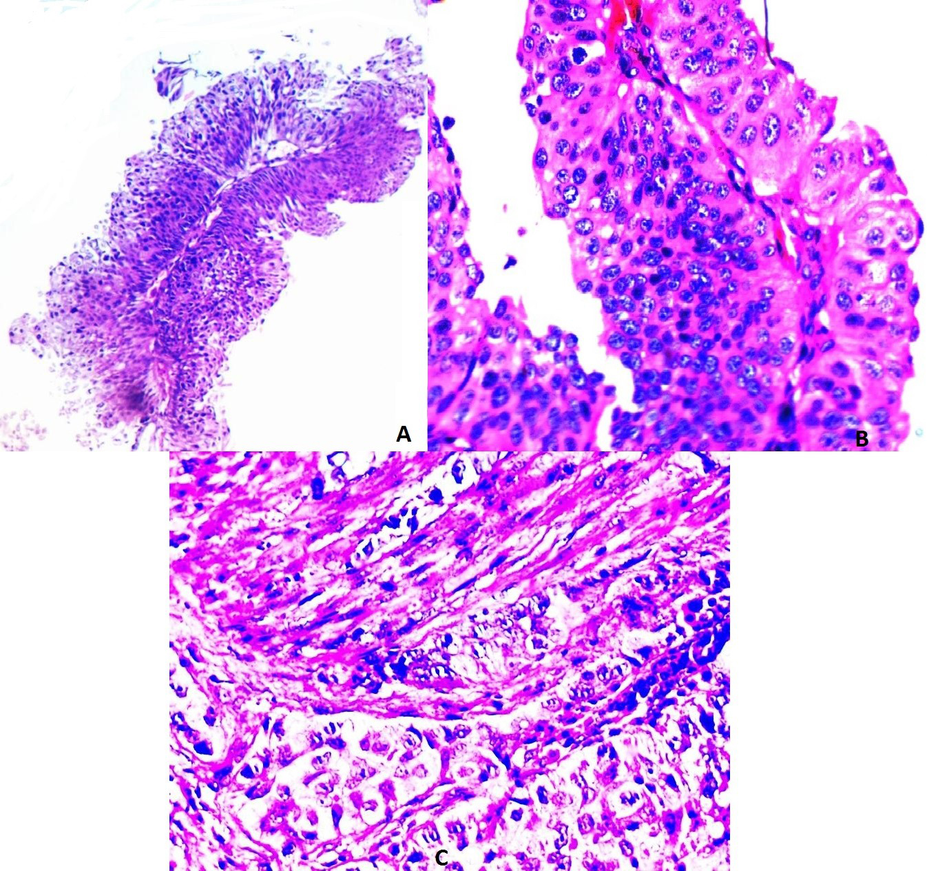
Quantitative data were expressed as the mean
Results
Patient’s characteristics
The patients’ mean age
Clinicopathologic variables of 50 cases of bladder cancer
Clinicopathologic variables of 50 cases of bladder cancer
SATB-1 was highly expressed in 62% (31/50) of cases with a negative expression in adjacent healthy mucosa. A significant association between high SATB-1 expression and the grade of tumor differentiation (
Relation between clinicopathological features and immunohistochemical staining for SATB-1 in 50 patients with bladder carcinoma
Relation between clinicopathological features and immunohistochemical staining for SATB-1 in 50 patients with bladder carcinoma
Positive Her2 expression (score3+) was detected in 32% (16/50) of specimens with equivocal expression (score2+) in 34 % (17/50) of cases (Table 2). Adjacent healthy mucosa showed negative expression. A significant association between positive Her2 expression and the depth of invasion (
Relation between clinicopathological features and immunohistochemical staining for Her-2 in 50 patients with bladder carcinoma
In bladder tissues, the mean values of mRNA fold expressions of SATB-1 and ERBB2 relative to the control adjacent tissues were (2.9
Upregulation of SATB-1 and ERBB2 mRNA expressions was significantly associated with the tumor stage, the depth of tumor and the lymph node involvement (
Relation between clinicopathological features and SATB-1 gene expression in 50 patients with bladder carcinoma
Relation between clinicopathological features and SATB-1 gene expression in 50 patients with bladder carcinoma
Relation between clinicopathological features and ERBB2 gene expression in 50 patients with bladder carcinoma
There was a significant association between SATB-1 and Her2 immunohistochemical expression (
The correlation between the variables
The correlation between the variables
Concerning the Receiver Operating Characteristic (ROC) curve analysis, SATB-1 gene expression showed high specificity and sensitivity as an early predictor of urothelial cancer (93.3% &85.5% respectively) at cutoff value of 4.1. ERBB2 gene expression has specificity (80%) and sensitivity (91.4%) at a cutoff level of 4.6. Interestingly, the combination of the two markers raised the specificity to 93.3% and the sensitivity to 94.3% (Table 8A and B, Fig. 4).
Validity of SATB-1 and ERBB2 gene expression as an early predictor for LN involvement in bladder carcinoma: ROC curve analysis
The test result variable(s): ERBB2, SATB-1gene has at least one tie between the positive actual state group and the negative actual state group. Statistics may be biased.
a. Under the nonparametric assumption. b. Null hypothesis: true area
Validity of SATB-1 and ERBB2 gene expression as an early predictor for LN involvement in bladder carcinoma: ROC curve analysis
ROC curve: Receiver Operating Characteristic curve; PPV: Positive Predictive Value; NPV: Negative Predictive Value: AUROC: Area Under Receiver Operating Characteristic curve;
SATB-1 immunohistochemical expression in bladder urothelial cell carcinoma: A: negative expression in adjacent healthy mucosa; B: low nuclear expression in low-grade papillary urothelial cell carcinoma; C: high nuclear expression in high-grade papillary urothelial cell carcinoma; D: high nuclear expression in high grade urothelial carcinoma with muscle invasion (IHC, X400).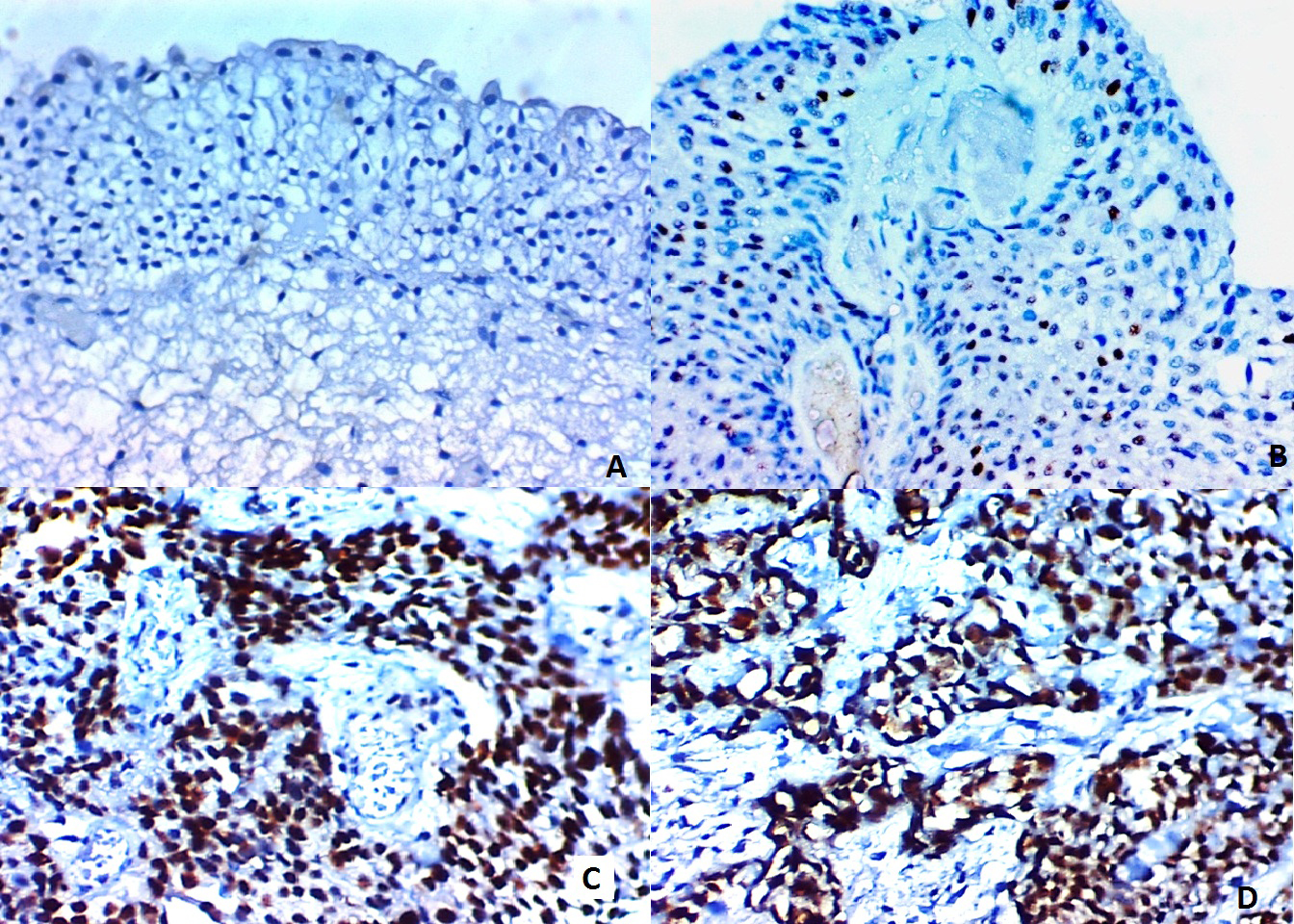
Her2 immunohistochemical expression: A: faint incomplete membranous staining (score 1+) in low-grade urothelial carcinoma; B: moderate complete membranous staining (score2+) of high-grade urothelial carcinoma, C: strong complete membranous staining (score 3) of high-grade papillary urothelial carcinoma (X 400).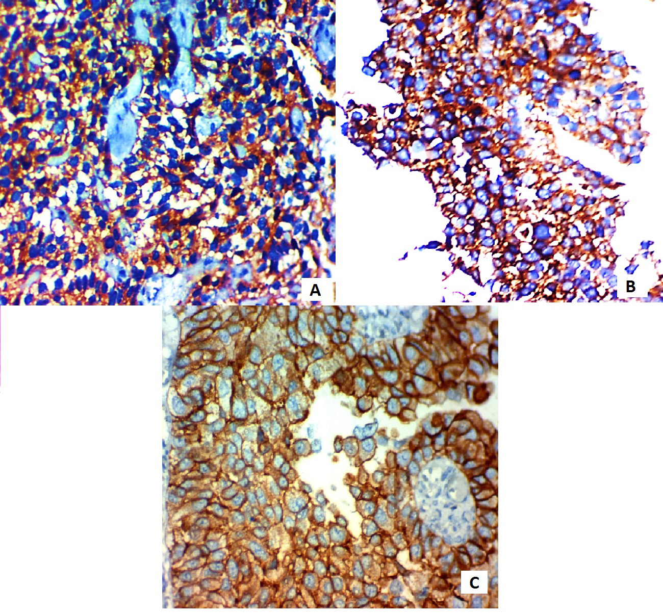
Roc curve: Validity of SATB-1 and ERBB2 gene expression as an early predictor for LN involvement in bladder carcinom.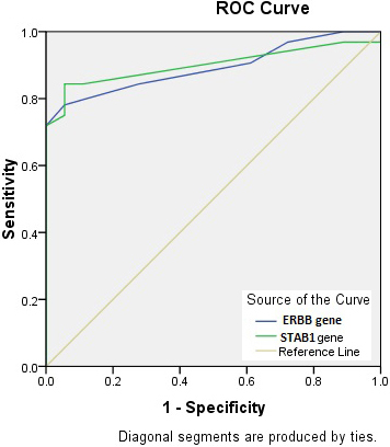
Bladder cancer is the 7th most common cancer worldwide representing about 3.2% of all cancers. Studying bladder cancer molecular biology revealed the presence of genetic alterations. So, detection of molecular biomarkers that help in monitoring the disease, evaluating patient’s prognosis, and response to therapy is needed [19].
SATB-1 is involved in chromosome remodeling, histone acetylation, and methylation, and can regulate the transcriptional activity of multiple genomes. It acts as a genome organizer by affecting the expressions of multiple tumors associated genes. So, SATB-1 can permit the tumor occurrence and metastasis facilitating its progression [20].
Our study showed higher SATB-1 expression at protein and mRNA levels in BC compared to the adjacent healthy mucosa. Similar results were reported by a previous study in non-muscle- invasive and muscle-invasive urothelial BC [6]. This suggests the role of SATB1 in the carcinogenesis and the progression of urothelial BC. Therefore, we also investigated the correlation between clinicopathological features and SATB1 expression in urothelial BC to validate its prognostic significance.
Concerning the grade of tumor differentiation, high SATB-1 protein expression was significantly associated with the tumor grade (
Previous studies found a correlation between up-regulated SATB-1 expression and advanced stages of malignant tumors such as breast cancer and laryngeal squamous cell carcinoma [21, 22]. The underlying mechanism of the SATB-1 role in affecting the behavior of the malignant tumors could be attributed to its ability for binding to the regulatory regions of hundreds of genes that directly affect the promoter activity and the expression of genes associated with tumor invasion and metastasis [8]. Moreover, SATB-1 overexpression was found to promote epithelial-mesenchymal transition processes by affecting E-cadherin down-regulation and E-cadherin repressors (Snail, Slug, and vimentin) upregulation. Thus, it increases the invasive and the metastatic capability of these lesions [6]. So, SATB-1 can be considered as an independent prognostic factor as well as a potential therapeutic target in human malignancies including urothelial BC.
Her2 is a transmembrane single subunit glycoprotein that has an extracellular ligand-binding domain, a transmembrane domain, and an intracellular tyrosine kinase catalytic domain. On ligand activation, the receptors dimerize forming heterodimers. This is followed by transphosphorylation which activates several intracellular signaling pathways such as Ras/mitogen-activated protein kinase pathway, the phosphatidylinositol 3 kinase (PI3K)/Akt pathway, the Janus kinase/signal transducer and activator of transcription pathway, and the phospholipase C pathway, which ultimately affects cell proliferation, survival, motility, and adhesion [12].
The current study detected Her2 positive expression in 32% of cases and equivocal expression in 34% of cases. However, negative expression was noted in 34% of cases. Her-2 positive expression was noted in 35% of low-grade transitional cell carcinoma and 30% of high grade but without statistical significance. This matched the result of Kumar et al. [23]. On the contrary, other studies had observed a significant association between Her2 overexpression and high-grade tumors (24-26). Different results could be attributed to the difference in region and ethnics.
Concerning lymph node involvement, no statistically significant correlation with Her2 protein expression was detected in our study (
Our study showed a significant association between high SATB-1 mRNA and protein expression and each of the depth of invasion of the primary tumor and TNM stage (
Like previous studies, no significant relation between Her2 protein expression and age, gender, or primary tumor size was detected [29, 30].
There was a statistically significant association between SATB-1 and Her2 expression at both mRNA and protein expression. Therefore, a positive regulation and interaction between SATB-1 and Her2 that affect the behavior of urothelial BC could be suggested. Similarly, direct up-regulation of the ERBB2 gene by SATB1 was reported in breast cancer [8, 9] and in gastric carcinoma [7].
In cancer, abnormal nuclear organization and alterations in the amount and the distribution of heterochromatin are hallmarks [31]. In eukaryotes, the genome is divided into chromatin domains by the attachment of chromatin to a supporting structure called the nuclear matrix. The interactions between chromatin and the nuclear matrix occur via AT-rich DNA sequences called matrix attachment regions (MARs). The MARs function in organizing chromatin loops, augmenting gene expression, and facilitating replication [32]. Not all MARs are bound to the nuclear matrix or participate in the organization of loop attachment regions. MAR binding is a dynamic event that is cell type and/or cell cycle-dependent and can allow the regulation of distant genes in a coordinated manner [33]. The expression of MAR-binding proteins is correlated with aggressive tumor phenotypes. Moreover, a modification in the interactions between nuclear matrix proteins and MARs is related to chromatin reorganization observed during carcinogenesis. SATB-1 is the best characterized MAR-binding protein, and its roles in the higher order of chromatin loop organization and global transcriptional regulation have been documented [34]. Interestingly, the level of interaction of SATB-1 with MAR sequences could be more important than its expression level [35].
SATB-1 acts as a “genome organizer” that functions as a platform to regulate tissue-specific gene expression. It regulates the expression of more than 10% of genes by binding to the upstream regulatory regions which directly influence the promoter activity and gene expression [21]. Increased transcription of Her2 protein can occur due to the activation of the Her2 promoter by transcriptional factors, including activating protein-2 (AP-2) and Yin Yang protein-1 (YY1) [36]. YY1 is a zinc-finger DNA binding phosphoprotein transcriptional factor that regulates initiation, activation, and repression of transcription for several genes connected with cell growth, development, and differentiation [37]. YY1 stimulates AP-2 transcriptional activity, as well. YY1 may direct histone deacetylases and histone acetyltransferases to a promoter to activate or repress the promoter. Ten thousands target genes of the YY1 transcription factor were predicted using the transcription factor binding site motifs. SATB-1 is one of these genes [38].
In conclusion, there is a positive correlation between the expression of both SATB-1 and Her2 in urothelial cell carcinoma of the bladder at mRNA and protein levels. They also could predict patients with poor prognosis that could benefit from targeted therapy.
Footnotes
Abbreviations
| RT-PCR: | Reverse transcription-polymerase chain reaction; |
| H&E: | Hematoxylin and eosin; |
| ANOVA: | Analysis of variance; |
| GADPH: | Glyceraldehyde 3-phosphate dehydrogenase; |
| ROC: | Receiver Operating Characteristic; |
| SATB-1: | Special AT-rich sequence binding protein-1; |
| MAR: | matrix attachment region. |