
Abstract
BACKGROUND:
MicroRNAs (miRNAs) have been reported to play an important role in tumor progression by regulating the expression of target genes.
OBJECTIVE:
This study attempted to verify the role of miR-711 in gastric cancer (GC) progression by in vitro and in vivo assays.
METHODS:
The expression of miR-711 in tumor tissues and cells was detected by real-time quantitative PCR (qRT-PCR). Expression of MiR-711 in NCI-N87 and SNU-1 cells was detected by FISH. We transfected GC cells with miR-711 mimics or inhibitors. The effects of miR-711 on the proliferation and metastasis of GC cells were detected by CCK-8, wound healing and transwell assays. Dual-luciferase reporter gene assay was used to verify the targeting relationship between miR-711 and CD44. Xenograft assays was used to verify the regulatory effect of miR-711 on tumor growth.
RESULTS:
In GC tissues and cell lines, the expression of miR-711 was down-regulated when compare with adjacent tissues or normal epithelial cells. The results indicated that overexpressing of miR-711 could suppress the GC cell proliferation, migration, and invasion through targeting CD44. The knockdown of CD44 showed similar effects as miR-711 overexpression in GC cells. Moreover, we confirmed these effects in the in vivo assays. Furthermore, we found that miR-711 could play a role by influencing tumor cell stemness.
CONCLUSION:
MiR-711 plays vital roles as a tumor-suppressor by targeting CD44 and may be a therapeutic target for GC treatment.
Introduction
Gastric cancer (GC) is the fourth most common cancer with poor prognosis and the fourth most common cause of cancer death [1, 2]. Currently, the main method for GC treatment is surgery, combined with chemotherapy, radiotherapy as well as other comprehensive treatments [3, 4]. Despite the relevant treatment, there were still many patients with recurrence or poor prognosis [5, 6]. In general, the occurrence of GC can be seen as a multi-step process that includes environmental factors, genetic changes, and other risk factors that cause cells to gradually transform into cancer [7]. So far, little was known about the regulatory mechanism of GC progression [2].
MicroRNAs (miRNAs) is a class of small noncoding RNA with a length of about 22 nucleotides, which plays as an important regulatory factor in disease progression [8, 9]. It is reported that about 30% of all genetic pathways and human genes are affected by miRNAs induced gene silencing either by translational suppression or by messenger RNA (mRNA) degradation [10]. In the background of GC, some miRNAs were discovered to be closely associated with cell growth, migration, invasion, and recurrence prediction [11, 12, 13]. Our research group used miRNA microarray screening to find that miR-711 was downregulated in GC cells. However, the current research on miR-711 mainly focuses on its expression disorder after tissue and organ injury. There are few reports on tumor related literature. Current studies have found that miR-711 regulates tumor progression by regulating cell proliferation, migration and invasion [14, 15, 16]. However, its mechanism in GC is unclear. Therefore, it is very important to further explore the role and mechanism of miR-711 in GC.
The theory of cancer stem cells (CSCs) is based on the findings of scientists that tumors are mutated from somatic cells, and tumor cells can grow without restriction [17, 18]. With the deepening of research on CSCs, CSCs markers have been discovered one after another, such as CD133 [19], Sox2, and OCT-4 [20]. A recent study found that CD44
Taken together, we hypothesized that miR-711 and CD44 may be key regulators GC progression. The in-depth discussion of them provides the basis for the screening of gastric cancer biomarkers and targeted therapy.
Methods and materials
Bioinformatic analysis
The Gene Expression Omnibus (GEO) database was used to obtain the miRNA expression data of GC and healthy control. For GSE93415, there were 40 samples, each pair included resected primary tumor and corresponding healthy gastric mucosa. For GSE63121, there were 30 samples (15 paired of gastric cancer vs. normal tissues). For GSE2379, there were 80 samples, which included 40 cancer tissues from Singapore national cancer center and Singapore Singhealth tissue bank and 40 non-cancer tissues. R package was used for analyzing the different expression of miRNAs. Overall survival (OS) was calculated using StarBase (
Clinical specimens
GC tissues were isolated from 30 GC patients at the Second Affiliated Hospital of Xinjiang Medical University (Xinjiang, China). All participating patients understood the purpose of this study and the content of the test and obtained informed consent from all patients. The diagnosis of GC was confirmed by more than two pathologists, and all included patients did not receive local or systemic therapy before surgery. Samples of tissue were obtained at surgery. Samples were immediately frozen using liquid nitrogen and stored at
Cell culture
GC cell lines, KATO3, HGC-27, MKN-45, NCI-N87 and SUN-1 were cultured in RPMI-1640 (Hyclone, USA) contained 10% fetal-bovine serum (FBS, Gibico, USA), and human gastric epithelial cell strain GES-1 was cultured in DMEM (Hyclone, USA) with 10% FBS as control. All the cell lines were obtained from BeNa and incubated at 37
Cell transfection
NCI-N87 and SNU-1 cell lines were inoculated 1
qRT-PCR
Total RNA was extracted from samples with TRIzol reagent according to the kit protocol (Invitrogen, USA) and subsequently quantified using NanoDrop 2000 (Thermo Fisher, USA). 1
Western blot
Proteins were extracted by RAPI lysis buffer (Beyotime, Shanghai, China) and then quantified with DC protein assay (Bio-Rad, USA) according to the manufacturer’s protocol. 30
Fluorescence in situ hybridization (FISH)
MiR-711 expression in NCI-N87 and SNU-1 cells was detected by FISH. The miR-711 probes were designed and purchased from BioSENSE (Guangzhou, China). Slides were hybridized overnight with probes and washed with 50% formamide/2
Dual-luciferase reporter assay
The miR-711 binding site in CD44 3’ UTR was predicted using the TargetScan 6.0. The wild-type vector (pmiR-RB-REPORT-h-CD44 WT) and mutant vector (MUT) of the CD44 3’ UTR were constructed by RiBoBio (Guangzhou, China). Cells were seeded in 24-well plates (1
Cell Counting Kit-8 (CCK-8) assay
CCK-8 (Biotechwell, Shanghai, China) was used in cell growth assay. Resuspended NCI-N87 and SNU-1 cells were seeded at 1
Wound healing assay
Seeded the cells in 12-well plates and grown to about 90% in complete medium. Then a 200-
Migration and invasion assay
For the migration assay, 5
For invasion assay, transwell chambers coated with Matrigel were used. 5
Abnormal expression of miR-711 in GC tissues and cell lines. (A) miR-711 relative expression level in GC tissues and matched adjacent tissues. (B) Comparison of miR-711 expression in GC tissues and normal gastric mucosal tissues in the GEO databases. (C) Overall survival analysis of patients with high or low miR-711 expression in the TCGA databases. (D) Relative expression of miR-711 in all five GC cell lines and in the normal human gastric epithelial cell strain GES-1. (E) Represent FISH images of miR-711 expression. Scale bars, 50 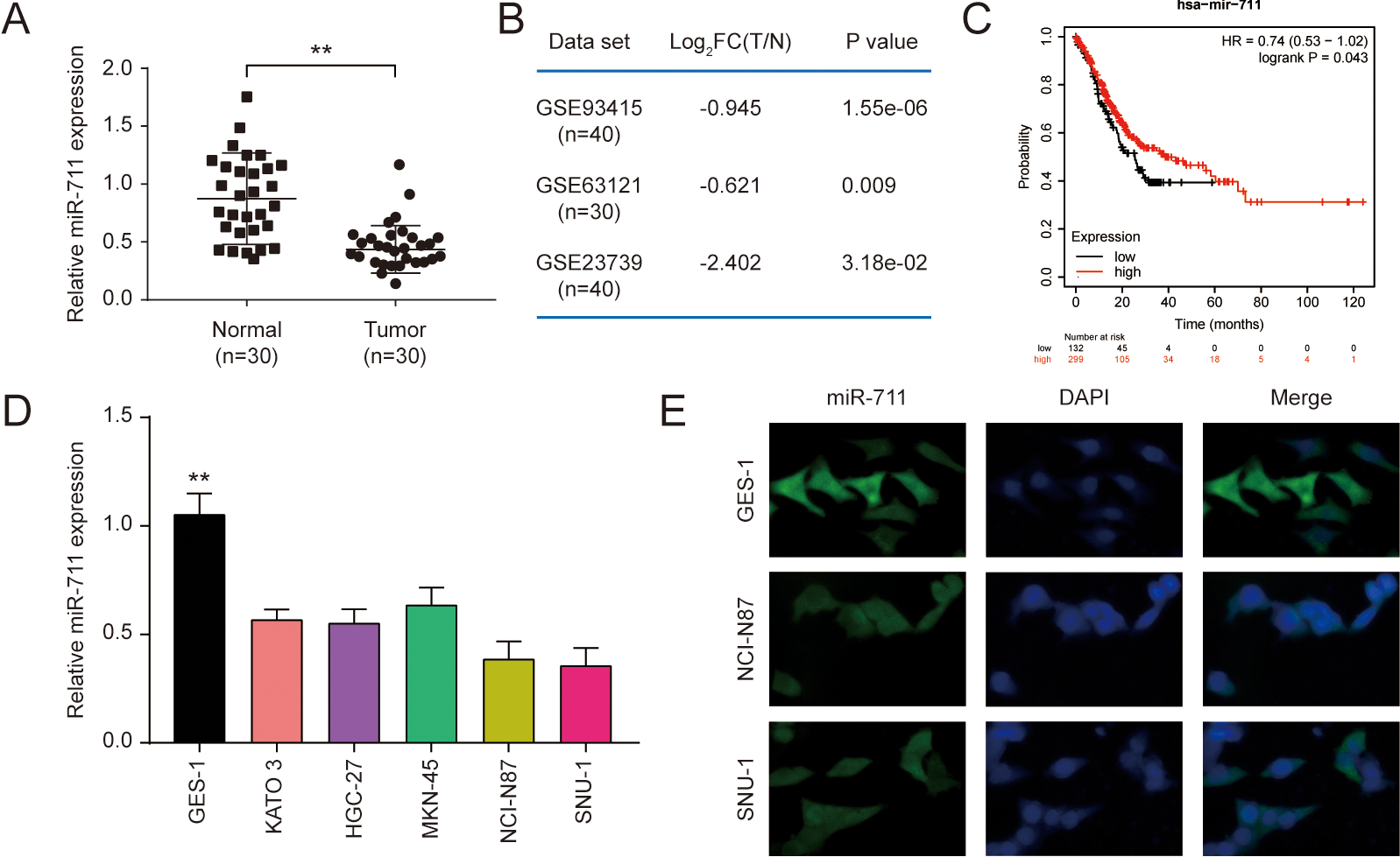
For examining spheroid formation potentials, a total of 1,000 cells were suspended in stem-cell medium for 7 days and the number of spheroids larger than 50
The effect of miR-711 expression on proliferation and metastasis of gastric cancer cells. (A) Relative expression of miR-711 in GC cell lines SNU-1 and NCI-N87 transfected miR-711 mimics, miR-711 inhibitor and empty vector respectively. (B) The OD value of GC cells proliferation in GC cell lines SNU-1 and NCI-N87 transfected miR-711 mimics, miR-711 inhibitor and empty vector respectively. (C) Wound-healing assay and (D) Transwell assay of SNU-1 and NCI-N87 cell lines treated with miR-711 mimics, miR-711 inhibitor and empty vector respectively (
Identification of CD44 as an miR-711 target in GC. (A) miR-711 targeting sites in the 3’-UTR of targets genes and corresponding mutations. (B) CD44 relative expression level in GC tissues and matched adjacent tissues. (C) Relative expression of CD44 in GC cell lines SNU-1, NCI-N87 and normal human gastric epithelial cell strain GES-1. (D) Pearson correlation was used to study the correlation between miR-711 and CD44 (
The effect of CD44 expression on proliferation and metastasis of GC cells. (A) The mRNA and (B) Protein levels of CD44 in GC cell lines SNU-1 and NCI-N87 treated with si-CD44 and pc-CD44 were examined. (C) The OD value of GC cells proliferation, (C) Wound-healing assay and (D) Transwell assay of SNU-1 and NCI-N87 cell lines treated with si-CD44 and pc-CD44 (
miR-711 inhibits the effect of CD44. (A) Relative expression of miR-711 in GC cell lines SNU-1 and NCI-N87. (B) Relative expression of CD44 in GC cell lines SNU-1 and NCI-N87. (C) Protein levels of CD44 in GC cell lines SNU-1 and NCI-N87. (D) The OD value of GC cells proliferation. (E) Wound-healing assay and (F) Transwell assay of SNU-1 and NCI-N87 cell lines. (
Effects of miR-711 and CD44 on CSCs. (A) The quantification of sphere diameter (A) and numbers (B) of SNU-1 CSCs and NCI-N87 CSCs with indicated transfection for 14 days were analyzed. (C) The mRNA levels of stem cell genes including SOX2 was analyzed by QRT-PCR. (
In vivo functional analysis of miR-711. (A) The tumor weight in each group. (B) Growth curve of tumors in the nude mice in response to miR-711-agomir. (C) HE and KI67 staining of tumor tissues of each treatment group. (D) Protein levels of CD44 in vivo

Males C57BL/6 mice (7-week-old) were obtained from Xinjiang Medical University (Xinjiang, China). Each mouse was injected subcutaneously with 2
Statistical analysis
All quantitative data was presented as the mean
Results
Expression of miR-711 in GC tissues and cell lines
QRT-PCR results showed that miR-711 was significantly down-regulated in all 30 paired GC tissues relative to the normal tissues (Fig. 1A,
The effect of miR-711 expression on proliferation of GC cells
To investigate its role in proliferation and metastasis, we transfected miR-711 mimics or miR-711 inhibitor into GC cell line and verified the transfection efficiency by qRT-PCR. The results showed that the operations to down-regulated and overexpressed miR-711 were successful, the expression of miR-711 had a significantly different in both two groups compared with the control (Fig. 2A,
Identification of CD44 as an miR-711 target in gastric cancer
To test whether the predicted miR-711-binding sites with CD44 3’-UTR regions are subjected to regulation by miR-711, we cloned their wild-type (WT) and mutated 3’-UTR regions into pGL3 downstream from a luciferase reporter gene (Fig. 3A,
The effect of CD44 expression on proliferation of GC cells
To test whether overexpression of CD44 affects cancer progression, the si-CD44 and pc-CD44 were introduced into SNU-1 and NCI-N87 cells and verified the transfection efficiency by qRT-PCR and western blot (Fig. 4A-B,
miR-711 inhibits the effect of CD44
In order to verify whether the effect of miR-711 in the regulation of proliferation and invasion is through targeting CD44 in GC. Si-CD44 was used to silence CD44 in SNU-1 and NCI-N87 cells transfected with miR-711 inhibitor (Fig. 5A-B,
Effects of miR-711 and CD44 on CSCs
To test the functional significance of miR-711 in GC cell stemness through regulating CD44, we found that CD44 overexpression facilitated tumor-spheres formation (Fig. 6A-B,
In vivo functional analysis of miR-711
To investigate whether miR-711 function as a tumor suppressor on GC cells in vivo, we used a mouse xenograft model to detect tumor formation and metastasis. The results showed that tumors produced by the miR-711-agomir cells displayed the slowest growth and the size reached 800 mm
Discussion
Increasing studies have reported that miRNAs play important roles in GC development [23]. We confirmed the inhibitory effect of miR-711 on GC in vitroand in vivo, including inhibition of cell proliferation, migration and invasion. CD44 was predicted as a target gene of miR-711, and luciferase reporter detection validated that miR-711 can target gene CD44. Moreover, in GC cells, the mRNA and protein levels of CD44 were both negatively correlated with miR-711. We also found that knock down of CD44 partially weakened miR-711 inhibitor-mediated effect on SNU-1 and NCI-N87 cells proliferation.
In addition, this study verify that miR-711 regulates CSCs through CD44 through the ball formation experiment. Studies have shown that CD44 can participate in the regulation of gastric cancer stem cells [22, 24]. CD44
In conclusion, our study showed the important role of miR-711 in GC. Our findings indicated that miR-711 expression was significantly downregulated in GC, while CD44 expression was inversely trending. And we found that miR-711 can play a role by influencing CSCs. These studies on miR-711/CD44 in tumorigenesis will contribute to the clinical therapy and serve as effective therapeutic targets.
Author contributions
Conception: Liang Li.
Interpretation or analysis of data: Jie Gao, Jiangang Li.
Preparation of the manuscript: Liang Li.
Revision for important intellectual content: Jun Wang.
Supervision: Jun Wang.
Funding
Natural Science Foundation of Xinjiang Uygur Autonomous Region (No: 2021D01C375).
Supplementary data
The supplementary files are available to download from http://dx.doi.org/10.3233/CBM-210213.