



Abstract
This in vitro study was designed to examine the effect of some newly synthesized aminoalcanolic derivatives of xanthone (I, II) and aroxyalkyl derivatives of 2-methoxyphenylpiperazine (III, IV) having cardiovascular activity on the haemorheological parameters of RBCs from healthy individuals and patients with chronic venous disease. Additionally, the influence of compounds I-IV on some RBCs associated enzymes such as acetylcholinesterase (Ache), glucose-6-phosphate dehydrogenase (G6PD) and glutathione reductase (GR) as well as glutathione (GSH) content were determined in vitro in RBCs from healthy subjects. The study showed that compounds I, III and IV significantly increased RBCs deformability. Moreover, both xanthone derivatives reduced RBCs aggregation and diminished RBCs aggregates strength in all RBCs groups. Compounds II and III significantly improved Ache activity, whereas compounds I and II increased G6PD and GR activity and GSH level. In conclusion, compounds I, III and IV, which significantly improved RBCs deformability in vitro, may facilitate the passage of blood in the vascular system. Additionally, compounds I and II which inhibit RBCs aggregates formation in vitro may contribute to more rapid degradation of red blood cell aggregates in circulating blood.
Keywords
Introduction
Erythrocytes (red blood cells, RBCs) ability to deform is vital for their circulation in blood vessels, as it enables their passage through capillaries having very small lumen diameter. This amazing property results from the coupling and interactions between membrane lipid bilayer and spectrin network [1–3]. Impaired RBCs deformability reduces cell orientation in blood flow, and consequently leads to elevated blood viscosity [4]. Another factor that plays a crucial role in the pathophysiological behavior of blood circulation is erythrocytes aggregation, that is a reversible phenomenon influenced by both RBCs properties (including surface charge, shape, membrane and cytoskeletal composition) and plasma factors (such as fibrinogen concentration) [1, 6]. Pronounced RBCs aggregation may contribute to increased whole blood viscosity at low shear [4].
Abnormal deformability and aggregation of RBCs were reported previously in various clinical states including coronary heart disease, myocardial infarction and stroke, diabetes mellitus, chronic venous disease (CVD), hypertension, obesity and chronic renal failure [7–12].
Erythrocytes haemorheological properties may be influenced by various pharmacological agents, that may be useful in improving both RBCs deformability and aggregation. Among the most popular agents with previously defined beneficial influence on RBCs rheological properties there are pentoxifylline and buflomedil [13–18]. Moreover, other compounds such as drotaverine, vinpocetine, dipyridamole, nicotine, high and low molecular weight heparins, L-carnosine and propofol were tested in the field of RBCs rheology [13, 19–21]. There is still, however, the need for searching of new compounds that may positively affect rheological behavior of red cells. Additionally, the influence of compounds with already defined biological activity on RBCs rheology should always be considered.
The objective of the present study was to investigate the effect of some newly synthesized aminoalcanolic derivatives of xanthone (I, II) and aroxyalkyl derivatives of 2-methoxyphenylpiperazine (III, IV) (Fig. 1), demonstrating some cardiovascular activity on deformability and aggregation of erythrocytes under in vitro conditions. In rheological studies RBCs from both healthy individuals (antecubital vein) and patients with chronic venous disease (CVD) (antecubital and varicose vein) were used, as it was shown formerly that these RBCs may exhibit some rheological alterations compared to normal red cells [17]. Additionally, the influence of compounds I-IV on some RBCs associated enzymes such as acetylcholinesterase (Ache), glucose-6-phosphate dehydrogenase (G6PD) and glutathione reductase (GR) as well as glutathione (GSH) content were determined in vitro in RBCs from healthy subjects.
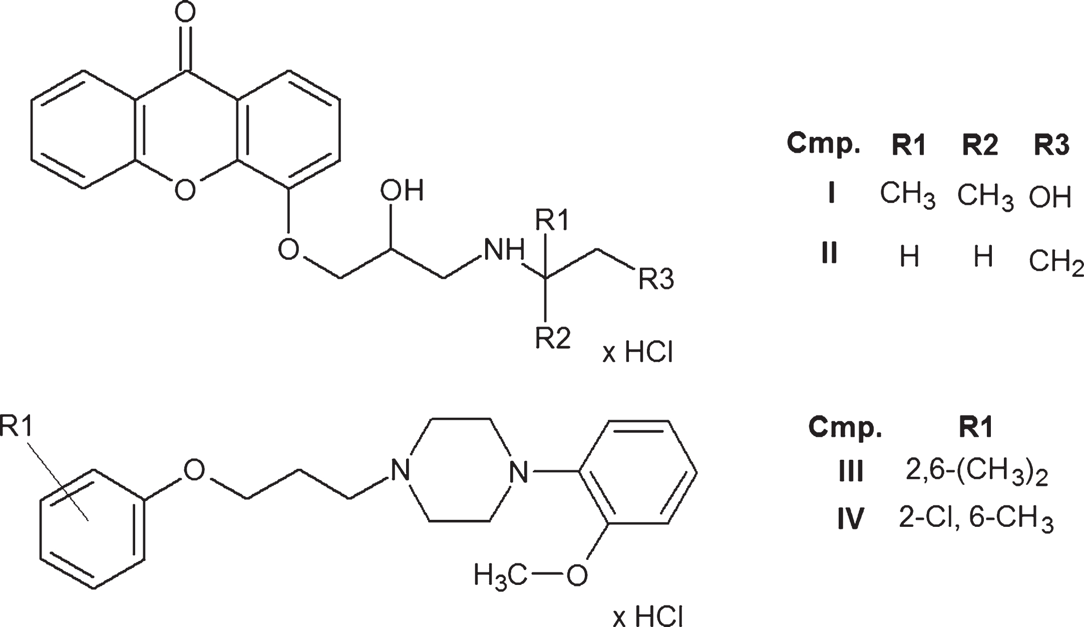
Chemical structures of compounds I-IV.
A series of both xanthone and piperazine derivatives were synthesized previously [22, 23] and examined with respect to their potential cardiovascular activity (antiarrhythmic and hypotensive activity) [22–26]. The most promising compounds (I-IV) were directed to further in vitro rheological studies. Among selected compounds there are two xanthone derivatives (I, II) possessing aminoalcanolic moieties, a well defined structural element of β-blockers. In previous studies compound II exhibited antiarrhythmic activity and β1-adrenoceptor affinity in rats [22, 26]. Compounds III and IV, belonging to derivatives of 1,4-disubstituted piperazine displayed significant affinity for α1-adrenoceptors with antagonistic properties confirmed in binding tests, functional bioassays and in vivo experiments [23, 24]. Moreover, compounds III and IV showed strong antiarrhythmic effects both in vitro and in vivo [25].
Blood samples
Whole blood was collected from healthy participants (n = 20; mean age 40±15 years) and CVD patients with varicosis (n = 10; mean age 45±15 years) into tubes containing EDTA as anticoagulant (1.6 mg/ml). The patients were rated as having lesions of CVD levels II and III according to the CEAP (clinical, etiological, anatomical and pathological elements) clinical classification. The study enrolled patients who attended 2-nd Chair of General Surgery Jagiellonian University Medical College in Krakow for the management of venous disease. The diagnosis of primary varicose vein was based on the clinical and duplex scanning examination. The patients had a positive family history of CVD and the symptoms of the disease were observed for more than one year. Moreover, the patients presented normal range of plasma fibrinogen. The subjects were not allowed to take any medication two weeks preceding blood withdrawal. Blood was sampled from antecubital veins of healthy subjects and from both antecubital veins and varicose veins of CVD patients. The Commission of Bioethics of the Jagiellonian University approved the research proposal (No. KBET/47/B/2007).
Test compounds
Test compounds (I-IV) were designed and synthesized in the Department of Bioorganic Chemistry Chair of Organic Chemistry Faculty of Pharmacy Jagiellonian University Medical College in Krakow and described previously [22, 23]. All compounds were dissolved in phosphate buffered saline (PBS) as stock solutions and added to the samples up to a desired final concentration of 100 μM and 10 μM.
Incubation with test compounds
Blood samples were centrifuged (1400×g, 5 min, 4°C) to separate RBCs. Then, blood plasma was removed and the remaining erythrocytes were washed three times with PBS. Subsequently, RBCs were resuspended in autologous plasma at a hematocrit of 40%. Next, RBCs suspensions were divided into aliquots and exposed to test compounds (final concentration of 100 μM and 10 μM) at 37°C for 30 min in constant agitation. Control samples received PBS only. After the incubation RBCs morphology was assessed microscopically (Leica, Japan). Then, RBCs deformability, aggregation as well as Ache, G6PD and GR activity and GSH content were evaluated. All measurements were completed within 4 h of blood sampling.
Deformability measurement
RBCs deformability was determined with Laser-assisted Optical Rotational Cell Analyser (LORCA, Mechatronics, The Netherlands). 0.025 ml of blood was diluted 200 times in a solution of 0.14 mM polivinylpyrrolidone (PVP, osmotic pressure 300 mOsm/kg, viscosity 30 mPa, Sigma) in PBS. Elongation Index (EI) was the parameter used to express RBCs deformability at increasing shear stress (0.30–59.97 Pa). EI was calculated using the formula: EI = (A–B)/(A+B), where A and B are the vertical and horizontal axes of the ellipse, respectively. The higher the value of EI, the deformation of blood cells is greater [27].
Aggregation measurement
Erythrocytes aggregation was examined using LORCA system described above. Firstly, 1 ml of blood was oxygenated for 15 min through the slow rotation in a glass vessel. The aggregation measurement was based on the detection of laser back-scattering from the sheared (disaggregated) to unsheared (aggregated) blood. RBCs aggregation parameters were determined with a syllectogram, which is a curve illustrating the change in the light intensity of scattered light during 120 s corresponding to the process of aggregation. The instrument provided the following parameters of erythrocyte aggregation: aggregation index (AI), threshold shear rate (THR) and aggregation halftime (t1/2), which correspond to the extent of RBCs aggregation, the tendency towards the formation of aggregates and of their stability and aggregation kinetics, respectively. The increase in THR provides information on the enhanced tendency towards aggregates formation and of their stability, whereas t1/2 decrease suggests the faster rate of the aggregation process [27].
Ache, G6PD and GR activity and GSH content assessment
Activities of three enzymes, namely Ache, G-6-PD, and GR as well as GSH content and haemoglobin concentration were assessed spectrophotometrically according to the methods described by Beutler [28].
Statistical analysis
Results are given as means ±SD of n experiments. Comparisons of deformability, aggregation, enzymes activity and GSH content for the investigated compounds were made by statistical one-way analysis of variance (ANOVA), followed by Dunnett’s test, p < 0.05 was considered significant. Tests were performed using GraphPad Prism 5.0 software.
Results
RBCs deformability
In vitro studies showed that compounds I, III and IV at both concentrations increased RBCs elongation significantly (Figs. 2, 4 and 5). Graphs depict only those shear stress values for which significant EI changes were observed.
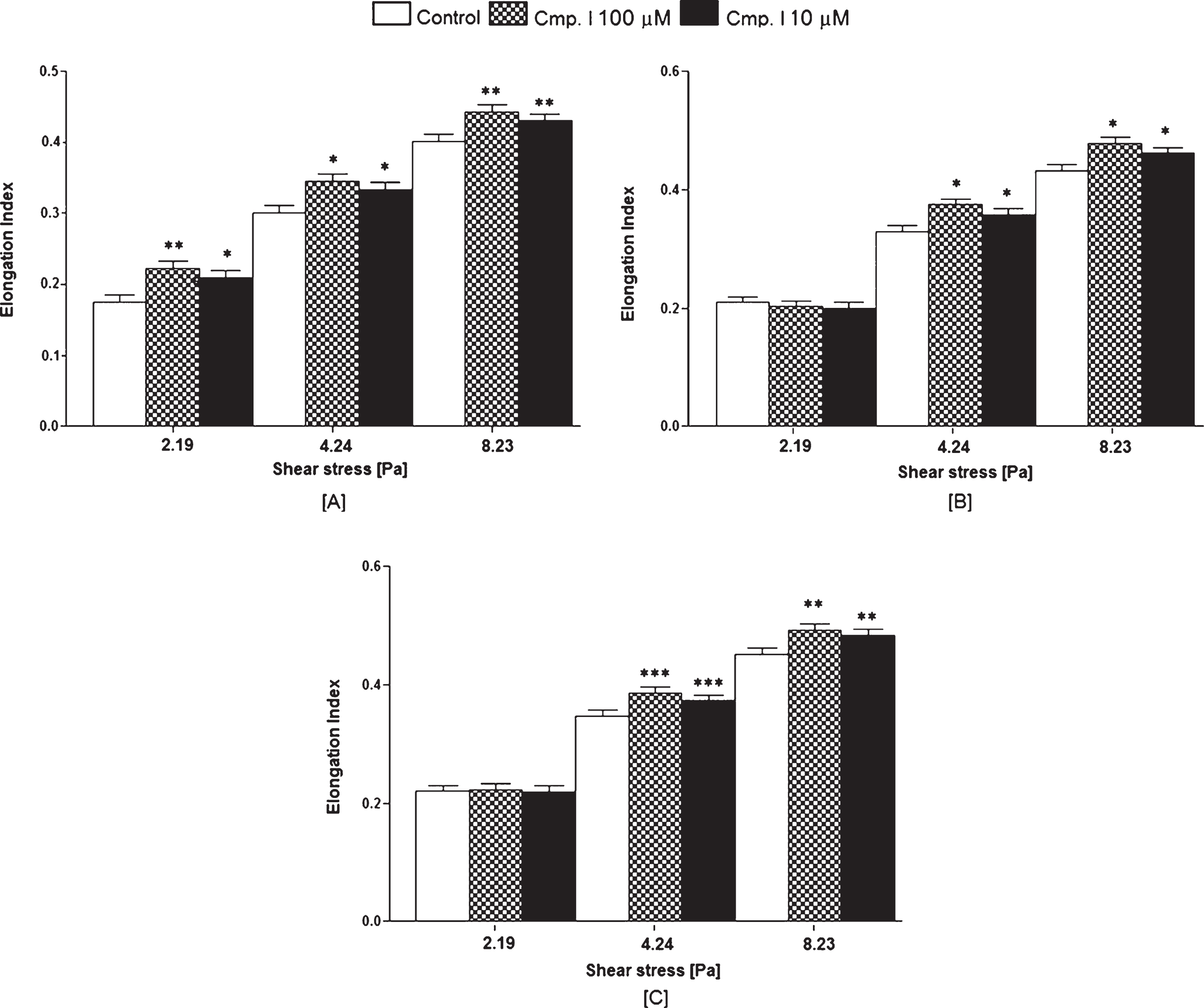
Effect of compound I on RBCs deformability; [A] – healthy adults RBCs, antecubital vein, [B] – CVD patients RBCs, antecubital vein, [C] – CVD patients RBCs, varicose vein (mean ±SD of n = 10). Significance vs. control: *p < 0.05, **p < 0.01 and ***p < 0.001.
In case of compound I during its incubation with healthy adults erythrocytes, a marked improvement of EI was found for shear stresses: 2.19 (+27.4% at100 μM, p < 0.01; +19.4% at 10 μM, p < 0.05), 4.24 (+14.6% at 100 μM, p < 0.05; +10.6% at 10 μM, p < 0.05) and 8.23 Pa (+10.5% at 100 μM, p < 0.01; +7.2% at 10 μM, p < 0.01) (Fig. 2A). In case of RBCs from antecubital vein of CVD patients EI increase was observed for shear stress values: 4.24 (+13.7% at 100 μM, p < 0.05 and +8.5% at 10 μM, p < 0.05) and 8.23 Pa (+10.7% at 100 μM, p < 0.05 and +6.7% at 10 μM, p < 0.05) (Fig. 2B). In RBCs from varicose vein of CVD patients compound I enhanced EI for shear stresses: 4.24 (+11.6% at 10 μM, p < 0.001 and +7.5% at 10 μM, p < 0.001) and 8.23 Pa (+9.1% at 100 μM, p < 0.01 and +7.1% at 10 μM, p < 0.01) (Fig. 2C). Compound II did not modify erythrocytes deformability significantly in all tested RBCs types (Fig. 3).

Effect of compound II on RBCs deformability; [A] – healthy adults RBCs, antecubital vein, [B] – CVD patients RBCs, antecubital vein, [C] – CVD patients RBCs, varicose vein (mean±SD of n = 10). Significance vs. control: *p < 0.05, **p < 0.01 and ***p < 0.001.
Compound III incubation with healthy adults RBCs resulted in a significant improvement of RBCs elongation index for shear stress values: 4.24 (at 100 μM +14.3% vs. control, p < 0.01; at 10 μM +9.3% vs. control, p < 0.01), 8.23 (at 100 μM +11.2% vs. control, p < 0.01; at 10 μM +7.0% vs. control, p < 0.01) and 15.96 Pa (at 100 μM +9.6% vs. control, p < 0.05; at 10 μM +6.3% vs. control, p < 0.05) (Fig. 4A). In case of RBCs from antecubital vein of CVD patients EI increase was found for shear stress values: 4.24 (+12.5% at 100 μM, p < 0.001 and +8.5% at 10 μM, p < 0.001) and 8.23 Pa (+8.6% at 100 μM, p < 0.01 and +6.0% at 10 μM, p < 0.01) (Fig. 4B). In RBCs from varicose vein of CVD patients EI increase was noted for shear stresses: 4.24 (+11.3% at 100 μM, p < 0.01 and +7.8% at 10 μM, p < 0.01) and 8.23 Pa (+10.0% at 100 μM, p < 0.01 and +6.9% at 10 μM, p < 0.01) (Fig. 4C).
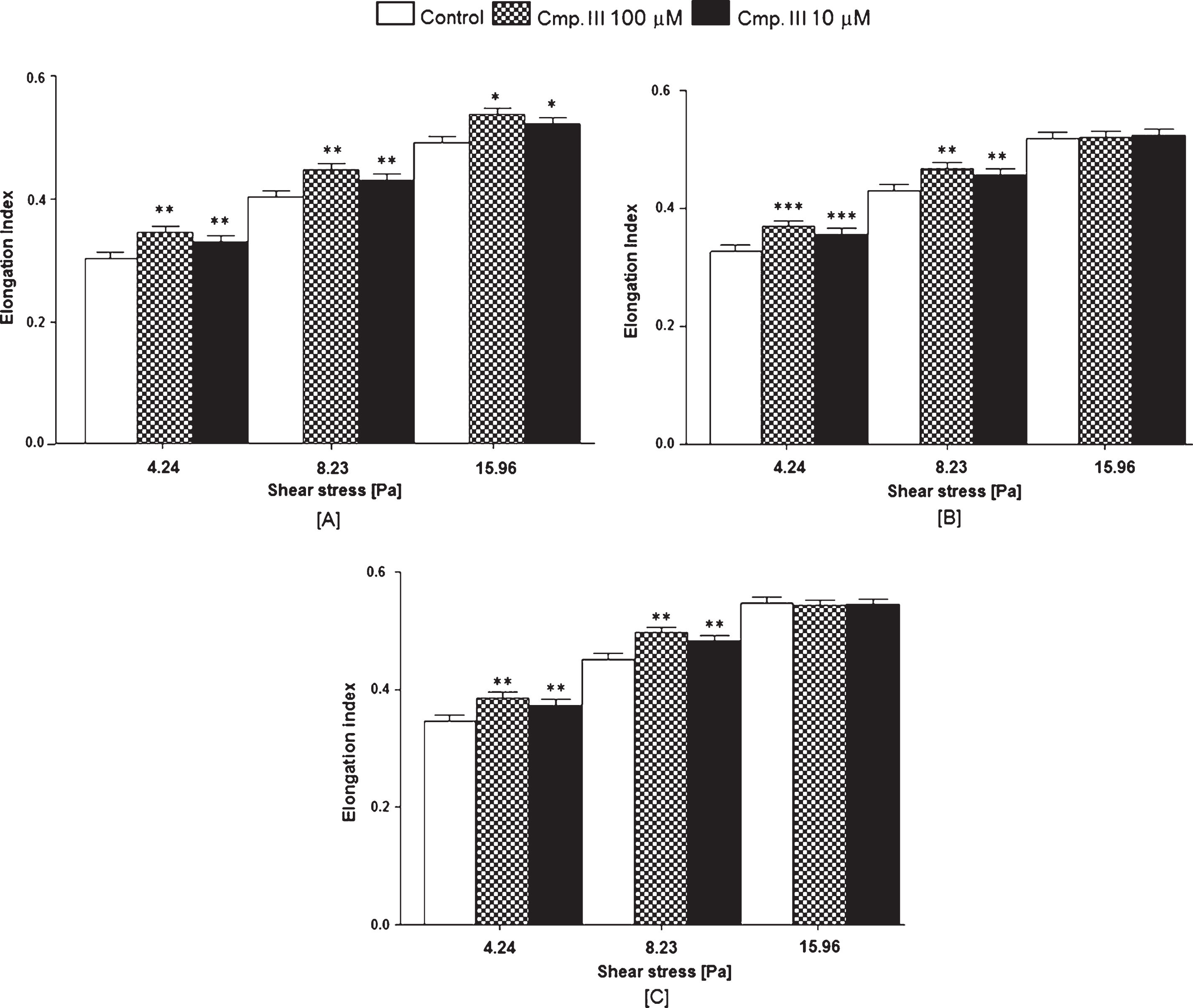
Effect of compound III on RBCs deformability; [A] – healthy adults RBCs, antecubital vein, [B] – VD patients RBCs, antecubital vein, [C] – CVD patients RBCs, varicose vein (mean ±SD of n = 10). Significance vs. control: *p < 0.05, **p < 0.01 and ***p < 0.001.
Compound IV in healthy adults RBCs produced a marked effect of EI for shear stresses: 4.24 (at 100 μM +15.0% vs. control, p < 0.01; at 10 μM +9.3% vs. control, p < 0.01), 8.23 (at 100 μM +10.0% vs. control, p < 0.01; at 10 μM +7.2% vs. control, p < 0.01) and 15.96 Pa (at 100 μM +7.4% vs. control, p < 0.05; at 10 μM +5.7% vs. control, p < 0.05) (Fig. 5A). In RBCs from antecubital vein of CVD patients an increase was observed for shear stress values: 4.24 (+11.3% at 100 μM, p < 0.01 and +8.8% at 10 μM, p < 0.01) and 8.23 Pa (+9.5% at 100 μM, p < 0.001 and +6.3% at 10 μM, p < 0.001) (Fig. 5B). In RBCs from varicose vein of CVD patients an increase was demonstrated for shear stresses: 4.24 (+11.9% at 100 μM, p < 0.01 and +8.4% at 10 μM, p < 0.01) and 8.23 Pa (+9.3% at 100 μM, p < 0.01 and +6.4% at 10 μM, p < 0.01) (Fig. 5C).

Effect of compound IV on RBCs deformability; [A] – healthy adults RBCs, antecubital vein, [B] – CVD patients RBCs, antecubital vein, [C] – CVD patients RBCs, varicose vein (mean ±SD of n = 10). Significance vs. control: *p < 0.05, **p < 0.01 and ***p < 0.001.
Considering aggregation measurement only, compounds I and II improved RBCs aggregation significantly (Table 1). In vitro treatment of healthy adults and CVD patients erythrocytes with compound I caused a marked decrease of AI (healthy adults: –20.2% at 100 μM, p < 0.05 and –14.7% at 10 μM, p < 0.05; CVD patients – antecubital vein: –16.6% at 100 μM, p < 0.05 and –12.9% at 10 μM, p < 0.05; CVD patients – varicose vein: –15.5% at 100 μM, p < 0.05 and –7.2% at 10 μM, p < 0.05). The decrease in THR was observable after healthy adults and CVD patients RBCs treatment with both concentrations of compound I (healthy adults: –11.0% at 100 μM, p < 0.05 and –8.4% at 10 μM, p < 0.05; CVD patients – antecubital vein: –22.3% at 10 μM, p < 0.01 and –11.8% at 10 μM, p < 0.05; CVD patients – varicose vein: –25.1% at 100 μM, p < 0.01 and –18.6% at 10 μM, p < 0.05). In case of t1/2 a significant effect of this parameter was observed after RBCs treatment with both concentrations of compound I only in RBCs from varicose vein of CVD patients (+21.9% at 100 μM, p < 0.05 and +6.3% at 10 μM, p < 0.05) (Table 1).
Effect of compounds I-IV on RBCs aggregation parameters: aggregation index (AI), threshold shear rate (THR), aggregation halftime (t1/2); A – healthy adults RBCs, antecubital vein; B – CVD patients RBCs, antecubital vein; C – CVD patients RBCs, varicose vein
Effect of compounds I-IV on RBCs aggregation parameters: aggregation index (AI), threshold shear rate (THR), aggregation halftime (t1/2); A – healthy adults RBCs, antecubital vein; B – CVD patients RBCs, antecubital vein; C – CVD patients RBCs, varicose vein
Data are presented as mean ±SD, n = 10. Difference from control: *p < 0.05, **p < 0.01 and ***p < 0.001.
It was observed that incubation of healthy adults and CVD patients erythrocytes with compound II caused a marked decrease of AI (healthy adults: –23.6% at 100 μM, p < 0.01 and –19.0% at 10 μM, p < 0.05; CVD patients – antecubital vein: –21.7% at 100 μM, p < 0.05 and –12.7% at 10 μM, p < 0.05; CVD patients – varicose vein: –24.6% at 100 μM, p < 0.001 and –13.9% at 10 μM, p < 0.05). The decrease in THR was observable after healthy adults and CVD patients RBCs treatment with both concentrations of compound II (healthy adults: –9.2% at 100 μM, p < 0.05 and –7.2% at 10 μM, p < 0.05; CVD patients – antecubital vein: –21.1% at 100 μM, p < 0.05 and –11.4% at 10 μM, p < 0.05; CVD patients – varicose vein: –22.0% at 100 μM, p < 0.05 and –16.2% at 10 μM, p < 0.05). In case of t1/2 parameter a significant effect was noticed after RBCs treatment with compound II at 100 μM only in RBCs from varicose vein of CVD patients (+28.1% at 100 μM, p < 0.05) (Table 1). Compounds III and IV did not alter RBCs aggregation significantly (Table 1).
The study showed that compound II and III significantly increased Ache activity. Compound II at the concentration of 100 μM increased enzyme activity by 29.2% (p < 0.01), whereas at the concentration of 10 μM by 16.9% (p < 0.05). Compound III in higher concentration augmented Ache activity by 24.0% (p < 0.01), whereas in lower concentration by 13.9% (p < 0.05). Compounds I and IV did not affect significantly Ache activity (Table 2).
Effect of compounds I-IV on healthy adults RBCs: acetylcholinesterase (Ache), glucose-6-phosphate dehydrogenase (G6PD) and glutathione reductase (GR) activity, and glutathione (GSH) content
Effect of compounds I-IV on healthy adults RBCs: acetylcholinesterase (Ache), glucose-6-phosphate dehydrogenase (G6PD) and glutathione reductase (GR) activity, and glutathione (GSH) content
Data are presented as mean ±SD, n = 10. Difference from control: *p < 0.05, **p < 0.01 and ***p < 0.001.
It was demonstrated that incubation of healthy adults RBCs with compounds I, II and IV resulted in an increase in the activity of G6PD when compared with control. Compound I at higher concentration increased enzyme activity by 21.7% (p < 0.01) and by 10.8% (p < 0.01) at lower concentration. Compound IV augmented G6PD activity by 15.7% (p < 0.01) and by 13.3% (p < 0.01) at 100 μM and at 10 μM, respectively. After RBCs treatment with compound II, enzyme activity was increased only at higher concentration by 18.1% (p < 0.01). Compound III did not modify significantly G6PD activity (Table 2).
Healthy adults RBCs treatment with compounds I and II resulted in a marked increase in GR activity in comparison with the control group. Conversely, compound IV led to a significant decrease in GR activity. In case of compound I in higher concentration the enzyme activity was increased by 26.1% (p < 0.05). Compound II augmented GR activity by 35.1% at 100 μM and by 27.9% at 10 μM. Compound IV at higher concentration led to a decrease in GR activity by 14.4% (p < 0.05). Compound III had no effect on GR activity (Table 2).
Finally, with respect to GSH, compounds I and II led to a marked increase in GSH content, whereas compound IV significantly reduced this parameter. At the concentration of 100 μM compound I increased GSH level by 14.1% (p < 0.01). Compound II increased GSH level by 38.4% (p < 0.01) and 22.0% (p < 0.01), at higher and lower concentration, respectively. In contrast, compound IV at the concentration of 100 μM diminished GSH level by 11.4% (p < 0.05) and at the concentration of 10 μM by 10.3% (p < 0.05). Compound III had no effect on this parameter (Table 2).
Rheological factors including RBCs deformability and aggregation are important determinants of blood flow and tissue perfusion. In the present study we compared RBCs rheological behavior of CVD patients (antecubital vein and varicose vein) and healthy subjects (antecubital vein) at baseline and after in vitro treatment with four (I-IV) cardiovascularly active compounds. Two of them are xanthone derivatives, whereas another two belong to appropriate piperazine derivatives. Moreover, the influence of test compounds on RBCs Ache, G6PD and GR activity and GSH content were determined with healthy controls erythrocytes.
Circulating erythrocyte aggregates are dissolved by shearing usually in the precapillary arterioles where the shear rates are the highest in the normal circulation [6]. The study showed that compounds I, III and IV significantly increased RBCs deformability when there was less pressure on RBCs (shear stress between 2.19–15.96 Pa). Such changes were not, however, noted for higher shear stress levels. This is mainly due to the fact that various RBCs deformability abnormalities and improvements may be visible only at particular shear rates levels. Interestingly, in case of RBCs from CVD patients this beneficial modification was observed for two shear stress values, whereas in healthy controls RBCs this effects was found in three shear stress levels.
As regards RBCs aggregation, both xanthone derivatives positively altered this process, this was seen by analyzing both AI and THR indices. An observed decrease in AI may suggest that erythrocytes aggregates formation was slowed. Compound I reduced RBCs aggregation in all RBCs groups but this reduction was greater in healthy controls. This tendency was not seen for compound II, which at the higher concentration comparably diminished RBCs aggregation both in healthy controls and patients with CVD. THR parameter reflects red cells ability to aggregation and aggregates stability. An increase in this factor suggests that observed RBCs aggregates are stable and its difficult to break them in the circulation. Based on THR analysis it can be noticed that both xanthone derivatives diminished RBCs aggregates strength in the three groups of erythrocytes but with a higher magnitude in RBCs from varicose vein of CVD patients.
Positive influence of xanthone derivatives on RBCs aggregation can be also seen based on the analysis of t1/2, which reflects aggregation kinetics. The growth of this parameter argues for a reduction in the rate of formation of RBCs aggregates. Compound I caused a marked decrease in the rate of RBCs aggregates formation in red cells from varicose veins. Interestingly, these particular cells were characterized by the highest rate of aggregation process in comparison with cells from the antecubital vein blood of patients and healthy donors. Compound II also reduced the rate of aggregates formation in varicose veins erythrocytes, but only at higher concentration tested.
According to Muravyov et al. [13] drugs may affect RBCs rheological behavior in three ways: neutral effect is related to compounds that do not exert significant effect on red cells deformability and aggregation; negative effect is observed when an agent diminishes RBCs deformability and increases their aggregation; and finally, positive effect is stated when a drug increases erythrocytes deformability and decreases their aggregation. Thus, one of the tested xanthone derivatives (I) may be classified as agent positively influencing RBCs rheological properties, whereas other compounds were able to positively influence only one RBCs rheological property such as compound II which had beneficial effect only on RBCs aggregation with no effect on RBCs deformability. None of the compounds had negative effect on RBCs haemorheological behavior. Such differences in haemorheological properties of compounds I-IV may be attributed primarily to the chemical structure of compounds.
Erythrocyte acetylcholinesterase (Ache) associated with cell membrane is one of the essential proteins that ensures the membrane integrity. Ache location on the outer side of RBCs membrane makes it susceptible to the action of various factors present in the circulation, particularly those which may affect the structure of the membrane. The enzyme is regulated by both the membrane lipid environment, as well as a variety of phenomena occurring at the cell surface [29]. Ache hydrolyzes acetylcholine, that is able to alter red cells rheology [27]. Among four compounds examined within the present work, only compounds II and III significantly improved Ache activity in healthy controls RBCs, and may therefore exert positive influence on the integrity of RBCs membrane. Compound III enhanced RBCs deformability, whereas compound II inhibited RBCs aggregation and facilitated the breakage of existing RBCs aggregates using in vitro conditions. This has led to speculation that the beneficial effect of these two compounds on RBCs rheology may, in part, be derived from elevation in Ache activity. On the other hand, compound I which positively affected both deformability and aggregation of RBCs did not, influenced Ache activity. Nowak et al. [30] who evaluated in vitro the influence of toxic 2,3,7,8-tetrachlorodibenzo-p-dioxin (TCDD) on RBCs deformability and erythrocytes Ache activity, observed that TCDD treatment resulted in a decrease in enzyme activity, and in the same time no changes in RBCs deformability were stated. Thus, membrane Ache may be more sensitive marker than erythrocytes deformability indices for compounds unfavourable effect on RBCs membrane.
Reactive oxygen species (ROS) and oxidative stress can negatively affect erythrocytes deformability and aggregation, mainly due to their influence on membrane cytoskeletal proteins and lipids, surface properties and cation permeability [31]. Therefore, when analyzing compounds action on RBCs rheological properties, an important aspect of further studies may be to assess the impact of these substances on some antioxidant parameters. In the present study in vitro effect of compounds I-IV on G6PD and GR activity as well as GSH content were determined in vitro in RBCs from healthy subjects. G6PD catalyses the first reaction of pentosephosphate cycle. This reaction is the major source of NADPH that is used by GR. GR is involved in the reduction of oxidized glutathione (GSSG) to its reduced form (GSH) [32]. The above mentioned reactions play crucial role in the proper functioning of RBCs membrane, as they protect the thiol groups of membrane proteins from oxidation [33].
The study demonstrated that compounds I and II significantly increased G6PD activity (compound II only at concentration of 100 μM), GR (compound I only at concentration of 100 μM) and GSH level (compound I only at the higher concentration). In the same time RBCs treatment with these compounds led to a decrease in RBCs aggregation, and the resulting blood cell aggregates underwent rapid disaggregation. Additionally, compound I enhanced RBCs deformability. We can therefore suggest that compound I effect on RBCs deformability may be partially explained by its effect on antioxidant parameters. Similar observations were made by Lipovac et al. [34], who stated that rheological effect of creatinine is, at least in part, related to its antioxidant activity. This is mainly due to the fact that efficient antioxidant defense mechanisms ensure the proper functioning of RBCs membranes and are one of the most important factors determining the proper formability and the interaction of cells with each other [35–37]. Compound IV significantly increased G6PD activity, and in the same time reduced GR activity (at the higher concentration) and GSH level. This compound had no marked effect on RBCs aggregation, however, it significantly increased erythrocytes deformability in vitro. The latter effect may not directly result from compound IV antioxidant effect, but may involve additional mechanisms of action, which would require further study. As compound IV decreased GR activity and GSH levels while improving RBCs deformability, it is expected that the unfavourable effect of compound IV on antioxidant defense is likely to be compensated by the cells and apparently does not interfere with the proper functioning of the cell membrane.
Conclusions
Summing up, all these results could indicate that pharmacological agents may significantly affect mechanical properties of RBCs. Compounds I, III and IV, which significantly improved RBCs deformability in vitro, may facilitate the passage of blood in the vascular system, and particularly in the microcirculation. Additionally, compounds I and II which inhibit RBCs aggregates formation in vitro may contribute to more rapid degradation of red blood cell aggregates in circulating blood. Particularly interesting seems to be compound I, which had beneficial influence both on RBCs deformability and aggregation. Although in some cases the observed changes in RBCs deformability and aggregation were slight, these variations could be significant in pathologies such as chronic venous disease, where an alternation in RBCs haemorheological parameters has been reported.