
Abstract
BACKGROUND:
Meniscus regeneration is observed within the peripheral, vascularized zone but decreases in the inner two thirds alongside the vascularization. Within this avascular area, cell-based tissue-engineering-approaches appear to be a promising strategy for the treatment of meniscal defects.
OBJECTIVE:
Evaluation of the angiogenic potential of cell-based tissue-engineering-products for meniscus healing.
METHODS:
Evaluation of angiogenesis induced by rabbit meniscus-pellets, meniscus-cells (MC) or mesenchymal stem-cells (MSC) in cell-based tissue-engineering-products within a rabbit meniscus-ring was performed using a transparent dorsal skin fold chamber in nude mice. Observations were undertaken during a 14 days period. Cell preconditioning differed between experimental groups. Immunohistochemical analysis of the regenerated tissue in the meniscus-ring induced by cell loaded composite scaffolds for differentiation and anti-angiogenic factors were performed.
RESULTS:
Meniscus-pellets and MSC-/MC-based tissue-engineering-products induced angiogenesis. An accelerated vascularization was detected in the group of meniscus-pellets derived from the vascularized zone compared to avascular meniscus-pellets. In terms of cell-based tissue-engineering-products, chondrogenic preconditioning resulted in significantly increased vessel growth. MSC-constructs showed an accelerated angiogenesis. Immunohistochemical evaluation showed a progressive differentiation and lower content for anti-angiogenic endostatin in the precultured group.
CONCLUSIONS:
Preconditioning of MC-/MSC-based tissue-engineering-products is a promising tool to influence the angiogenic potential of tissue-engineering-products and to adapt these properties according to the aimed tissue qualities.
Introduction
The meniscus has a decisive functional and biomechanical relevance for an intact knee joint [1, 2]. The importance of the intact meniscus for the load bearing, shock absorption, stability of the knee joint, lubrication and proprioception has already been documented in many clinical and experimental studies [3–6]. Injury to meniscus continuity negatively influences the biomechanics and function of the meniscus and predisposes the development of osteoarthritic changes [4, 7]. Thus, meniscus injuries have a high socioeconomic impact [4, 7].
However, the treatment of meniscus lesions is a challenging condition. Currently, the location of the meniscus defect influences treatment options [4, 7]. Endogenous meniscus regeneration is limited to the vascularized, peripheral zone [8]. Meniscal tears within the avascular zone show a poor endogenous regeneration potential. Thus, meniscus-preserving techniques are not suitable for tears within this zone. Hence, they are treated by partial meniscectomy, which enhances the risk of osteoarthritis [9].
One treatment approach is to utilize the healing potential of meniscus lesions within the vascular zone, to stimulate the angiogenesis and vascularization of meniscus tissue within the avascular zone. In a clinical approach, rasping and trephination are performed to induce perfusion within the defect site and enhance the healing potential of meniscus lesions within the vascularized area prior to suturing. Furthermore, the association of meniscus regeneration and vascularization has been documented by Verdonk et al., who described a complete vessel ingrowth up to the avascular meniscus apex, to demonstrate a successful meniscus replacement [10]. Further data describe a beneficial effect of cell-based strategies for the regeneration of meniscus tissue including the avascular zone. In this context, mesenchymal stem cells (MSCs) and meniscus cells (MCs) have been shown to improve the regeneration potential of meniscus tissue [5, 11–19].
However, there is no information about the influence of rabbit MSC- and MC- cell based tissue engineering products on angiogenesis during meniscus tissue healing. Therefore, the aim of this study was to investigate the angiogenic capacities of MSC- and MC-loaded tissue engineering products and see whether the angiotactic properties of these cell based tissue engineering products can be influenced by chondrogenic pre-cultivation in a nude mice dorsal skin fold chamber model.
Material and methods
Composite scaffolds
The composite scaffolds were manufactured from 70 % derivatized hyaluronan-ester and 30 % gelatin as described previously [20–22]. The hyaluronan component was obtained from the commercially available product Jaloskin (Fidia Advanced Biopolymers, Abano Terme, Italy), which is manufactured from hyaluronate, highly esterified with benzyl alcohol on the free carboxyl groups of glucuronic acid within the polymer. The gelatin component was hydrolyzed bovine collagen type I (Sigma, Taufkirchen, Germany) [20].
Porous scaffolds discs were manufactured by a mix of both solved components in combination with NaCl-crystals. After air drying salt was washed out. The porosity of the discs sized 350 –450 μm. Scaffolds had a diameter of 2.0 mm and a height of 1.3 mm. Before cell seeding scaffolds were sterilized with 25kGy beta rays (Beta-Gamma-Service GmbH, Saal, Germany).
Cells
The local government’s animal rights protection authorities approved this study in accordance with the National Institutes of Health guidelines for the use of laboratory animals (621-2531.1-18/02).
Bone marrow derived stem cells – harvest and culture
The bone marrow harvest and MSC cell isolation was performed as previously described [20, 23]. Bone marrow derived mesenchymal stem cells were harvested by puncture of the iliac crest of New Zealand White Rabbits on both sides and collected into a heparinized syringe. Dulbecco’s modified Eagle’s medium (DMEM), low glucose concentration, with 10 % fetal bovine serum, 1 % penicillin, and 1 % Hepes was added to the aspirate and nucleated cells (20×106) were plated in 75 cm2 culture dishes and cultivated at 37°C incubator. The medium was changed after one week of adhesion twice a week until the adherent cells (MSCs) reached 80 % confluence.
Meniscus derived cells – harvest and culture
Meniscus tissue was harvested from New Zealand White Rabbits. Following sacrifice of the rabbits, the whole menisci were carefully removed after surgical luxation of the knee joint. The complete meniscus was directly processed after explantation by mechanical dissociation with a maximum size of 1 mm3 and following enzymatical dissociation with collagenase (1 mg/ml), hyaluronidase (0.1 mg/ml) and deoxyribonuclease (0.15 mg/ml). RPMI 1640 medium with 10 % fetal calf serum, 1 % penicillin and 1 % Hepes was added to the isolated meniscus derived cells (MCs) to stop the enzymatical dissociation. Following centrifugation, viable nucleated cells (1×10⁶) were plated in 75 cm2 culture dishes and cultivated at 37 °C, 5 % CO2, 95 % air humidity. The medium was changed after one week adhesion twice a week until the adherent cells reached 80 % confluence. Cells were split at a 1:4 ratio, plated in 75 cm2 culture dishes and were cultivated as described above.
Cell seeding of the composite scaffolds
The hyaluronan collagen composite matrices were seeded with a pure cell concentrate (0.8×10⁶ nucleated cells) of MSCs or MCs formed in an aggregation culture medium (DMEM, high glucose concentration (4500 mg/l), with 10 % pyruvate, 10 % ITS, 10 % dexamethasone, 10 % TGFβ1 and 10 % ascorbic acid).
Depending on the group, the composite matrices were cultivated at 37°C, 5 % CO2, 95 % air humidity for the period of 1 or 14 days. In the “14 days group” the medium was changed three times a week.
Meniscus tissue - meniscus pellets and meniscus rings
The menisci were harvested from New Zealand White Rabbits as described above (see 2.2). Following explantation, the samples were aseptically stored in serum free DMEM high glucose medium at 37 °C, 5 % CO2, 95 % air humidity and proceeded directly before implantation.
Prior to implantation, fat and connective tissue was removed from the menisci and were cut axially into discs of 1 mm thickness.
The meniscus pellets were punched out of these discs with a 2 mm biopsy punch. The origin of the pellets out of the vascular or avascular zone of the meniscus were strictly differentiated and then implanted in the nude mice dorsal skin fold chamber.
To prepare a meniscus ring, a hole of 2 mm diameter was punched out of the disc by a 2 mm biopsy punch near to the avascular zone of meniscus discs. Afterwards, these rings were filled with different cell loaded composite scaffolds, one day before implantation in the nude mice dorsal skin fold chamber (see Fig. 1).
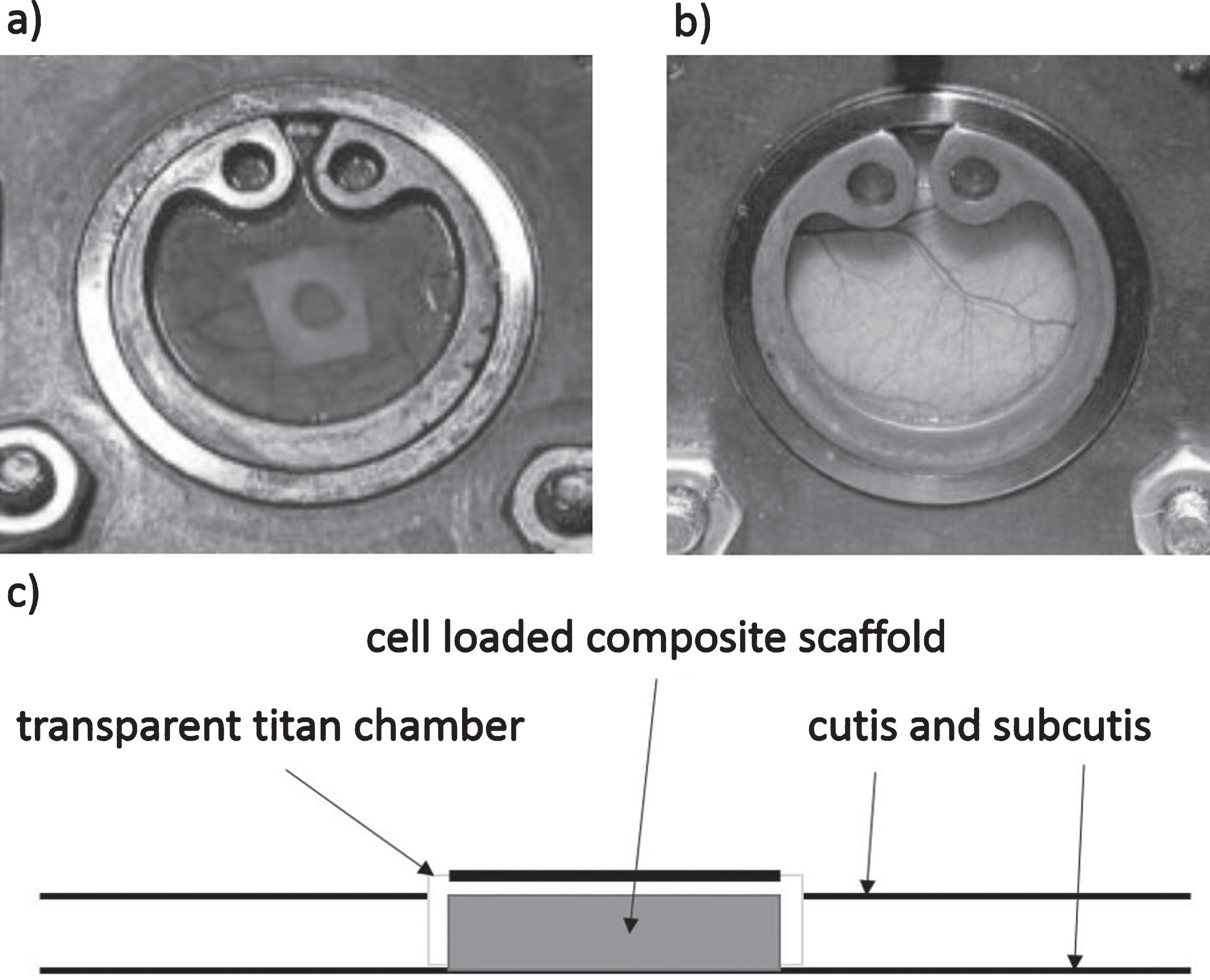
a + b) intravital view on a transparent titan dorsal skin fold chamber before and after sample loading with a meniscus ring (4x enlargement); c) schematic profile of the transparent titan dorsal skin fold chamber.
The composite matrices were seeded with the isolated MCs and MSCs. According to the subsequent treatment (1 vs. 14 days culturing) the cell based tissue engineering products were matched to the precultured group and the non-precultured group (see Table 1).
Description of the different treatment groups
Description of the different treatment groups
The meniscus pellets were differentiated according to their origin within the vascularized and avascular meniscus (see Table 1).
Isolation of mouse erythrocytes was performed using venous blood samples harvested by cannulation of the vena cava inferior from Balb/c nu/nu mice and mice were sacrificed afterwards. Following centrifugation of venous blood samples and a PBS wash, the harvested erythrocytes were labelled with the fluorescent marker PKH67 (Sigma) according to manufacturer’s instructions.
Intravital microscopy – the dorsal skin fold chamber model
In vivo evaluation of the angiogenesis of tissue engineering products was performed using intravital microscopy. This new procedure in orthopedic research is based on cancer and microcirculation research [24–27]. The transparent titan dorsal skin fold chamber is implanted into the spine of immunodeficient male Balb/c nu/nu nude mice. The dorsal skin fold chambers were filled with MSC- and MC-cell based tissue engineering products (as shown in Fig. 1). Following fixation of the mice, the development of angiogenesis was detected and documented via the transparent dorsal skin fold chamber using a microscope (Zeiss Axiotech, Zeiss, Jena, Germany) and SHVS video recorder (MD 835, Panasonic, Hamburg, Germany).
Mean vascular density
The neo-angiogenesis within the implanted composite scaffolds was documented on the basis of the development of vessels. The development of the vasculature and the vascular density were calculated based on the intravital microscopical documentation at 7, 10 and 14 days after implantation. The maximal vessel length and vascular density was measured using ImageJ software (ImageJ 1.34 s, Wayne Rasband, National Institutes of Health Bethesda, USA) (see Figs. 2 and 4).
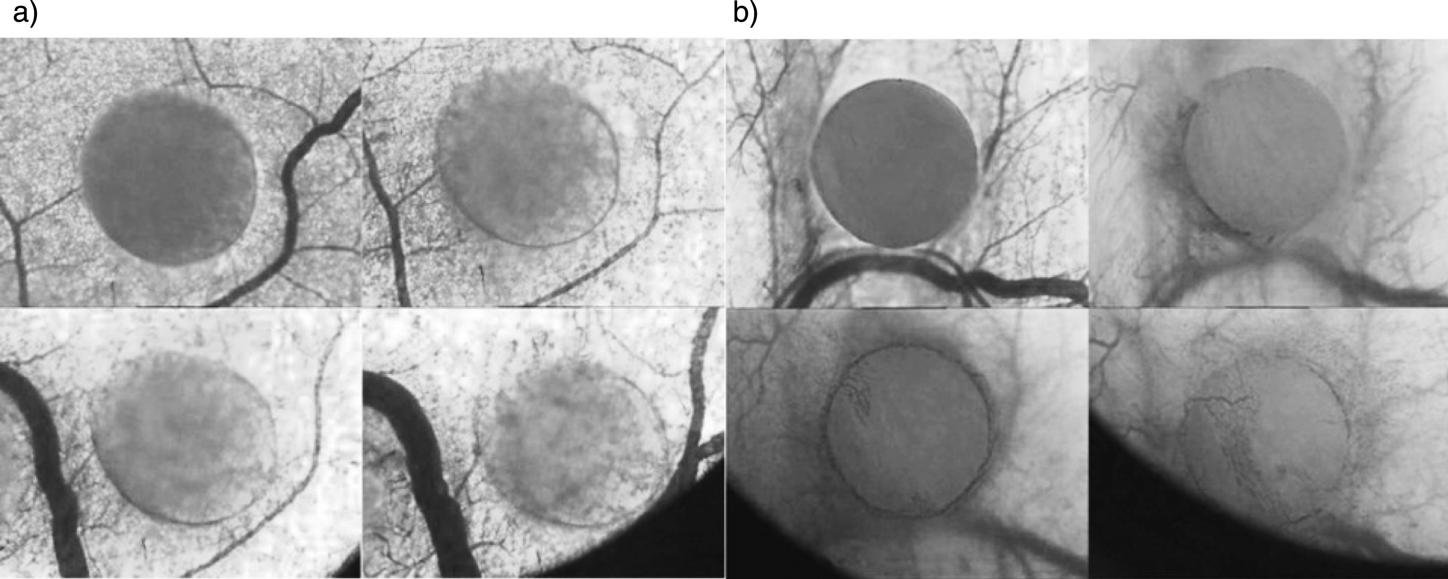
Intravital microscopically view (10x enlargement) on meniscus pellets inside of the transparent titan dorsal skin fold chamber at day 0 top left and clockwise at day 7, 10 and 14. a) shows meniscus pellets out of the avascular zone; b) shows meniscus pellets out of the vascular zone.
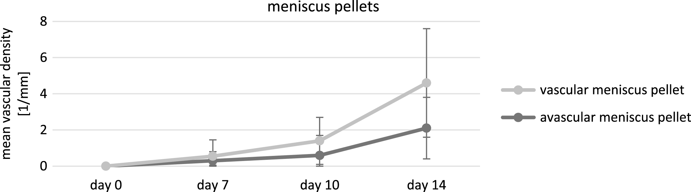
Mean vascular density induced by the meniscus pellets out of the vascular and avascular zone at day 0, 7, 10, 14 calculated in relation to the area of the pellets. There are no significant differences until day 14.
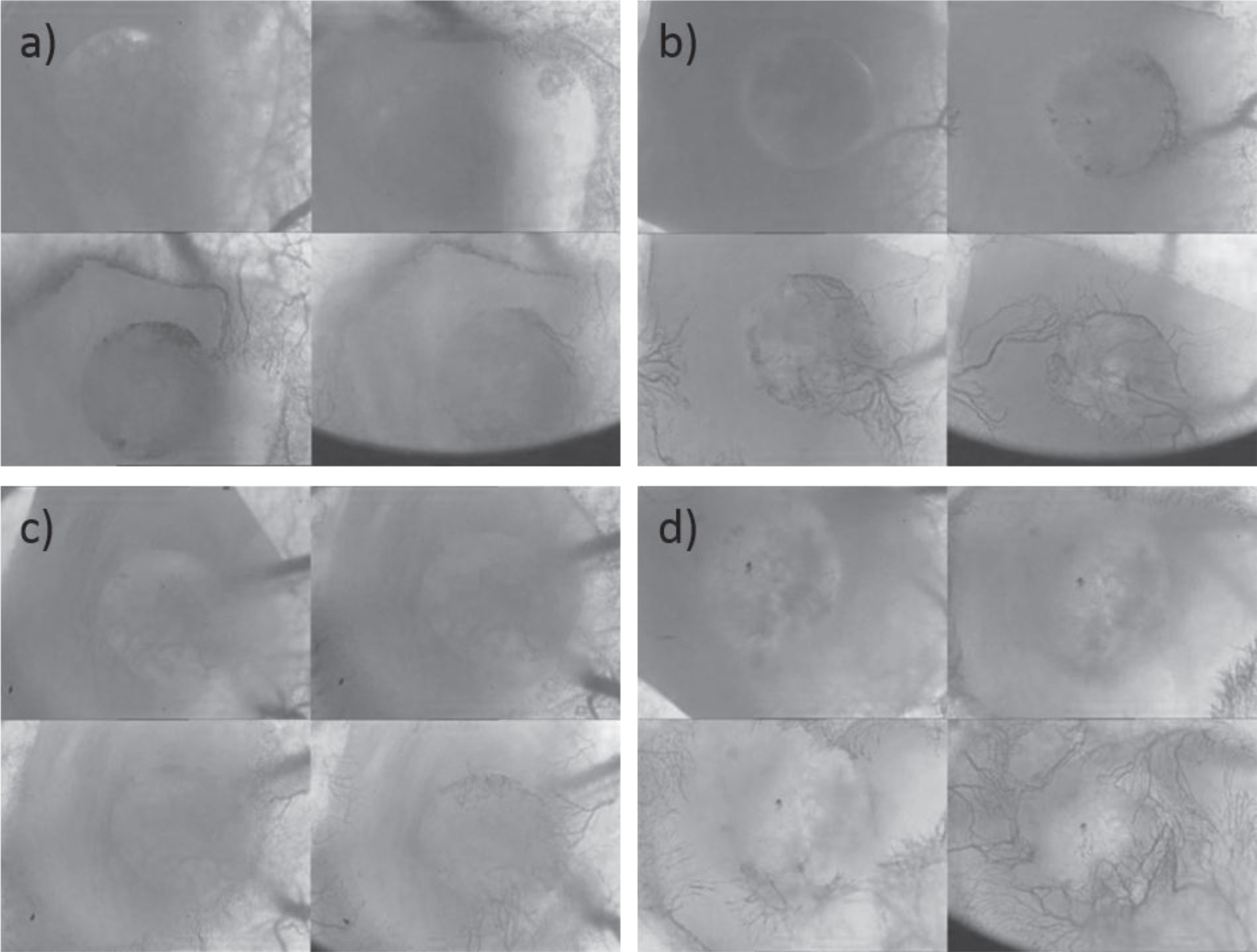
Intravital microscopically view (10x enlargement) on meniscus rings loaded with MSC tissue engineering products a) and b) as well as MC tissue engineering products c) and d) inside of the transparent titan dorsal skin fold chamber at day 0 top left and clockwise at day 7, 10 and 14. a) and c) show meniscus rings with one day precultured tissue engineering products; b) and d) show meniscus rings with 14 days precultured tissue engineering products.
The functionality of the newly developed vessels was evaluated by the detection of fluorescently labelled erythrocytes. Labelled erythrocytes (n = 100×106) were injected into the tail vene of the Balb/c nu/nu mice. The perfusion of the implanted composite scaffolds was detected using a fluorescent microscope (Zeiss Axiotech, Zeiss, Jena, Germany) and a fluorescent light (EBQ 100 dc-z, Leistungselektronik Jena GmbH, Germany).
Histological analysis
Histological evaluation of neovascularization within the composite scaffolds was assessed on mice sacrificed after 14 days. The tissue engineering products as well as the surrounding meniscus and mouse tissue were harvested out of the dorsal skin fold chamber and were directly proceeded afterwards, except samples used for viability analysis. Following dehydration, the tissue samples were fixated in Tissue Tek (Sakura) and stored at –80°C. The histological cut (10 μm thickness) was performed by a cryostat cutter (Microm) and fixated on object plate before toluidine blue and diaminobenzidin staining were performed. Afterwards, the histological sections were analyzed microscopically (Nikon Eclipse TE 2000-U, Nikon, Düsseldorf, Germany).
Viability testing
The viability testing of tissue samples was performed using Live/ Dead Viability/ Cytotoxicity Kit (Molecular Probes, Eugene, USA). In contrast to histological analysis, the meniscus-matrix-scaffolds were directly proceessed. Prior to fixation in Tissue Tek (Sakura), the menisci-matrix-constructs were cultivated in a 4 μM calcein-am and 2 μM ethidiumbromid containing medium for 22 hours. Subsequently, the samples were processed and cut into 40 μm slices before fixation on an object plate. The dead negative controls were dipped into fluid nitrogen before handled in the same manner as described above. In fluorescent microscopy (Nikon Eclipse TE 2000-U, Nikon, Düsseldorf, Germany) calcein-am marks vital cells and stains these green. In contrast ethidiumbromid labels dead cells and signs these red.
Immunohistochemically analysis – endostatin
Endostatin is a decomposition product of collagen 18 and acts as a strong endogenous inhibitor of angiogenesis [28, 29]. Tissue samples were prepared as previously described. The endostatin staining was performed by adding a primary antibody Goat-Anti-Mouse-Endostatin-IgG (50 μg/ml) (R&D Systems, Minneapolis, USA) on the samples for a duration of 10 hours before a secondary antibody Donkey-Anti-Sheep-IgG 1:500 (1,2 mg/ml) (Dianova, Hamburg, Germany) was applied. Evaluation was performed microscopically (Nikon Eclipse TE 2000-U, Nikon, Düsseldorf, Germany).
Statistics
Statistical analysis was performed using SPSS software version 23.0 (SPSS, Chicago, IL, USA) to determine relationships between variables. The Mann-Whitney-U test was used for qualitative data analysis. The significance level was set at a P-value of 0.05. The histologically and immunohistochemically analysis were evaluated descriptively.
Results
Meniscus pellets
The meniscus pellets were grouped according to their vascular and avascular origin. The evaluation of the vascular density at different time points showed that both groups induced vascularization within 14 days with vascular density increasing over the time. At day 10 onwards, an accelerated growth could be detected for the vascular and avascular meniscus pellets. In total, the rate of growth did not differ significantly during the whole study period, although the vascular pellets showed a trend to work a stronger induction of vascularization (p = 0.07) (see Fig. 3 and Table 2).
Mean vascular density induced by meniscus pellets at day 0 / 7 / 10 and 14
Mean vascular density induced by meniscus pellets at day 0 / 7 / 10 and 14
The matrix composite scaffolds were seeded with MSCs and MCs before implantation into the meniscus rings. The scaffolds differed concerning the used cells (MSCs/ MCs) and the state of preconditioning (1 or 14 days). All groups induced angiogenesis within 14 days, but they varied in their growth rate and the extent of growth. The highest rate of growth showed the group of pre-cultured MSCs (MSC +). In this group, a significantly accelerated start of angiogenesis was detected at day 7 in comparison to the non-pre-cultured MSCs (MSC –) (p = 0.02) and pre-cultured MCs (MC +) (p = 0.01). At subsequent time points, the significantly enhanced growth of vessels remained compared to the non-precultured group (day 10: p = 0.0.1; day 14: p = 0.004). In contrast, the group of pre-cultured MCs had a similar mean vascular density at day 14 (p = 0.8). The comparison of the non-pre-cultured and pre-cultured MCs showed significant differences at day 10 (p = 0.002) and day 14 (p = 0.001). Besides the accelerated growth rate of pre-cultured MSCs in comparison to pre-cultured MCs, the group of non-precultured MSCs showed an earlier angiogenic boost compared to the non-precultured MCs (day 10: p = 0.005). Similar to the precultured groups, the non precultured groups showed no significant differences at day 14 (see Fig. 5 and Table 3).
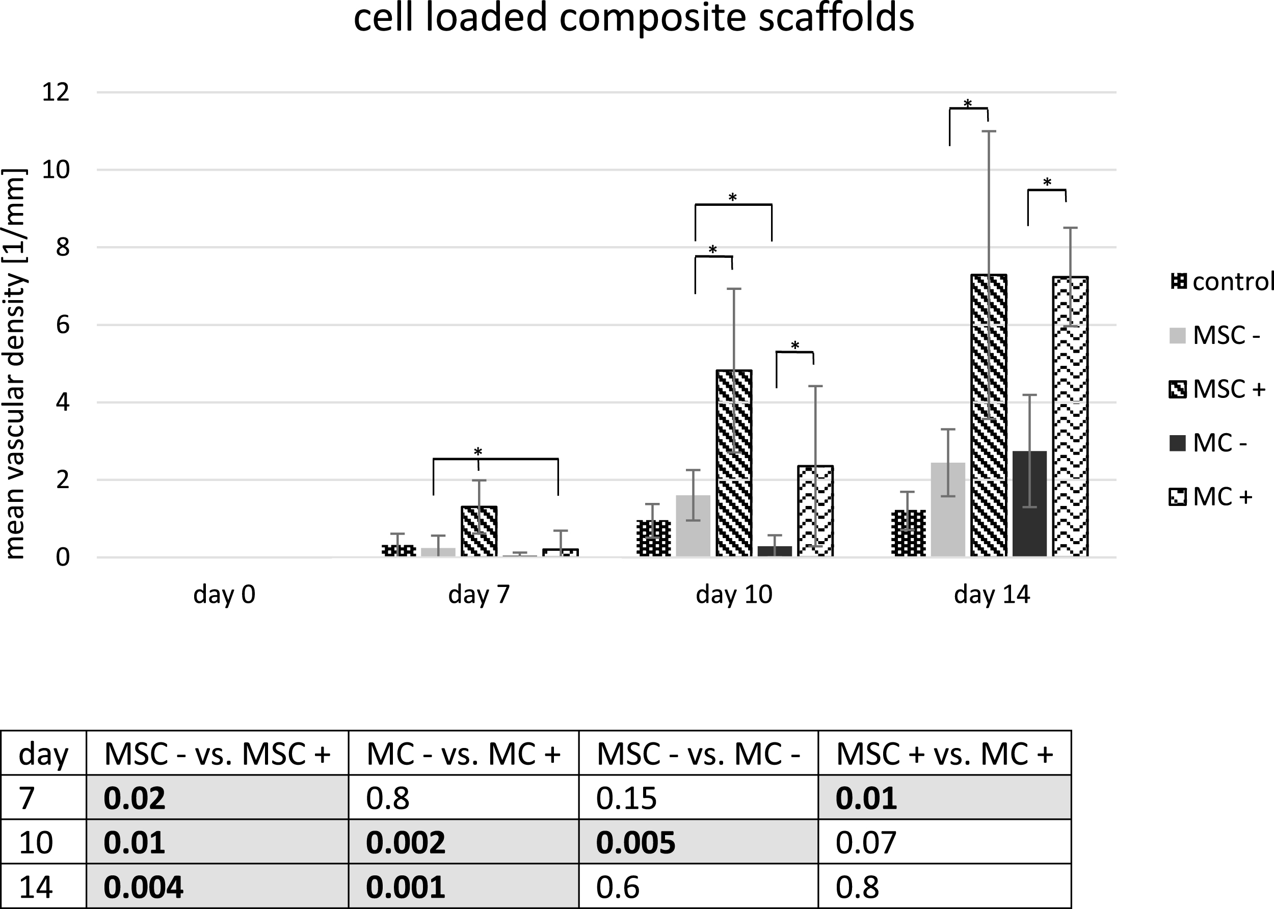
Mean vascular density induced by the MSC and MC loaded composite scaffolds at day 0, 7, 10 and 14. [–] means one day precultured cell loaded tissue engineering products; [+] means 14 days precultured tissue engineering products. P-values < 0.05 were assessed as significant and marked with a [*]. The table below shows the absolute p-values. Significant differences were printed fat and are coloured grey in the background.
Mean vascular density induced by cell seeded matrix composite scaffolds at day 0 / 7 / 10 and 14
PKH67-labelled erythrocytes were injected to demonstrate the functionality of newly developed vessels. The fluorescent signal was detected daily, initially along the meniscus surface to the composite scaffolds and then at the connection of the composite matrix to the surrounding meniscus. The precultured matrices showed an earlier start of angiogenesis and perfusion compared to the non-precultured scaffolds as shown in Table 4.
Documentation and evaluation of the start of angiogenesis and perfusion of the MSC and MC loaded tissue engineering products by detecting labeled erythrocytes via intravital microscopy. [–] means one day precultured cell loaded tissue engineering products; [+] means 14 days precultured tissue engineering products
Documentation and evaluation of the start of angiogenesis and perfusion of the MSC and MC loaded tissue engineering products by detecting labeled erythrocytes via intravital microscopy. [–] means one day precultured cell loaded tissue engineering products; [+] means 14 days precultured tissue engineering products
The MSC- and MC-based tissue engineering products were histologically evaluated by toluidine-blue staining and unspecific erythrocyte peroxidase detection (diaminobenzidin = DAB). The toluidine-blue staining was used to analyze the state of differentiation of the composite scaffolds. Using DAB staining, the existence of vascularity was detected.
All cell based tissue engineering products showed vascular cuts with erythrocytes within the whole matrices (see Fig. 6). Concerning the staining for toluidine blue, differences between the precultured and non-precultured groups were detected. The precultured constructs showed a stronger metachromatic staining caused by a higher content of proteoglycans in terms of a higher grade of differentiation (see Fig. 7).
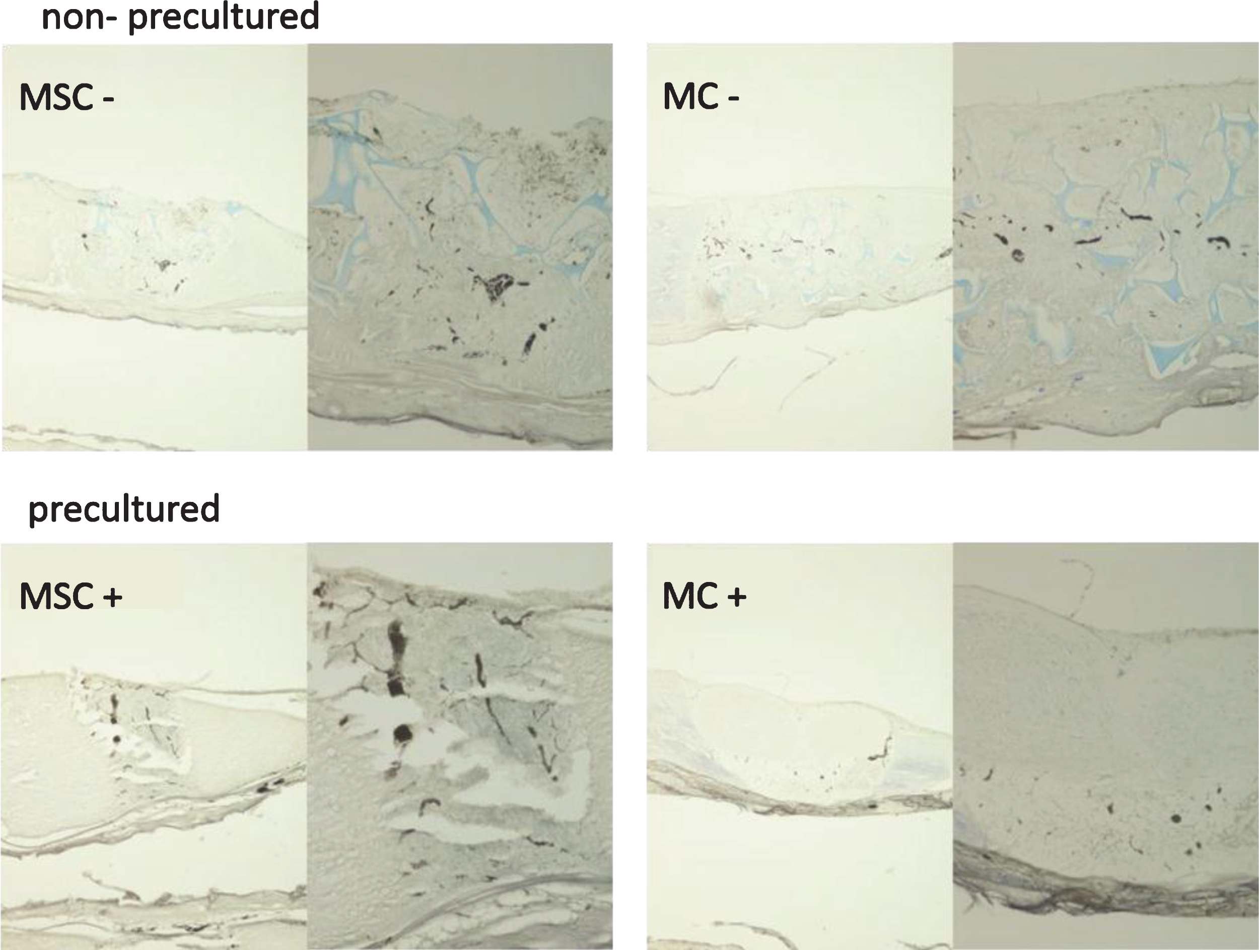
Unspecific erythrocyte peroxidase detection (diaminobenzidin = DAB), meniscus ring plus MSC or MC loaded tissue engineering products, 4x enlargement left, 10x enlargement right. [–] means one day precultured cell loaded tissue engineering products; [+] means 14 days precultured tissue engineering products. Vascular cuts with erythrocytes were detected in all groups.
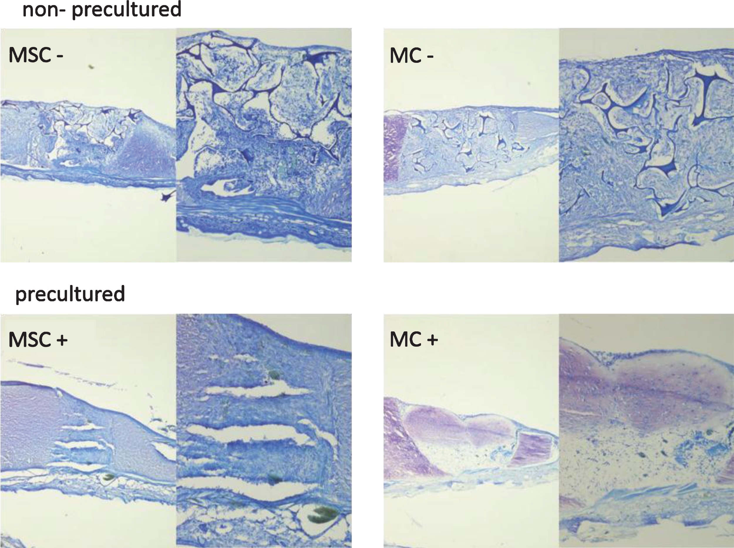
Toluidine blue staining, meniscus ring plus MSC or MC loaded tissue engineering products, 4x enlargement left, 10x enlargement right. [–] means one day precultured cell loaded tissue engineering products; [+] means 14 days precultured tissue engineering products. Stronger metachromatic staining was detected in the precultured groups.
In total, the MSC-based tissue engineering products seemed to show a stronger staining for endostatin compared to equivalent MC-based tissue engineering products. Additionally the content of endostatin of the precultured groups appeared to be less than the non-preculturedgroups.
In many of the precultured MSCs, a few necrotic areas were detected. Limited to these zones, a strong staining for endostatin was described according to their low grade of differentiation (see Fig. 8).

Immunohistological endostatin staining, meniscus ring plus MSC or MC loaded tissue engineering products, 4x enlargement left, 10x enlargement right. [–] means one day precultured cell loaded tissue engineering products; [+] means 14 days precultured tissue engineering products. Areas of necrosis were seen in many of the precultured MSC-based tissue engineering products according to the lower grade of differentiation.
The meniscus-matrix-constructs showed a high viability within the peripheral zone and along the surface. The red signal of ethidium bromide was only detected in the border areas of the viable meniscus sample and in the dead negative control (see Fig. 9). In summary, the meniscus rings were viable and fully functioning during the whole study period.

a) Fluorescence microscopic calcein-am staining of a meniscus after 14 days in a transparent titan dorsal skin fold chamber, 10x enlargement; on the left side is the vascularized, peripheral zone; on the right side is the avascular meniscus apex; b) + c) Fluorescence microscopic ethidiumbromid staining of a meniscus after 14 days in a transparent titan dorsal skin fold chamber, 10x enlargement; b) after explantation immediately proceeded meniscus (viable control); c) after explantation devitalized sample before viability testing (dead control).
The current study showed that cell based tissue engineering products were able to induce vascularisation within a meniscus defect model in a nude mice dorsal skin fold chamber. MSC and MC loaded composite scaffolds implanted in a meniscus ring model in a transparent dorsal skin fold chamber developed an ingrowth of functional vessels with a traceable perfusion within 14 days.
Regarding the fact that endogenous meniscus regeneration is associated with the vascularization of the meniscus tissue [7, 30], the role of angiogenesis and induction of vascularization by cell-based treatment strategies for meniscus healing needs to be evaluated.
MSCs and local MCs showed properties to improve meniscus regeneration. Recent studies analysed that MSC- and MC-cell-based strategies induced meniscal healing in critical meniscus defects in various animal models [5, 30].
As progenitor cells, MSCs act in a multifactorial way to enhance the regeneration of injured tissues. A positive effect for the repair of meniscus tears and cartilage defects can be achieved by MSC differentiation into repair cells according to the surrounding tissue [20]. MSC differentiation involves a multi-step process controlled by bioactive factors out of the local microenvironment. Additionally, MSCs positively influence the regeneration in an indirect way by the release of bioactive mediators (e.g. cytokines and growth factors) inducing both paracrine and autocrine activities [31, 32]. These indirect bioactive factors have an immunosuppressive effect, enhance the differentiation into cells of the surrounding tissue and reduce the development of scar tissue formation [31, 33]. Furthermore, these mediators influence the angiogenesis by survival and maturation of vascular cells, and stimulating neovascularization i.e. via the release of vascular endothelial growth factors (VEGF) [32, 34]. These direct and indirect effects of angiotactic interaction of the MSCs with the surrounding tissue were also shown by Tang et al. [35, 36]. Following MSC implantation into an ischemic area of cardiac muscle tissue after myocardial infarction, VEGF level, vascular density and perfusion grade increased, whilst the rate of apoptosis was reduced due to the secretion of bioactive mediators [35] and the direct conversion of MSCs into endothelial cells [36]. A similar effect was described by Murphy et al., who investigated a fractional regeneration of the lost meniscus tissue with vascularization of the neo-meniscus after application of hyaluronic acid together with labeled MSCs after partial meniscectomy. Furthermore, labeled MSCs were detected in the repair tissue [37].
A multi-lineage differentiation capacity has been described for MCs [19, 38]. All MCs isolated from the outer (vascular), inner (avascular) and horn (mixed vascular and avascular) parts of the meniscus showed a potential for mesenchymal differentiation. However, MCs out of the outer region were more plastic and differentiated along all three mesenchymal lineages [38]. In the current study, a tendency for higher vascularization was induced by meniscus tissue pellets derived from the vascularized outer part compared to meniscus tissue from the avascular zone. Despite no significant differences concerning the vascular density, the group of meniscus pellets derived from vascularized zone, clearly showed an earlier and more enhanced vascularization (p-value = 0.07). Nevertheless, meniscus pellets of both, the vascular and avascular area, induced vessel ingrowth within a period of 14 days in the transparent dorsal skin fold chamber. These observations are confirmed by previous results of Hoberg et al., who described an inhibitory effect in a co-culture of MCs and endothelial cells on the proliferation of endothelial cells mediated by an increased release of endostatin, a strong anti-angiogenic factor, which interferes with the VEGF pathway [39]. These data based on the effect of MCs isolated out of meniscus tissue from the inner two-thirds of elderly patients. Specifically these more hyaline cartilage like cells are responsible for the avascular environment within the avascular zone. Additionally, further cell-biological studies showed that these cells differ from the cells of the vascular area, where mainly fibroblast-like cells are located [5].
Beside this influence of cell origin within the meniscus, the current study also detected an earlier induction of angiogenesis induced by MSCs compared to MCs, showing a potentially higher angiogenic capacity. However, besides the cell type, multiple factors appear to influence the potential for angiogenesis.
Interestingly, this study showed that the grade of differentiation of the cell based tissue-engineering products also influenced the angiotactic capacity of the tissue engineering products. A 14 day chondrogenic preconditioning period for cell based tissue engineering products significantly accelerated vascularization and increased the extent of vessel ingrowth.
The comparison between MSCs and MCs showed significantly more angiogenesis at an earlier time point in the group of pre-cultured MSCs. However, after 14 days the mean values of the induced vascular density were equivalent. Nevertheless, the faster vascularization and stronger angiogenic activity of the pre-cultured MSCs could be of advantage for clinical application. Cells die due to the lack of oxygen, nutrients and inadequate elimination of waste products. A slow vascularization of the meniscus defects inhibits the tissue regeneration in certain pathological conditions [40]. It has been shown that the initial supply of in vivo implanted tissue engineered repair tissue with endogenous cells, nutrients and growth factors by a fast connection to a functional vascular system is crucial for its integration, maturation and survival at the defect site [41, 42].
To analyze meniscus regeneration with an emphasis on the vascularization a nude mice dorsal skin fold chamber model was chosen. Live-dead staining showed a viable meniscus over the whole study period of 14 days suggesting an appropriate environment for evaluation of vascularization patterns following meniscus injury.
The influence and mechanisms of angiogenesis for cell based tissue engineering products in in vivo meniscus repair models and regeneration are still unclear. Becker et al. detected an increased expression of VEGF especially in the avascular area after meniscus injury [43]. However, further studies failed to improve any meniscal healing by the application of VEGF into the meniscal defects via a VEGF coated suture material [8, 45]. Besides the special release kinetics of VEGF the missing effect could be caused by the high content of endostatin within the avascular meniscus tissue as described above. As part of collagen 18, endostatin acts as a strong angiostatic factor via inter alia interference with the VEGF signaling pathway and downregulation of the VEGF expression to maintain the antiangiogenic environment within the avascular zone [28, 47]. So, an increased angiogenesis might not be singularly based on the presence of VEGF. Beside the presence of these pro-angiogenic factors, it could also be induced by a reduced existence of angiostatic factors like endostatin. According to this, the cell loaded tissue engineering products in this study were immunohistochemically evaluated concerning their staining for endostatin. Whereas non-pre-cultured samples strongly stained for endostatin, pre-culturing of the cell based tissue engineering products resulted in a reduced endostatin content in comparison to the non-precultured group, whereby an angiotactic effect of VEGF could be facilitated.
Meniscus regeneration underlies complex multifactorial mechanisms. The isolated evaluation of single factors, as angiogenic and angiostatic mediators, does not reflect the complexity of the agonists and antagonists in the meniscus healing process as seen with the single application of regeneration mediating factors, which failed to result in sufficient repair [8, 20].
The tissue requirements for a successful regeneration of the meniscus tissue are high. Regarding the different vascularized areas, the partial vascularized zone and especially the avascular zone, where a higher content of endostatin is described [39], have a special requirement. However, the actual impact of the preconditioning on the regeneration within the partial vascularized and avascular area cannot be exclusively determined.
Overall, the question remains, does the fast induction of angiogenesis and vascularization within a naturally avascular tissue (i.e. the central parts of the meniscus tissue) is adjuvant or does it impair the low endogenous regeneration potential. Zellner et al. evaluated the relevance of MSCs for meniscus regeneration in the avascular area. They described a completely integrated meniscus like repair tissue after implantation of a non-precultured MSC-loaded tissue engineering product into an avascular meniscus defect [15]. Similar results were described by Pabbruwe et al., who inserted MSC-seeded collagen scaffolds between two ovine fibrocartilage discs and subsequently detected significantly less integration with the ovine meniscal surface after chondrogenic differentiation in comparison to the undifferentiated MSCs [30, 48]. This contrary impact of the proangiogenic preconditioning on the integration of cell based tissue engineering products within the inner two thirds of the meniscus could be explained by the observations of Esparza et al.. The vascular and avascular areas of the meniscus show differences in terms of the repair and expression of different factors by the located cells. In addition, the growth factors act in various ways in each meniscal area [49].
In summary, pre-culturing of cell based tissue engineering products influences the angiogenic capacity on the one hand, whilst non-precultured constructs enabled more cell-cell-interactions.
Therefore, further studies are necessary to investigate the angiotactic and angiostatic interaction in context of regeneration within different vascularized tissues like the meniscus tissue.
Footnotes
Acknowledgments
The authors thank Daniela Drenkard and Richard Kujat for their excellent technical assistance.