
Abstract
Mesenchymal stem cells (MSCs) are targeted as vehicles for cell mediated gene therapy. Here we report on a macromolecular carrier, which was designed aiming at successful targeted gene delivery into MSCs through the mediation of folate receptor and reduced cytotoxicity compared to established cationic polymer vector – polyethylenimine with a weight average molecular weight (Mw) of 25,000 Dalton (PEI25K). The carrier PHPA-PEI1800-FA was synthesized in a two-step procedure. PHPA-PEI1800 was prepared by grafting polyethylenimine with a Mw of 1800 Dalton (PEI1800) onto the α,β-poly(N-3-hydroxypropyl)-D,L-aspartamide (PHPA) backbone. PHPA-PEI1800-FA was obtained by chemically conjugating folic acid onto PHPA-PEI1800. The grafting degree of PEI1800 was 3.9±0.2% in relation to the CH groups of PHPA and the molar ratio of folic acid conjugated to PEI1800 (χFA) was 1.8±0.1 as calculated by NMR spectroscopy. The copolymers were biodegradable and exhibited lower cytotoxicity than PEI25K. Compared to PHPA-PEI1800, PHPA-PEI1800-FA led to a significantly higher transfection efficiency in human MSCs, which could be attributed to the mediation of folate receptor during the transfection process as confirmed by folic acid competition assay. Both marker gene (GFP) and therapeutic gene (VEGF) were delivered into human MSCs from multi-donors using PHPA-PEI1800-FA. The percentage of GFP+ MSCs showed an average value of 2.85±1.60% but a large variation for different samples. The VEGF expression level of the PHPA-PEI1800-FA transfected cells was significantly higher than that of either untransfected or naked DNA transfected cells. Conclusively, PHPA-PEI1800-FA is a suitable vector to deliver genes into human MSCs through the interaction with folate receptor.
Keywords
Introduction
Mesenchymal stem cells (MSCs) have demonstrated an effective therapeutic paradigm over the past decade for a variety of diseases due to several beneficial properties [1, 2]. They distribute widely throughout the body and can be easily isolated from diverse tissue sources [3]. They can be expanded to a large scale of cell numbers through in vitro or ex vivo culture in appropriate conditions without losing their stem cell properties [4, 5]. As cellular therapeutics, their multi-lineage differentiation potential and paracrine activity endow them with high efficacy in regenerative medicine. They are immunoprivileged and immunosuppressive, and hence can be used for immunomodulation [6, 7]. In addition, they can serve as gene/drug delivery vehicles as they are able to home to damaged tissues and solid tumors following in vivo administration [8].
In recent years, genetic modification of MSCs has become an effective strategy to further improve the clinical benefits of MSCs [9]. For example, the introduced functional genes could enhance the MSC survival after transplantation [10], guide the MSC differentiation towards desired lineage [11], and modulate the MSC secretion to promote tissue restoration [12, 13]. MSCs modified with anticancer genes could be used as efficient tumor target vehicles in cancer gene therapy [14]. However, as MSCs are difficult to be efficiently transfected via commonly used non-viral methods and are relatively sensitive to the surrounding environment, successful genetic manipulation of MSCs is still a challenge [13, 15]. Therefore, there is an urgent need for developing an effective delivery carrier for MSC genetic modification, which should at least present the advantages such as high transfection efficiency, low cytotoxicity, degradability and targeted delivery for both in vitro and in vivo applications.
The folate receptor (FR) is a single chain cell-surface glycoprotein that binds folic acid and mediates uptake of the vitamin. As FRs are significantly overexpressed in the majority of cancer cells, they could remarkably enhance the efficiency of FR-targeted gene/drug delivery into cancer cells [16, 17]. For cationic polymer-mediated gene delivery, the modification of a polymer with folic acid could drive the polymer/DNA polyplex to bind onto the surface of FR-bearing cells and subsequently to be internalized by the cells via endocytosis, which resulted in enhanced transfection efficiency [18–20]. In this study, we hypothesized that the conjugation of folic acid might promote the transfection efficiency of cationic polymer-mediated gene delivery into human MSCs through FR-mediated cell surface binding and uptake of polyplexes. Two copolymers, PHPA-PEI1800 and PHPA-PEI1800-FA, were synthesized by grafting branched polyethylenimine with a Mw of 1800 Dalton (PEI1800) onto poly(N-3-hydroxypropyl)aspartamide (PHPA) and further chemically conjugating folic acid onto the PHPA-PEI1800 copolymer. The PHPA serves as a biodegradable backbone, which is expected to reduce the cytotoxicity and avoid the copolymer retention in the cells. The selection of PEI1800 as polycation for DNA condensation was based on the consideration of both transfection efficiency and cytotoxicity. As a highly effective polymeric gene carrier, branched PEI contains primary, secondary and tertiary amino groups, which makes it partially protonated at physiological pH ranges and further protonated at endosomal pH levels. This will induce the rupture of endosomes through “proton sponge effect” and consequently facilitate the release of DNA into cytosol [21]. However, the transfection activity and cytotoxicity of PEI is highly dependent on its molecular weight. Low molecular weight PEI normally presented low cytotoxicity but poor transfection efficiency due to its low DNA condensing capability [22, 23]. In contrast, high molecular weight PEI showed high transfection efficiency but significant cytotoxicity as well [24–26]. In addition, the utilization of high molecular weight PEI may bring up safety concerns since PEI is not degradable in physiological environment. To assemble low molecular weight PEI into large molecules through biodegradable cross-linkers or polymer backbone is an effective approach to combine the beneficial features of both, keeping the biodegradability and low cytotoxicity while still preserving the DNA condensing capability and transfection efficiency [27–29]. The aim of this study is to develop a novel non-viral vector with lower cytotoxicity and biodegradability for effective gene delivery into human MSCs. Therefore, the PHPA and low molecular weight PEI1800 were used for copolymer synthesis. The copolymers were evaluated with respect to the biodegradability, DNA condensing capability, cytotoxicity and transfection efficiency. Particularly, the capacity of PHPA-PEI1800-FA to deliver genes into human bone marrow-derived MSCs was investigated.
Materials and methods
Materials
Branched polyethylenimine with molecular weight Mw of 1800 Dalton (PEI1800) and 25,000 Dalton (PEI25K) were purchased from Polysciences (Warrington, PA, USA) and Sigma-Aldrich (St. Louis, MO, USA), respectively. 3-Amino-1-propanol, triethylamine (Et3N), DL-aspartic acid and 3-(4,5-dimethyl-2-thiazolyl)-2,5-diphenyl-2H-tetrazolium bromide (MTT) were purchased from Sigma-Aldrich. Folic acid, N,N-dimethylformamide (DMF) and dimethyl sulfoxide (DMSO) were purchased from Sinopharm Chemical Reagent (Shanghai, P. R. China). 1,1’-Carbonyldiimidazole (CDI) was from Pierce (Rockford, IL, USA). All chemical reagents were of analytical grade, unless otherwise stated.
Polymer preparation
Fig. 1 shows the synthesis scheme of PHPA-PEI1800 and PHPA-PEI1800-FA. The polymer α,β-poly(N-3-hydroxypropyl)-D,L-aspartamide (PHPA) was prepared following the method described in our previous work [30]. The precursor PHPA-PEI1800 was synthesized by CDI-mediated activation of the hydroxy group from PHPA and subsequent grafting of PEI1800 according to the procedure described before [31]. For the preparation of PHPA-PEI1800-FA, folic acid (0.1 mmol) dissolved in 2 mL DMSO was activated by CDI (0.12 mmol) under nitrogen atmosphere and magnetic stirring for 1.5 hours. The activated folic acid solution was added to PHPA-PEI1800 solution (120 mg in 5 mL DMSO) and stirred for 12 hours. The crude product was purified by dialysis (Cut Off Mw = 14,400 Da) against deionized water for 48 hours. Finally, PHPA-PEI1800-FA was obtained as a pale yellow powder after freeze-drying.
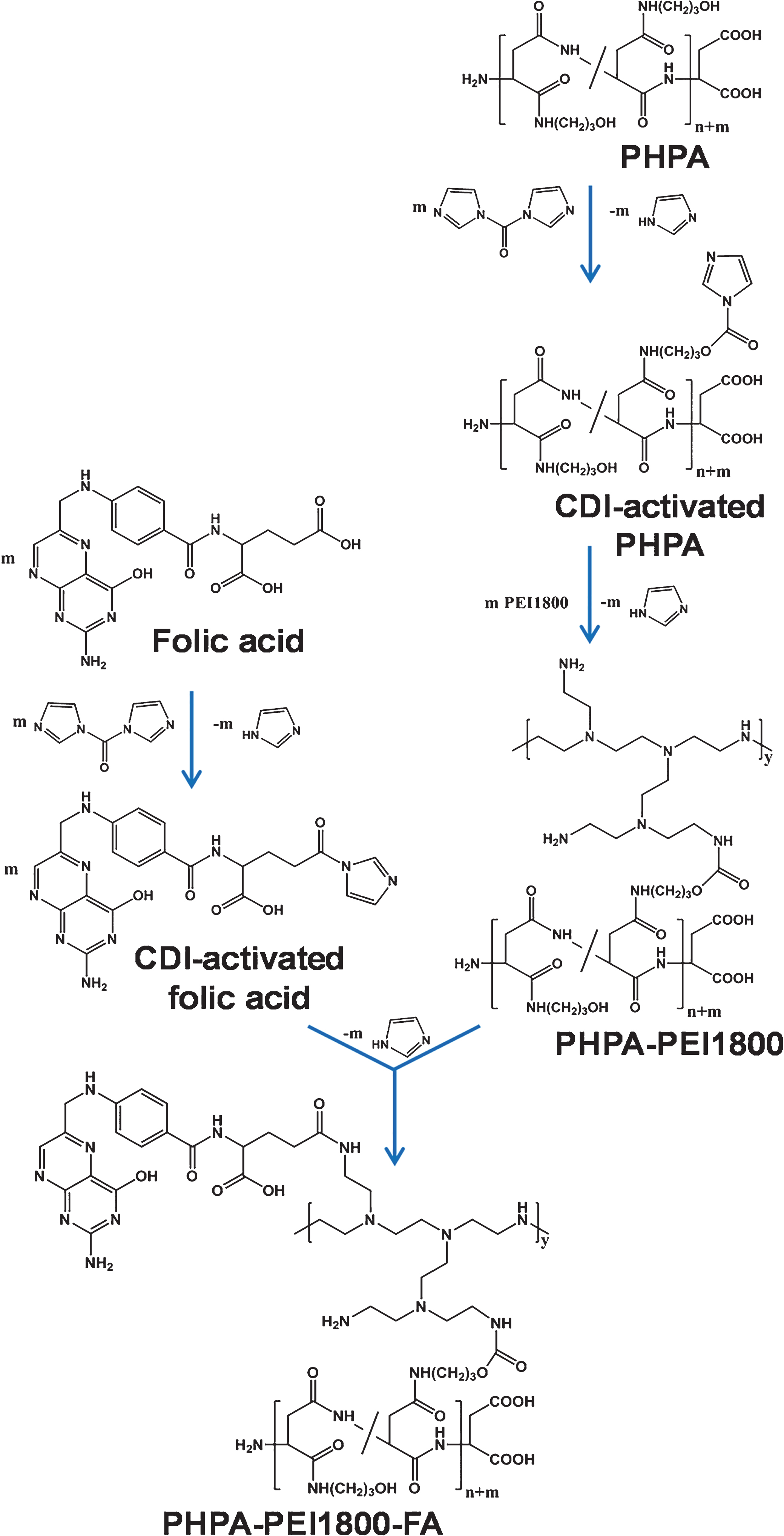
Synthesis route of PHPA-PEI1800 and PHPA-PEI1800-FA copolymers.
The chemical structure of the synthesized copolymers was characterized using nuclear magnetic resonance spectroscopy (1H NMR). Samples (8 mg) were dissolved in deuterium oxide (D2O, 0.5 mL) and were scanned on a Bruker Avance DMX-400 NMR spectrometer at 400 MHz at room temperature. Chemical shifts were referenced to the solvent peaks of D2O (δ= 4.70 ppm). The degree of grafting (dg) of PHPA with PEI1800 and the molar ratio of folic acid (χFA), which was linked to PEI1800 in PHPA-PEI1800-FA, was calculated by NMR according to the following equations, where I is the integral peak area of the corresponding signals from the polymer (see Fig. 2) and Mw is the molecular weight of PEI1800.

1H NMR spectra of PHPA, PHPA-PEI1800 and PHPA-PEI1800-FA.
To evaluate the degradation of the prepared copolymer, PHPA-PEI1800 was dissolved in PBS (phosphate buffered saline), serum free DMEM (Dulbecco’s Modified Eagle Medium) and DMEM containing 10% fetal bovine serum (FBS), respectively. The solution was incubated at 37 °C and the relative remaining molecular weight (Mwrel) of the copolymer at time points was measured with gel permeation chromatography (GPC), which was equipped with a Waters 600E pump, a Waters 2410 Refractive Index Detector and a set of columns (Phenomenex Polysep Guard S/n 70987G, Polysep GFC-P S/n 70977 and Polysep GFC-P S/n 70976). Double distilled water was used as mobile phase with a flow rate of 0.7 mL/min (33 °C). The instrument was calibrated using poly(ethylene glycol) standards with number average molecular weights ranging from 7100 to 56,000 Dalton.
Amplification and purification of plasmid DNA
Plasmids containing the gene of luciferase (pGL3-luciferase, a gift from the National Laboratory of Oncogenes and Related Genes of the Shanghai Cancer Institute, Shanghai Jiaotong University, Shanghai, China), enhanced green fluorescent protein (pEGFP-N3, Clontech, Palo Alto, CA, USA) and VEGF (pCEP4-VEGF165 [32], a gift from Prof. Renke Li, University of Toronto, Toronto, Ontario, Canada) were propagated in selective Luria-Bertani (LB) medium. Then, the amplified plasmid DNA was purified using the Endofree Mega Plasmid Kit (Qiagen, Hilden, Germany) according to the manufacturer’s instruction. The concentration and purity of the plasmid DNA were determined by measuring the absorbance at 260 nm and 280 nm using a UV spectrophotometer. Finally, the purified plasmid DNA was stored in aliquots at –20 °C prior to use.
Polyplexes preparation and characterization
PHPA-PEI1800/DNA and PHPA-PEI1800-FA/DNA polyplexes were prepared by adding polymer solution dropwise to DNA solution, followed by a vortex (30 seconds) and an incubation (30 minutes) at room temperature. The polymer/DNA ratio was defined as N/P ratio, where ‘N’ is the molar amount of nitrogen from the cationic copolymers and ‘P’ is the molar amount of phosphate from plasmid DNA. Gel electrophoresis was performed to examine the capability of PHPA-PEI1800 and PHPA-PEI1800-FA to condense plasmid DNA. The polyplexes with various N/P ratios were mixed with loading buffer and subsequently loaded onto 1% agarose gel containing ethidium bromide. Electrophoretic mobilization was carried out in tris-acetate-EDTA (TAE) buffer at 100 V in a Sub-Cell System (Biorad Laboratories, CA). The DNA bands were visualized using an UV illuminator and the images were recorded by GeneSnap imaging system (Syngene, Cambridge, UK). The particle size and zeta potential of the polyplexes were measured by dynamic light scattering (DLS) using a Zetasizer Nano ZS (Malvern Instruments, Southborough, MA, USA). Complex solutions (1 mL) containing 2 μg of DNA were prepared at various N/P ratios and measured at 25 °C in triplicate.
Cytotoxicity assay
The cytotoxicity of polyplexes was studied using cell lines derived from different tissues. HEK293 cells (human embryonic kidney 293 cell line) and B16 cells (mouse melanoma cell line) were seeded into 96-well plate at a density of 1×10⁴ cells/well and allowed to grow to reach 60–70% confluence. Then, the culture medium was replaced with 200 μL of serum-free medium containing polyplexes with various N/P ratios. After 4 hours of incubation, the medium was replaced with fresh serum-containing medium and the cells were incubated for 20 hours. At the end of the incubation, sterilized MTT stock solution (5 mg/mL in PBS) was added into the wells, reaching a final MTT concentration of 0.5 mg/mL. After 4 hours, the unreacted dye was removed by aspiration, and the purple formazan crystal in each well was dissolved with 100 μL of DMSO. The absorbance was measured using a microplate reader (Spectra Plus, TECAN) at 570 nm wavelength. The relative cell viability related to control cells, which were cultured in medium without polyplex, was calculated using the equation: Cell viability (%) = [A] sample /[A] control ×100%.
Luciferase expression assay
Luciferase expression was evaluated using FR-overexpressing HeLa cells (human cervical cancer cell line) and FR-deficient A549 cells (human lung adenocarcinoma epithelial cell line). Briefly, cells were separately seeded into 48-well plate. When cells had grown to 60–70% confluence, the medium was replaced with fresh serum-free DMEM (300 μL/per well) containing polyplexes with various N/P ratios and the DNA dosage of 1 μg pGL3-luciferase/per well. After incubation of 4 hours at 37 °C, the transfection medium was replaced with fresh DMEM containing 10% FBS and the cells were incubated for additional 44 hours. The luciferase expression level was measured using a luciferase assay kit (Promega, Madison, WI, USA) and the protein concentration in cell extracts was measured using a BCA protein assay kit (Pierce, Rockford, USA). The transgene expression was reported in terms of relative light unit (RLU)/mg cell protein.
Isolation, culture and characterization of human bone marrow derived MSCs
For studies involving human tissues we obtained ethical approval of the local ethical committees, Medical Ethics Commission II, Medical Faculty Mannheim, Heidelberg University and Heidelberg University Ethical Board. Bone marrow for research purposes was received according to the approval by the Heidelberg University Ethical Board; approval nos.: 251/2002 and S-076/2007. All samples were taken after written consent using guidelines approved by the Ethic Committee on the Use of Human Subjects at the University of Heidelberg.
The isolation and culture of MSCs was performed as described before [13]. Cell surface markers of MSCs were analyzed using flow cytometry. In brief, cells were blocked with human FcR Blocking Reagent (Miltenyi Biotec, Bergisch Gladbach, Germany) followed by 30 minutes of incubation at 4 °C with antibodies: CD29, CD44, CD45, CD73 (BD Biosciences, Heidelberg, Germany), CD105 (AbD Serotec, Düsseldorf, Germany) and corresponding isotype controls. Then, cells were washed with PBS/EDTA (2 mM) and analyzed using a Flow Cytometer (LSR II, Becton Dickinson, Heidelberg, Germany). Dead cells were excluded with staining kit (LIVE/DEAD®, Invitrogen). The FR expression of human MSCs was quantified by staining cells derived from 3 donors with phycoerythrin (PE) conjugated antibody (anti-human FOLR1-PE, R&D systems, Wiesbaden-Nordenstadt, Germany) and corresponding isotype control according to the given protocol, followed by the flow cytometric measurement. The data analysis was performed using Flowjo software.
GFP delivery into MSCs and folic acid competition assay
MSCs were pre-seeded into 6-well plate (3.5 mL medium/well) and cultured until the cell confluence reached 60–70%. The culture medium was replaced with new medium 30 minutes before the transfection experiment. For folic acid competitive binding assay, 1 mM free folic acid was included in the new culture medium. Polymer/pEGFP polyplexes were added with an optimized N/P ratio of 40 and a DNA dosage of 1 μg/cm2, followed by incubation without medium change. The transfection efficiency was quantified using flow cytometry 48 hours post transfection. In brief, the culture medium was removed and the cells were washed three times with PBS. Then, cells were harvested by trypsin-EDTA and washed twice with PBS. For each sample, at least 10000 cells were counted by flow cytometer. The untransfected cells from the same donors were used as control and the data were analyzed with FlowJo software.
VEGF delivery into MSCs
To study the capability of the synthesized copolymer to deliver therapeutic genes, MSCs were transfected using functional gene (human VEGF165). Cells were seeded into 24-well plates and the culture medium was replaced with fresh medium (700 μL/well) before transfection. The MSCs were divided into three groups: untransfected, naked DNA (1 μg DNA/cm2) transfected and PHPA-PEI1800-FA/DNA polyplex (N/P ratio 40, 1 μg DNA/cm2) transfected cells. 48 hours post-transfection, the culture medium was collected and the VEGF protein content in the culture medium was measured using a human VEGF ELISA kit (QuantiGlo, R&D Systems, Wiesbaden-Nordenstadt, Germany). The content of total protein in the cell extracts was measured using a BCA protein assay kit and was used as reference.
Statistics
The number of replication for the experiments was in the range from three to six, and the results were expressed as mean±standard deviation (SD) or standard error of the mean (SEM), as indicated respectively in the figure legend for each assay. Statistical analysis was performed using Paired-samples t test or Independent-samples t test depending on the experiments, and a significance level (Sig.) <0.05 was considered to be statistically significant.
Results
Polymer characterization
The 1H NMR spectra of the synthesized polymers are shown in Fig. 2. The characteristic peaks of the PHPA backbone were clearly observed and assigned (Fig. 2A). The obtained PHPA-PEI1800 copolymer exhibited a new multiplet at a chemical shift of 2.4–2.8 ppm. This signal was attributed to the ethyl groups (-CH2-CH2-NH-) in PEI1800 (Fig. 2B) as previously reported [29] illustrating in this way the successful grafting of PHPA with ethylene imine moieties. In order to determine the degree of grafting (dg), the ratio of the integral peak areas between PEI1800 ethyl groups and the CH groups at a chemical shift of 4.5 ppm (H1) from N-3-hydroxypropyl aspartamide units was calculated resulting in dg = 3.9±0.2%. The peaks corresponding to the hydrogen atoms of folic acid (δ= 6.6–8.5 ppm) were observed in the spectrum of PHPA-PEI1800-FA, demonstrating the successful conjugation of folic acid onto the PHPA-PEI1800 copolymer (Fig. 2C) [33]. Here, a molar ratio χFA (molar ratio between folic acid and PEI1800) of 1.8±0.1 was calculated by comparing the integral areas of signals from PEI1800 (-CH2-CH2-NH-) and FA (H6/7, H8/9, and H10).
As the biodegradability is a key function of polymeric gene vectors, degradation experiments were performed at 37 °C using tree different media: PBS, serum free DMEM, as well as DMEM containing 10% FBS. In order to evaluate the degradation behaviour of PHPA-PEI1800, the molecular weight of the copolymer was investigated by means of GPC measurements using poly(ethylene glycol) for standard calibration and the relative remaining molecular weight (Mw,rel) is illustrated in Fig. 3 as function of degradation time. Here, Mw,rel decreased to 74±7%, 62±6%, and 65±7% within 3 days in PBS, serum-free DMEM and serum-containing DMEM, respectively. After 7 days, Mw,rel was almost independent of the degradation media and about 50%.
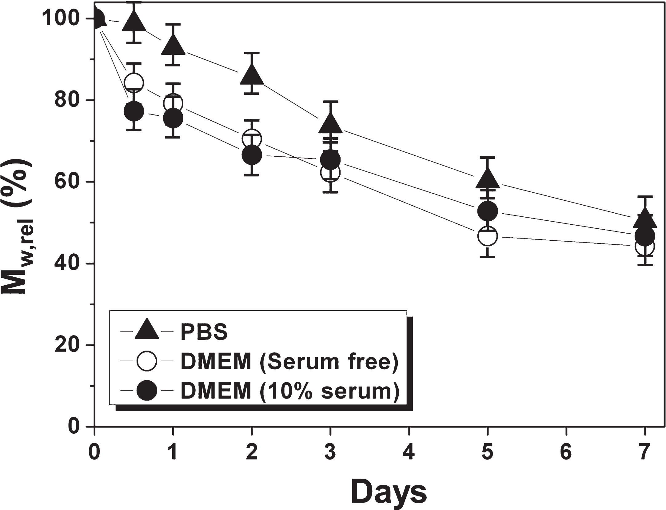
Degradation of PHPA-PEI1800 copolymer in different media.
The DNA condensation capacity of the synthesized copolymers was studied by gel electrophoresis. Both PHPA-PEI1800 and PHPA-PEI1800-FA showed increased capability to condense and retard DNA with increasing N/P ratio. DNA could be completely retarded by PHPA-PEI1800 at a N/P ratio of 3.0 and by PHPA-PEI1800-FA at a N/P ratio of 4.0 (Fig. 4A). The particle size and zeta potential of the polyplexes are crucial for efficient gene delivery as well as the polymer degradation. Both PHPA-PEI1800 and PHPA-PEI1800-FA could condense DNA to form nano-sized and positively charged polyplexes (Fig. 4B). With the increase of N/P ratio, the particle size of PHPA-PEI1800/DNA polyplex slightly varied, which was 191.70 nm, 188.83 nm and 199.38 nm at N/P ratios of 20, 30 and 40, respectively. The particle size of PHPA-PEI1800-FA/DNA polyplex first decreased from 209.13 nm at a N/P ratio of 20 to 178.03 nm at a N/P ratio of 30, and became close to constant after that (180.40 nm at a N/P ratio of 40). The surface charge of both polyplexes showed an increasing trend with the increase of N/P ratio. From N/P ratio 20 to 40, the zeta potential constantly increased from +11.60 eV to +15.70 eV for PHPA-PEI1800/DNA polyplex and from +11.06 eV to +17.35 eV for PHPA-PEI1800-FA/DNA polyplex.
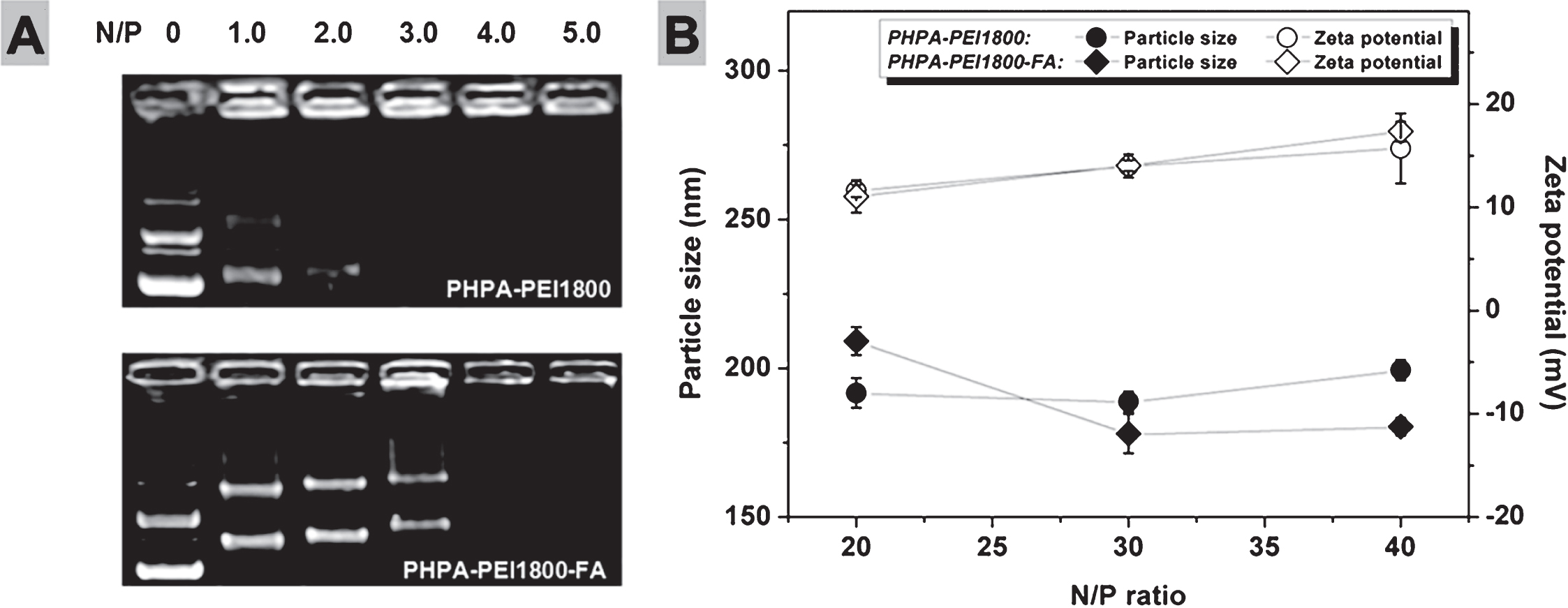
Characterization of polymer/DNA polyplexes. (A) Agarose gel electrophoresis of polyplexes prepared using PHPA-PEI1800 and PHPA-PEI1800-FA copolymers at various N/P ratios. (B) Particle size and zeta potential of polyplexes at different N/P ratios.
In order to evaluate the cytotoxicity of polyplexes prepared using the synthesized copolymers, an MTT assay was performed on HEK293 and B16 cell lines to measure the metabolic activity of the cells. Compared to the cells treated by PEI25K/DNA polyplex, the cells treated by PHPA-PEI1800/DNA and PHPA-PEI1800-FA/DNA polyplexes presented much higher viability at various N/P ratios (Fig. 5).
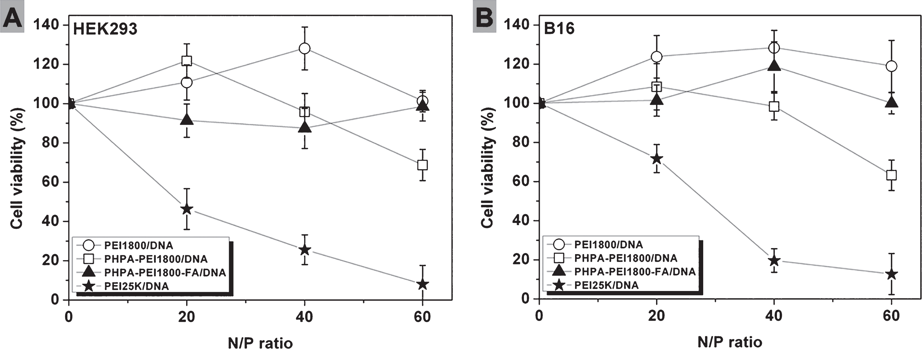
Cell viability of (A) HEK293 and (B) B16 cell lines after incubation with polyplexes at various N/P ratios (mean±SD, n = 4).
FR-overexpressing HeLa cells and FR-deficient A549 cells were used to study the FR-mediated gene delivery. PHPA-PEI1800-FA/DNA polyplex led to a significantly higher luciferase expression than PHPA-PEI1800/DNA polyplex in HeLa cells. On the contrary, no significant difference of luciferase expression was observed between PHPA-PEI1800 and PHPA-PEI1800-FA mediated transfections in A549 cells (Fig. 6).

Luciferase expression of (A) HeLa and (B) A549 cells transfected by polyplexes at various N/P ratios (mean±SD, n = 3, *Sig <0.05 by Independent-samples t test).
The immunophenotype and FR expression of human MSCs were analyzed with flow cytometry (Fig. 7). The cells showed characteristically positive mesenchymal markers (CD29, CD44, CD73 and CD105) and negative haematopoietic marker (CD45). A fraction of FRα positive MSCs was observed. For the MSCs derived from 3 donors, the percentage of FRα positive cells was 9.7±6.2% (mean±SD).
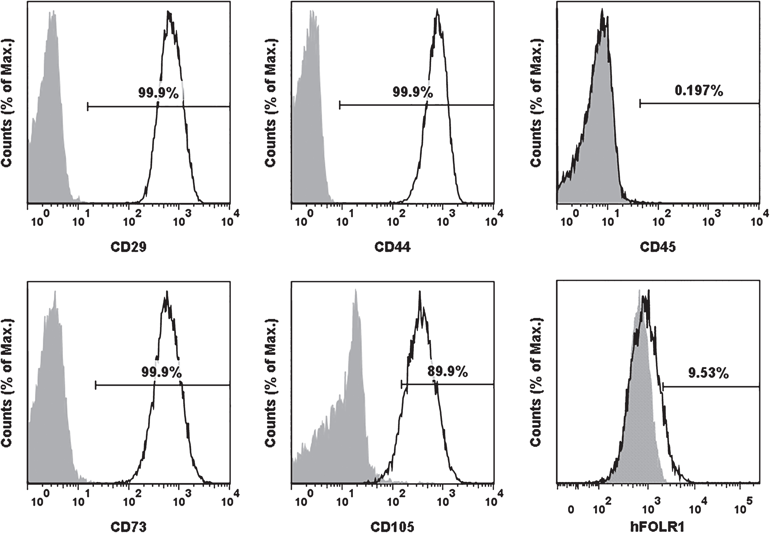
Expression of surface markers and FR of human MSCs was characterized using flow cytometry after staining the cells with antibodies (open histograms) and corresponding isotype controls (filled histograms). The cells express mesenchymal markers CD29, CD44, CD73, CD105 positively and haematopoietic marker CD45 negatively. The last graph shows the representative histogram of FRα expression of the cells.
To clarify whether the FR on human MSCs could mediate the transfection, we performed a folic acid competition assay using cells derived from 3 donors. As shown in Fig. 8, PHPA-PEI1800, which was not conjugated with folic acid, resulted in extremely low transfection efficiency at either the absence or presence of free folic acid molecules. In contrast, PHPA-PEI1800-FA showed a significant higher transfection efficiency (4.2±0.4%) at the absence of free folic acid than at the presence of free folic acid (1.5±0.3%).
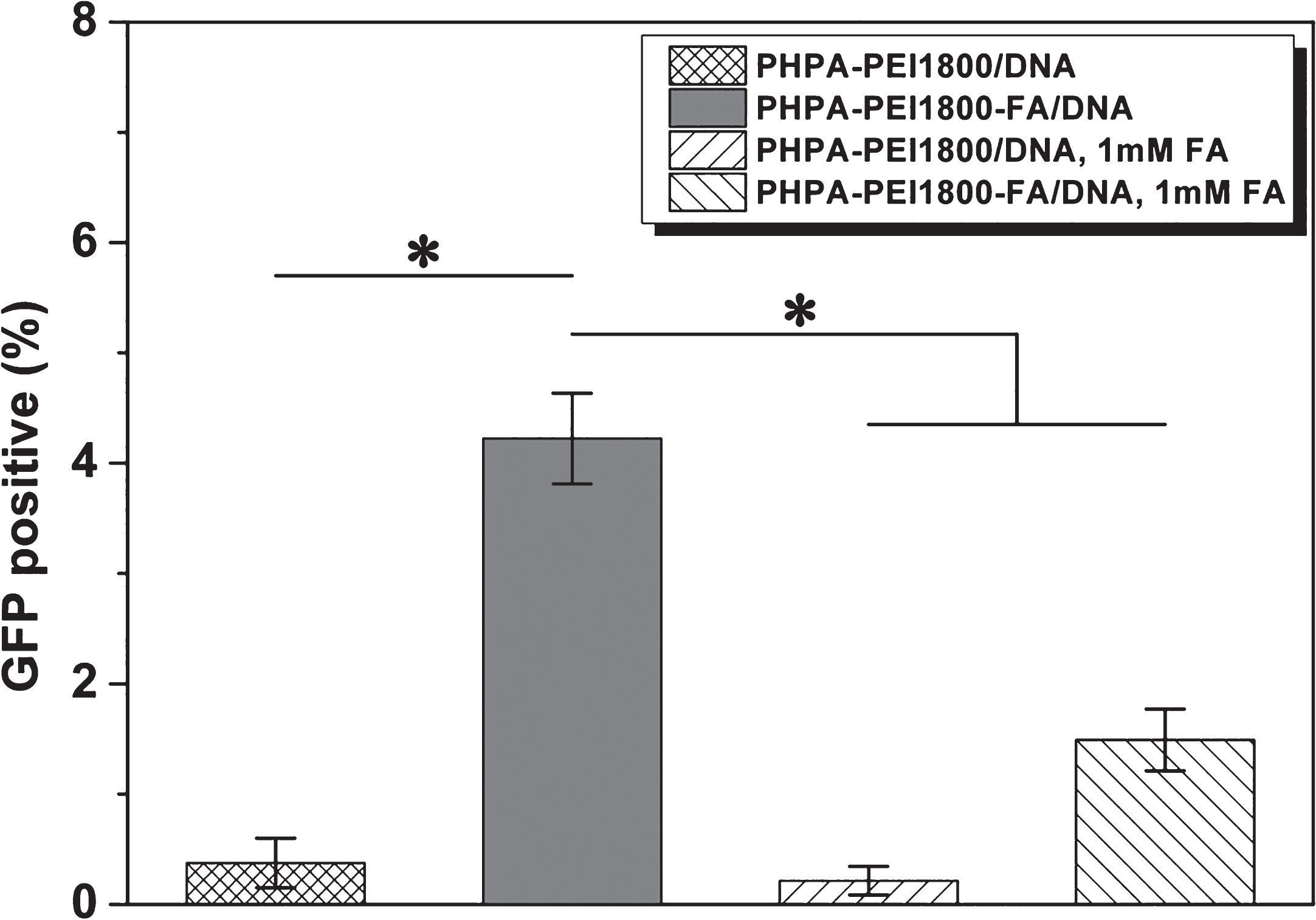
Transfection efficiency of cells transfected by PHPA-PEI1800 and PHPA-PEI1800-FA at the absence or presence of 1 mM free folic acid molecules (mean±SEM, n = 3, *Sig <0.05 by Paired-samples t test).
The MSCs derived from 11 donors were transfected with PHPA-PEI1800-FA/pEGFP-N3 polyplex to evaluate the multi-sample-based transfection. These cells presented a transfection efficiency of 2.85±1.60% (mean±SD; Fig. 9A). However, the variation of transfection efficiency for different samples was large. As a result, the transfection efficiency varied from 1.13% to 5.79%. Fig. 9B-D shows representative pictures of human MSCs transfected with PHPA-PEI1800-FA/pEGFP-N3 polyplex.

Transfection efficiency of human MSCs derived from 11 donors transfected by PHPA-PEI1800-FA/pEGFP-N3 polyplex. (A) Scattered plots represent the percentage of GFP+ cells 48 hours post transfection. (B-D) Representative images of human MSCs transfected by PHPA-PEI1800-FA/pEGFP-N3 polyplex: (B) fluorescent, (C) phase-contrast and (D) merged images (bar = 100 μm).
To investigate the capability of PHPA-PEI1800-FA to deliver therapeutic genes into human MSCs, we transfected the cells derived from 6 donors using plasmid DNA pCEP4-VEGF165, which encodes human vascular endothelial growth factor (Fig. 10). Compared to untransfected cells which intrinsically express VEGF 1.55±0.10 pg/μg total protein in the medium, the cells transfected by either naked DNA or polyplex showed an significant enhancement of VEGF expression. The VEGF expression level was increased to 1.69±0.07 pg/μg total protein through naked DNA transfection, and was enhanced to 2.12±0.11 pg/μg total protein via PHPA-PEI1800-FA mediated transfection.

VEGF expression level of human MSCs 48 hours post transfection was analyzed by ELISA (mean±SEM, n = 6, *Sig <0.05 by Paired-samples t test).
In this study, we prepared a novel cationic copolymer gene carrier PHPA-PEI1800-FA consisting of PHPA as a biodegradable backbone, PEI1800 as polycation for DNA condensation and folic acid as ligand that can target to FR on cell membrane. This copolymer has demonstrated the significantly enhanced transfection efficiency into human MSCs via FR-mediated cell surface binding and uptake of the polyplexes.
As a key function for polymeric gene vectors, biodegradability could generate the safety benefits in clinical treatment. Compared to the non-degradable counterparts, the biodegradable gene vectors could reduce the cytotoxicity and avoid the polymer accumulation in the body especially after repeated administration, since the degradation leads to low molecular weight products that are less- or non-toxic. Moreover, the degradation of the polymer can facilitate the release of DNA into the cytosol, and thereby improve the transfection efficiency [34]. Here, the PHPA-PEI1800 copolymer presented a half-life of degradation around 7 days in different media, showing the potential advantages of the copolymers as polycationic gene carriers. The grafting degrees of PEI onto PHPA seem to be a critical parameter to affect the transfection efficiency, as demonstrated in our previous work, in which PEI with a molecular weight Mw of 600 Dalton (PEI600) was grafted onto PHPA [31]. PHPA-PEI600 with 30% PEI600 grafting degree showed the highest transfection efficiency in different cell lines, as compared to PHPA-PEI600 with lower (14%) or higher (76%) grafting degrees. In the present study, considering that MSCs are relatively more sensitive to toxic compounds than immortal cell lines, we synthesized the cationic copolymers with a lower PEI grafting degree (3.9±0.2%). However, both PHPA-PEI1800 and PHPA-PEI1800-FA could still effectively condense DNA to form the nanoscale particles with positive charge. Both surface charge and particle size of the polyplexes are crucial parameters for an efficient gene delivery mediated by polycationic carriers. The surface charge of the polyplex can influence the electrostatic interaction between the anionic cell surface proteoglycans and the positively charged polyplex, while the particle size may play an important role for the polyplex with respect to their cellular internalization, cytoplasmatic transport, as well as nuclear entry through the nucleopore complexes [35]. Therefore, these results suggested that PHPA-PEI1800 and PHPA-PEI1800-FA are suitable to serve as gene delivery vectors in term of the surface charge and particle size of the polyplexes. Notably, PHPA-PEI1800 exhibited higher DNA retardation capability than PHPA-PEI1800-FA as measured by electrophoresis. This was perhaps due to the reason that the conjugation of folic acid molecules lowered the density of primary amino groups of the copolymer, which participate in forming polyplexes with DNA by an ionic interaction with phosphate groups [36].
The cytotoxicity of cationic polymers is caused by the interactions with the plasma membrane or with negatively charged cell components and proteins. The molecular weight as well as the charge density of the polymer are predominant factors influencing the cytotoxicity [37]. Compared to PEI25K which has been considered as a gold standard for polymeric carrier-mediated gene delivery, both PHPA-PEI1800 and PHPA-PEI1800-FA exhibited much lower cytotoxicity, which was even comparable to pure PEI1800 at lower N/P ratios. This could be explained by the reduced charge density in PHPA-PEI1800 and PHPA-PEI1800-FA as a result of the presence of PHPA backbone. In addition, the biodegradability of the copolymers may also play a role to reduce their cytotoxicity. This result was consistent with a previous report, in which low molecular weight PEI grafted chitosan showed much lower cytotoxicity than PEI25K [38]. Human MSCs are primary cells, which are normally more sensitive to cationic polymers than immortal cell lines. For example, they presented lower viability than COS7 cell lines when transfected by PEI25K at the same transfection conditions (N/P ratio and DNA dosage) [13, 39]. Here, the healthy spindle-shaped morphology of human MSCs derived from multi-donors was observed after transfection with PHPA-PEI1800-FA, indicating the relatively lower cytotoxicity of PHPA-PEI1800-FA for human MSCs.
Targeted delivery of nanoparticles at the tissue, cellular, or subcellular levels can increase the effect of the delivered therapeutics and importantly decrease the undesired side effects by reducing or eliminating the delivery into untargeted sites [40]. As an important tumor marker overexpressed in a large numbers of cancer cells, FR has been explored as the target molecule for delivering gene/drug into FR-bearing cancer cells [16, 17]. In addition to some cancer cells, FRs are also expressed at relatively low levels in normal tissues such as lung, thyroid and kidney [41–43]. Different FR isoforms have been identified. FRα and FRβ are attached to the cell surface through the glycosylphosphatidylinositol (GPI) anchor, while FRγ and its truncated version FRγ’ are constitutively secreted [44–46]. Here, the PHPA-PEI1800-FA copolymer containing folic acid as targeting ligand was expected to show advantages for targeted delivery of genes into FR-expressing cells. To assess the function of the conjugated folic acid, the transfection was first performed using FR-overexpressing HeLa cells and FR-deficient A549 cells. Compared to PHPA-PEI1800, PHPA-PEI1800-FA resulted in a markedly enhanced transfection efficiency in HeLa cells but the similar transgene expression in A549 cells, suggesting that folic acid could improve the transfection activity of the copolymer through the mediation of FR.
MSCs represent a heterogenous population of multipotent cells [47]. In this study, although only a fraction of human MSCs (9.73±6.18%) were FRα positive, one could still expect that the transfection efficiency of MSCs could be highly improved through the mediation of FR. As hard-to-transfect primary stem cells, human MSCs transfected with PEI25K showed an average transfection efficiency less than 5% [13]. Therefore, the transfection activity of human MSCs could be highly improved if the FRα positive subpopulation could be efficiently transfected. As expected, PHPA-PEI1800-FA led to a significantly higher transfection efficiency than PHPA-PEI1800 in MSCs, suggesting that gene delivery into human MSCs through the mediation of FR is an effective approach. This result was confirmed by the folic acid competition assay, as the blockage of FR with free folic acid significantly decreased the transfection efficiency of PHPA-PEI1800-FA. Notably, when PHPA-PEI1800-FA was utilized to transfect human MSCs derived from multi-donors, a large variation of transfection efficiency for different samples was observed. Human MSCs derived from 11 donors showed the transfection efficiency ranging from 1.13% to 5.79% with an average value of 2.85±1.60%. This result was in consistence with our previous finding, in which human MSCs from 30 donors transfected with PEI25K presented the highly different transfection efficiency [13]. Further study is still necessary to explain the large variation of different individuals. Based on the advantage of PHPA-PEI1800-FA for delivering gene into human MSCs, we transfected the human MSCs from 6 donors using therapeutic gene VEGF in order to evaluate the transfection capacity of PHPA-PEI1800-FA relevant to clinical applications. VEGF is the major growth factor secreted by MSCs, which serves as the predominant inducer and mediator during angiogenic processes and hence contributes to tissue regeneration [48]. Compared to untreated cells, the MSCs transfected using PHPA-PEI1800-FA showed a significantly higher VEGF expression level with a fold increase of 1.38±0.18. These results highlight the potential of PHPA-PEI1800-FA in clinical applications.
Conclusion
The folic acid-terminated copolymer PHPA-PEI1800-FA was prepared by conjugating folic acid onto a graft copolymer PHPA-PEI1800, which was created by grafting low molecular weight PEI (1800 Dalton) onto the PHPA backbone. The copolymer was evaluated as gene delivery vector with respect to DNA condensing ability, cytotoxicity and transfection efficiency. PHPA-PEI1800-FA led to significantly higher transfection efficiency in folate receptor bearing cells, such as HeLa cells and human MSCs, than the copolymer without folic acid groups. Both marker gene (GFP) and therapeutic gene (VEGF) were delivered into human MSCs derived from multi-donors using PHPA-PEI1800-FA, resulting in an average percentage of GFP+ cells of 2.85±1.60% and a VEGF protein expression level of 2.12±0.11 pg/μg total protein. Our results suggested that PHPA-PEI1800-FA could serve as an efficient vector for in vitro and in vivo targeted gene delivery into human MSCs, with high potential to improve the therapeutic efficacy of MSCs in clinical treatment.
Competing interests
The authors confirm that there are no known conflicts of interest associated with this publication.
Authors’ contributions
WW, WL, GT, AL, and NM designed the study. JW and QH performed polymer synthesis and characterization, polyplex characterization and test using cell lines. KB provided hMSCs, WW and WL performed hMSCs experiments. WW, WL, MB, KB, CS, FJ, GT, AL, and NM performed data analysis and interpretation. WW, MB, AL, and NM wrote manuscript.
Footnotes
Acknowledgments
This work was financially supported by the German Research Foundation (Collaborative Research Center 1112, project A03 and Z01), the Helmholtz Association through programme-oriented funding, and the Federal Ministry of Education and Research, Germany, for funding through the Programme Health Research (grant no. 13GW0098).