
Abstract
Laser tissue soldering (LTS) based on indocyanine green (ICG)-mediated heat-denaturation of proteins might be a promising alternative technique for micro-suturing, but up to now the problem of too weak shear strength of the solder welds in comparison to sutures is not solved. Earlier reports gave promising results showing that solder supported by carrier materials can enhance the cohesive strength of the liquid solder. In these studies, the solder was applied to the carriers by dip coating. Higher reliability of the connection between the solder and the carrier material is expected when the solder is bound covalently to the carrier material. In the present study a poly(ether imide) (PEI) membrane served as carrier material and ICG-supplemented albumin as solder substrate. The latter was covalently coupled to the carrier membrane under physiological conditions to prevent structural protein changes. As laser source a diode continuous-wave laser emitting at 808 nm with intensities between 250 mW and 1500 mW was utilized. The albumin functionalized carrier membrane was placed onto the tunica media of explanted pig thoracic aortae forming an overlapping area of approximately 0.5×0.5 cm2. All tests were performed in a dry state to prevent laser light absorption by water. Infrared spectroscopy, spectro-photometrical determination of the secondary and primary amine groups after acid orange II staining, contact angle measurements, and atomic force microscopy proved the successful functionalization of the PEI membrane with albumin. A laser power of 450 mW LTS could generate a membrane-blood vessel connection which was characterized by a shear strength of 0.08±0.002 MPa, corresponding to 15% of the tensile strength of the native blood vessel. Theoretically, an overlapping zone of 4.1 mm around the entire circumference of the blood vessel could have provided shear strength of the PEI membrane-blood vessel compound identical to the tensile strength of the native blood vessel. These in-vitro results confirmed the beneficial effects of solder reinforcement by carrier membranes, and suggest LTS with covalently bound solders on PEI substrates for further studies in animal models.
Keywords
Introduction
Micro-suturing is the gold standard for small blood vessel anastomosis. But this suturing procedure is time consuming and associated with an increasing risk of hypoxia and tissue damage, because during the suturing process the blood flow through the target vessel and the supply of the vessel with oxygenated blood, respectively, are stopped. Additionally, the quality of micro-suturing strongly depends on the skills of the surgeon. Various methods other than conventional suturing such as rings, clips, adhesives, and laser tissue fusion have been developed [1]. Among these methods, the laser assisted methods like laser tissue soldering (LTS) have more advantages and lower risks of stenosis and foreign body reactions since they require shorter time for surgery, are less traumatic to the surrounding tissues and limit anastomotic thrombogenicity [2]. Different laser light sources (CO2 - , argon-, diode-lasers) are in use, where the diode laser is the most popular at present. Typically, for tissue welding, deeply penetrating diode lasers (emitting at 800–810 nm) are used, in combination with a strong chromophore-enhanced protein solder containing the dye ICG. ICG has a maximum absorption coefficient at 805 nm and binds preferentially to proteins [3]. ICG mediates absorption of the laser light and the resulting thermal effects can contribute essentially to the fusion of local tissues.
Laser-soldered anastomoses form an immediate watertight connection. Furthermore, it has been shown that laser-soldered wounds have a better inflammatory response than sutured wounds [4–6]. One of the major drawbacks of LTS is the weak shear strength of the solder welds when compared to sutures. Earlier reports showed that carrier materials like poly[(DL-lactic acid-(co-glycolic acid)] (PLGA) or poly-(ɛ-caprolactone) can reinforce cohesive strength of the liquid solder [7, 8]. The carrier substrates enhanced the physical strength and prevented a draining of the liquid solder and a leakage of the solder into intraluminal spaces resulting in embolism [9]. Up to now the solders were applied to the carrier surfaces by dip coating [8]. However, in these studies the solder coagulum itself became the limiting factor in the strength of the repair [8]. This motivated the study presented here, to apply LTS with a solder covalently bound to a carrier membrane. The latter was made of poly(ether imide) (PEI) which can be steam sterilized and the membranes show good biocompatibility [10]. Such membranes can be covalently modified i.e. with amino-functionalized macromolecules, which react with the imide repeating group in the main chain of PEI [11, 12]. Additionally, the absorption coefficient of PEI at a wavelength of 808 nm, used for LTS in this study, is low (<1 cm-1) [13]. Thus the laser light is only minimally attenuated by the PEI membrane.
Materials and methods
Membrane
PEI (Ultem 1000, GE-Plastics, Fairfield, USA) flat sheet membranes were prepared by a continuous non-solvent induced phase inversion process using a solution of 17.5 wt.% PEI in 30 wt.% γ-butyrolactone and 52.5 wt.% dimethylacetamide and water as a coagulant. Membranes were prepared both on a polyester nonwoven support and as free-standing membrane.
Surface functionalization
PEI membranes were functionalized with albumin fraction V from bovine serum (MERCK, Germany) to provide a solder. Based on wet chemistry, the carbonyl groups of the imide backbone part of PEI were coupled with amine groups of the albumin. For this amination process the PEI membrane was incubated in a 2 wt.% aqueous solution of albumin in PBS (pH 7.4; ratio 1 : 1) at 40°C for 2, 5, 10, 30, 60, 180, 480, or 960 min. Functionalized membranes were thoroughly washed in phosphate-buffered saline (PBS) containing 150 mM NaCl, 5.8 mM NaH2PO4·H2O, and 5.8 mM Na2HPO4·12H2O (pH 7.4; Sigma Aldrich, Germany) and then stored in the wet state at 4°C until further investigations.
Characterization
Tests for surface characterization with the exception of the acid orange II assay were performed after stabilization of the backside of the carrier membrane with a polyester fleece (Histar 100, GMT, Germany).
Functionalization of membranes on the non-woven support were analyzed by Fourier transform attenuated total internal reflection infrared (FT-ATR-IR) spectroscopy (Nicolet MagnaIRTM 550, Thermo Fisher Scientific, Waltham, MA, USA). All spectra were normalized to the C-O stretching vibration of the ether bond at 1230 cm–1.
The functionalization of free-standing membranes, which leads to an increase of primary and secondary amine groups on the membrane surface, was evaluated with an acid orange II assay [14]. The modified membrane was washed three times with 2 ml of diluted hydrochloric acid (pH 3) for 10 minutes to protonate all the amines. Subsequently, it was incubated in a 500μM acid orange II solution (pH 3 adjusted with HCl) for 24 h at room temperature. Thereafter, the membrane was washed again three times with diluted hydrochloric acid (pH 3) for 10 minutes until the solution was colorless. With diluted sodium hydroxide solution (pH 12), the dye was again washed off by deprotonation of the amines from the membrane. The optical density of the solution was measured spectrophotometrically at 492 nm (SpektraFluor Plus, Tecan, Switzerland). The amount of amine groups on the membrane surface was determined by comparison with a standard curve of known concentrations of acid orange II. The assay was performed directly after PEI functionalization with albumin and also after desorption of the non-covalently bound albumin from the PEI membrane applying the detergent SDS (0.3 M NaOH/1% SDS) for 1 h at room temperature under continuous shaking.
Contact angle measurements based on the captive bubble method were performed on supported membranes to assess membrane wettability and hydrophobicity, respectively. For these tests a drop shape analyzer (DSA 100) from Krüss GmbH (Hamburg, Germany) was used.
Surface topography of the supported PEI membranes was characterized by using atomic force microscopy (AFM, tapping mode; NanoScope, MultiMode V, Bruker Nano, Germany). Only wet membranes were used which were incubated in PBS without calcium and magnesium ions at pH 7.4. The scanning area for each scan was 10.0μm2 and the mean square roughness (Rq) was determined by analyzing three samples at three different areas.
Tissue preparation
Thoracic aortas were obtained from pigs, which were sacrificed at the Charité-University Medicine Berlin, Research facilities for Experimental Medicine, Germany, in the context of animal experiments according to an approved study (LAGESO: AZ G 0298/07). The aortas were rinsed with PBS, wrapped in saline soaked gauze, and stored at 4°C until required. Before usage, aortas were cut into rectangular specimens having approximate dimensions of 0.5×2 cm2. Of each specimen the adventitia was stripped and the tunica media was trimmed to obtain a specimen thickness of approximately 1 mm.
Laserwelding
For near-infrared laser welding, the albumin coating was supplemented with the photosensitizer ICG. Therefore the albumin-functionalized free-standing PEI membranes were incubated for 12 h in an aqueous solution containing 0.02 mg·ml-1 indocyanine green (ICG, Sigma Aldrich, Germany). After this period, the PEI membrane was rinsed twice with PBS to remove unbound ICG from the surface. Based on the concentration of the unbound ICG in the rinsing solution, the concentration of ICG bound to the PEI membrane was calculated to be 2.8μg·cm-2. The functionalized membrane was air-dried before usage to limit laser absorption by water [4, 15].
A continuous wave diode laser emitting at 808 nm (Albers Laser GmbH, Germany) was used as light source. Laser radiation was delivered through a 190-μm-core silica fiber. Each fiber terminated in a hand piece, which enabled easy and precise handling. As operating diode power 250, 350, 450, 650, 850, 1050, 1250, 1450, or 1500 mW were applied.
Specimens with a size of 0.5×2.0 cm2 were cut from the membranes and placed onto the media of the blood vessels which were blotted before with cotton gauze to remove excess moisture. The overlapping area between the PEI membrane and the blood vessel had a size of approximately 0.5×0.5 cm2. The laser beam was evenly guided via a handpiece in a meandering way over the entire non-coated surface of the PEI membrane within 30 sec, maintaining a 90° angle between the beam and the membrane. A distance holder on the handpiece guaranteed a constant distance of 3.0 mm between the fiber tip of the handpiece and the membrane surface. Non-functionalized PEI membranes and functionalized PEI membrane after denaturation of the albumin layer at 80°C served as negative controls.
Shear strength analysis
Shear strength measurements of the resulting PEI membrane-blood vessel connections were carried out on a Instron 3345 shear tester, (Instron GmbH, Darmstadt, Germany). The membrane-blood vessel compound was fixed in the apparatus by the distal ends of the membrane and the blood vessel which were outside the overlapping area of both and not connected together by welding. Tests were performed at room temperature at a strain velocity of 10 mm min–1 until the polymer-tissue-compound disintegrated.
Histology
Immediately after finishing the laser welding process the membrane-blood vessel compounds were embedded in section medium (Neg-50, Thermo Scientific, Germany) and subjected to frozen sectioning. For assessment of tissue structural integrity, eight sections with a thickness of 4μm were made from each compound and hematoxylin-eosin (HE) stained according to the protocol of Romeis [16]. For dehydration and conservation respectively, the stained specimens were immersed in 70 vol% ethanol, 96 vol% ethanol, 100% ethanol, and Roti©-Histol (1.5 min each). All sections were finally mounted in Roti© Histokitt II (Carl Roth, Germany) on glass slides before analysis by transmitted light microscopy (phase contrast mode). Each slide was evaluated at five different fields of view using the image analysis software AxioVisio (Zeiss, Germany).
Results
Surface functionalization
In the FT-ATR-IR spectrum of the albumin functionalized PEI membrane symmetric and asymmetric C=O stretching vibration of the imide group caused strong absorption bands at 1720 and 1780 cm–1 (see Fig. 1). At 1360 cm–1 the C-N stretching vibration of the imide was detectable.
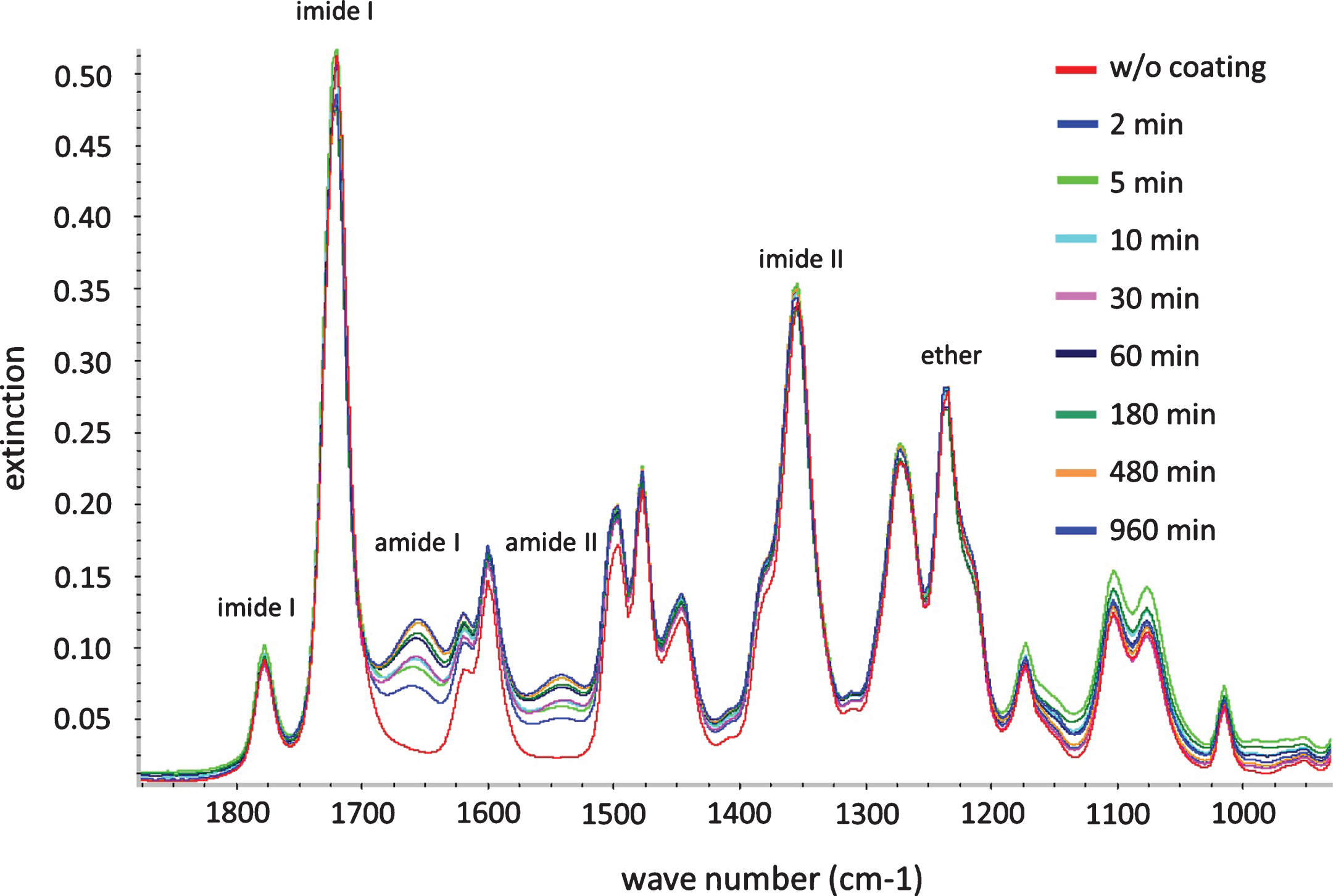
FT-ATR-IR spectra of a PEI membrane before (red colored line) and after albumin functionalization at different functionalization times (2 to 960 min) carried out at pH 7.4 and 40°C.
Functionalization with albumin led to an increase of the amide I-band (C=O stretching vibration) at 1660 cm–1 and of the amide II-band (C-N stretching vibration) at 1560 cm–1; the longer the reaction time (2 to 960 min) the stronger the increase.
PEI membrane functionalization for 960 min with albumin resulted in an amine content on the membrane surface of 175.7±7.2 nmol·cm2 (n = 6) which was 5.7-fold higher than without the albumin coating (31.0±3.4 nmol·cm2, n = 6). After desorption of the non-covalently bound albumin from the PEI membrane by using the detergent SDS (0.3 M NaOH/mol, 1% SDS), 171.4±5.1 nmol·cm–2 (n = 6) of the amines were left equivalent to a 2.5% reduction.
The advancing and receding contact angles (θadv and θrec) were 61.9±4.8° and 32.2±1.0° on the non-functionalized PEI membrane. PEI functionalization resulted in a significant decrease of the contact angle to θadv = 31.5±2.3° and θrec = 28.6±1.3. In accordance, the calculated hysteresis on the non-functionalized PEI was 29.7° and significantly higher than after functionalization with albumin (3.0°).
Surface roughness
The non-functionalized PEI membrane exhibited an almost homogenous surface topography characterized by a mean root mean square roughness of Rq = 6.47 nm (n = 9). Surface functionalization resulted in an increased Rq value of 9.92 nm (n = 9) showing a more heterogeneous surface topography (Fig. 2).
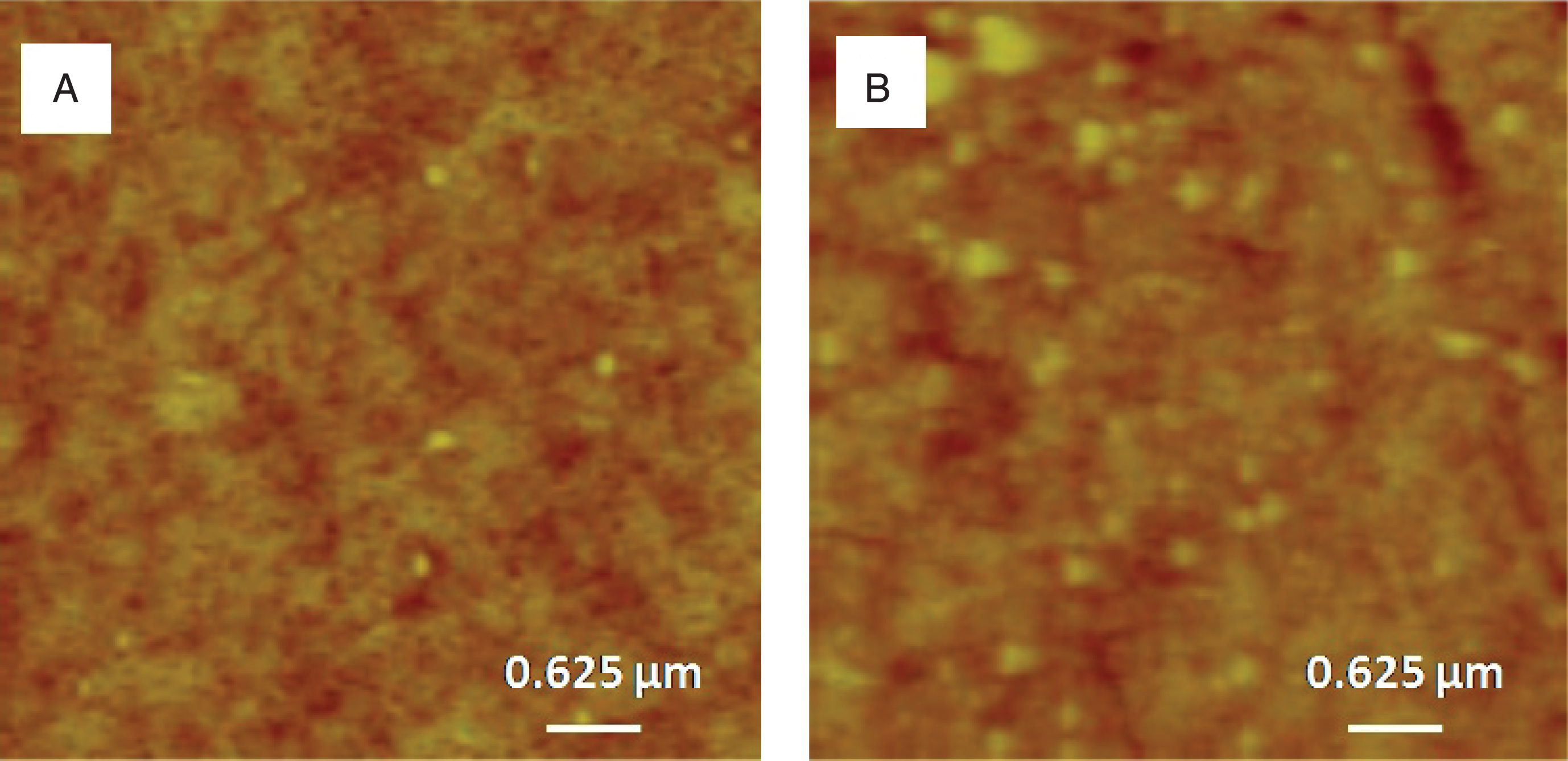
Surface roughness measured by Atomic Force Microscopy (AFM) of a PEI membrane (A) before and (B) after albumin functionalization at pH 7.4 and 40°C for 960 min. Brightness increased with increasing height (roughness); maximal height: 150 nm, n = 9.
Shear strength of PEI-blood vessel compounds (connecting area of 0.25 cm2) was strongly depending on the used power applied. At 250 mW the shear strength of the PEI-blood vessel connection measured 0.04±0.01 MPa. At a power of 450 mW the maximal shear strength (0.08±0.02 MPa) was reached (n = 7 each; Fig. 3). This shear strength corresponds to 11.4–22.2% (mean 15.1%) of the tensile strength of the native thoracic artery (native artery: min: 0.36 MPa, max: 0.70 MPa; mean: 0.53±0.24 MPa, n = 10). The use of higher laser powers did not result in higher shear strength. There was a decrease, rather, of the shear strength with laser powers surpassing 450 mW.
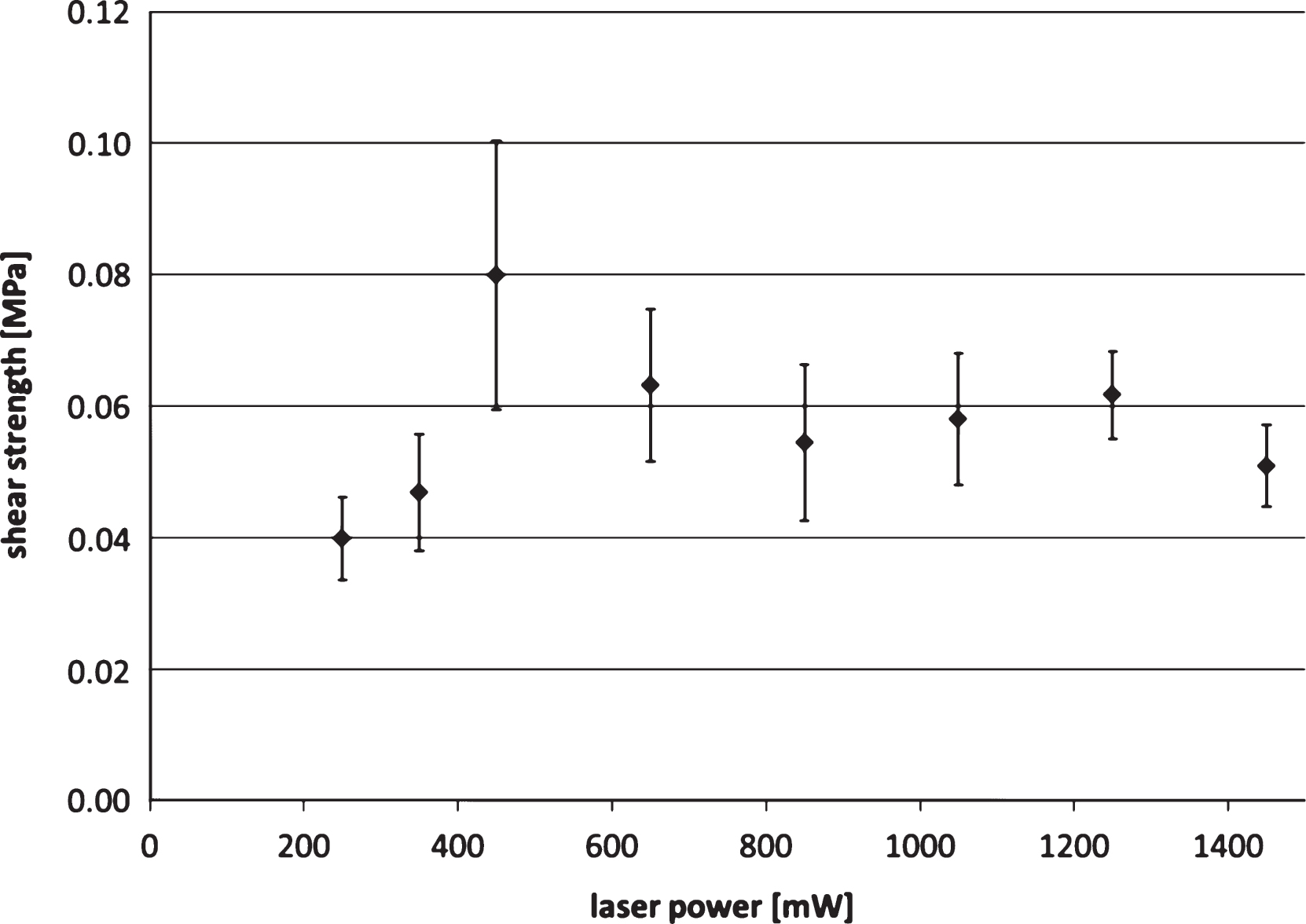
Shear strength of functionalized PEI membrane-blood vessel compounds coupled to one another by laser welding with a continuous wave diode laser emitting at 808 nm at different laser powers; means and standard deviation, n = 7.
The peripheral tunica media of the blood vessels changed due to the laser soldering process to a compact eosinophilic necrosis zone with clearly delineated collagen fiber bundles. The organization of the collagen fibers in this zone changed remarkably and the number of cell nuclei was drastically reduced (Fig. 4).
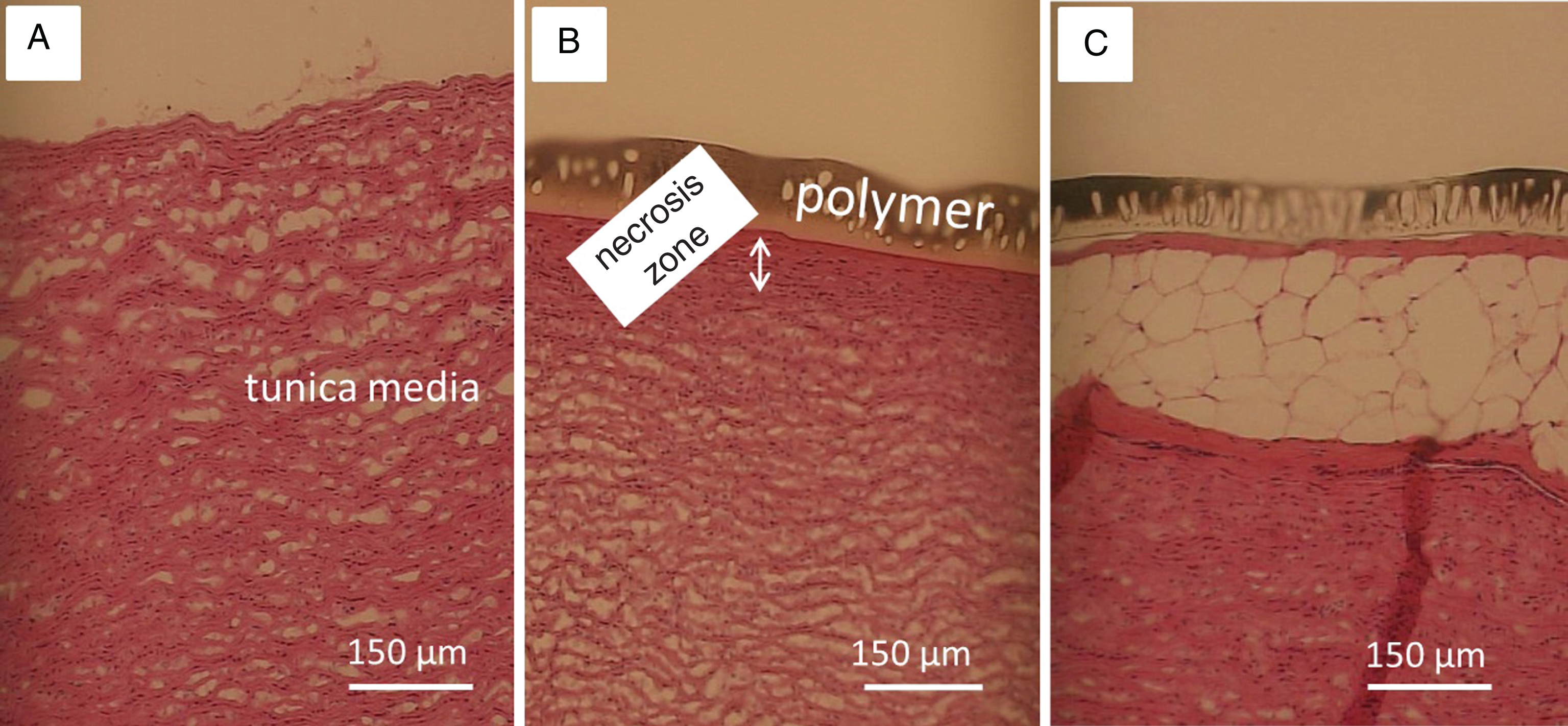
Histology of pig aorta wall alterations induced by thermal effects of laser soldering; HE staining of the extraluminal part (tunica media) of the thoracic artery without treatment (A); after welding together with an albumin-functionalized PEI membrane using a continuous wave 808 nm diode laser with a laser power of 450 mW (B) or 1500 mW (C).
Welding of the functionalized PEI membrane onto this dissected outer layer of the explanted pig aorta resulted in a more stable junction between both when the laser power did not exceed 450 nm (see Fig. 3). However, the outer layers of the tunica media were affected by the laser welding process, and a necrotic zone marked the interface between the PEI membrane and the blood vessel reaching from 36.4±9.0μm at 450 mW up to 122.2±24.8μm at 1500 mW (n = 7 each; see Fig. 5). Laser powers higher than 450 nm additionally caused non-connected focal areas which increased in size with increasing laser power.
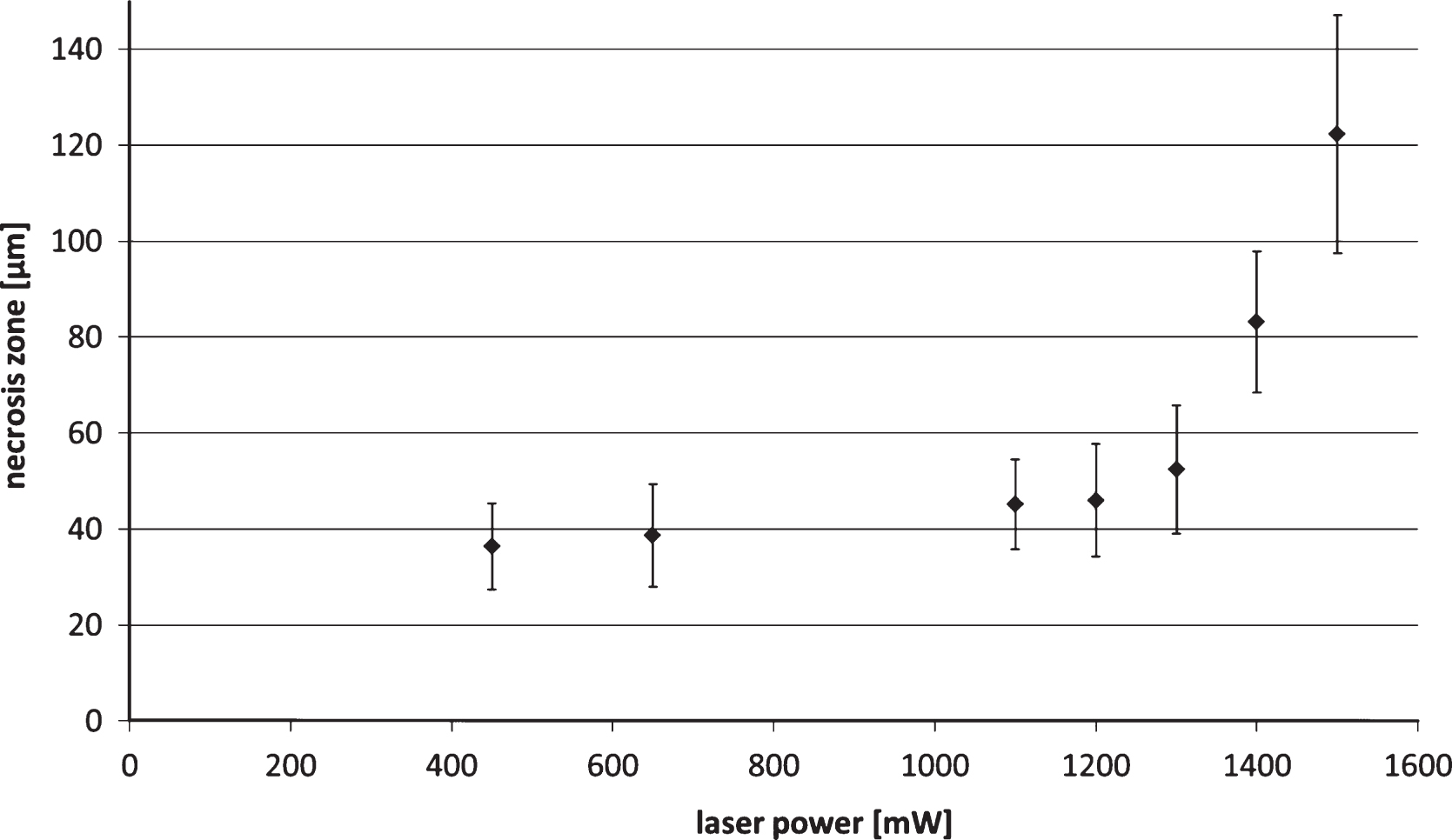
Necrosis zone of the outer tunica media after PEI-aorta junction generated by Laser welding using a continuous 808 nm diode laser with a laser power of 450–1500 mW; means and standard deviation, n = 7.
The PEI membrane itself with the characteristic finger pores was practically not affected by the laser welding process. With increasing laser power the size and the number of finger pores remained constant.
The conventional technique for blood vessel anastomosis is microsuturing, which can cause clot formation at the endoluminal perforation sites [1–5]. In addition, microsuturing requires major surgical skills and is often time consuming. Both aspects can adversely affect the therapeutic success especially under clinical conditions where temporary ischemia is an issue for the survival of anastomosed blood vessels. Another disadvantage of suturing is the necessity for the surgeon to have free moving space which limits the practicability of this technique for minimally invasive surgery. Laser tissue soldering (LTS) based on an ICG-mediated heat-denaturation of proteins might be a promising alternative technique. However, one of the major drawbacks of LTS is the weak shear strength of the solder welds when compared to sutures. In this study, it was tested whether sufficient shear strength of the solder welds can be achieved by a carrier membrane to which the chromophore-enhanced protein solder substrate is covalently bound and which can provide additional physical strength to the weld. The membrane also prevents the ICG to spread out over a large volume of tissue, thus failing to confine the welding procedure locally. Here the support membrane was made of the polymer PEI because this polymer allows steam sterilization, has shown good cell compatibility, and can be covalently functionalized through reaction of nucleophiles with the imide group [10, 12].
The study revealed that LTS using the albumin-functionalized PEI membrane was able to establish a polymer-blood vessel junction in an area of 0.25 cm2 whose shear strength corresponded to 15% of the shear strength of the native blood vessel. On the basis of an outer diameter of the thoracic artery of about 1.3 mm [17] it can be calculated that a length of an overlapping zone of 4.1 mm, which would span around the entire circumference of the blood vessel, could theoretically provide a shear strength of the PEI-blood vessel compound which would be identical to that of the native blood vessel.
The efficacy of the laser welding process is strongly dependent on the structure of the proteins which are used as soldering substrate. For this reason, PEI membrane functionalization with albumin was performed at a physiological pH of 7.4, and not at the commonly used alkaline pH range from 9–12, which favors protein deprotonation [11]. In addition, the temperature of the functionalization process was adapted to the need of an unaffected protein structure and set to +40°C, although higher temperatures (e.g. 70°C) would result in higher rates of amination [18, 19].
FT-ATR-IR analysis of the albumin functionalized PEI showed that the time period used for membrane functionalization had a strong impact on the intensity of carrier membrane amination. PEI functionalization with albumin caused an increase of the peaks of the amide I and II bands over time with peak maxima appearing after 960 min. Desorption of non-covalently bound albumin from the surface by a detergent resulted in a reduction of the amine content of the membrane of only 2.5%. This is the reason why it may be assumed that most of the albumin on the PEI membrane was bound by covalent bonding.
A significantly decreased advancing contact angle of θadv = 31.5±2.3° associated with a very low contact angle hysteresis of 3° was a further evidence for an effective functionalization of the PEI membrane. AFM investigation revealed an increase in surface roughness after albumin modification associated with a higher heterogeneity in surface topography, which might limit the reproducibility of LTS repaired tissues.
LTS induced fusion of the PEI-membrane-bound solder and adjacent tissue resulted in almost amorphous necrotic zones in the tissue characterized by a delineation of collagen fiber bundles and a reduced number of cell nuclei [4, 8]. LTS is not based on covalent binding between proteins of the solder and the adjacent tissue [18, 19]. LTS was described to cause heat-induced coagulation, instead, of the structural elements of the tissue through cell necrosis and denaturation. It is a rate process characterized by a linear proportionality to time and an exponential proportionality to temperature [20]. The temperature effect is strongly dependent on the laser absorption by the chromophore ICG. After light absorption, the ICG molecule can partially convert the absorbed energy to fluorescent light that is centered at 830 nm [21]. A further minor part of the absorbed light energy is transferred to ICG triplet T1 state that can generate reactive oxygen species [22]. About 85% of the absorbed energy can be converted to heat inside the ICG molecule by internal conversion [23]. Therefore, heat generation and heat transfer are the major processes after light absorption by ICG [24]. Since in the study presented ICG concentration on the PEI membrane was not changed, the primary determinant of temperature development in solder and tissue during LTS must have been laser power density.
Shear strength of the polymer-tissue compound showed a maximum at a laser power output of 450 mW. Higher laser powers can generate higher welding temperatures and deeper zones of protein denaturation as described by Poppas et al. [25] and McNally et al. [4], but they do not coincide necessarily with an elevation of compound shear strength, as laser powers higher than 450 mW resulted in this study in reduced shear strength of the PEI-tissue-compound. The main reasons for this phenomenon are assumed to be the blistered distensions which characterized the solder adjacent tissue if laser powers higher than 450 mW were applied. The size of the blisters increased with increasing laser power, and surpassing a power of 1200 mW the blistering effects resulted in a positive exponential correlation between the necrosis zone and the laser power. This reduced the shear strength to one third (36.3%) of the shear strength which was achieved with a laser power of 450 mW.
Conclusion
In summary, the results of this study showed that covalent bonding of liquid albumin solders on a polymer carrier can provide benefits with respect to solder allocation and to cohesive strength between solder and tissue. However, results are based on in-vitro tests and were performed in dried tissues. Before the clinical applicability of the method can be assessed, additional research is needed to determine the acute and chronic results under wet conditions and in an appropriate animal model.
Footnotes
Acknowledgments
This work was supported by the Helmholtz-Association through programme-oriented funding. The authors acknowledge the experimental assistance of Manuela Keller.
This paper is dedicated to the 70th birthday of Prof. Friedrich Alfons Jung.