



Abstract
Bioengineered cell substrates are a highly promising tool to govern the differentiation of stem cells in vitro and to modulate the cellular behavior in vivo. While this technology works fine for adult stem cells, the cultivation of human induced pluripotent stem cells (hiPSCs) is challenging as these cells typically show poor attachment on the bioengineered substrates, which among other effects causes substantial cell death. Thus, very limited types of surfaces have been demonstrated suitable for hiPSC cultures. The multilayer coating approach that renders the surface with diverse chemical compositions, architectures, and functions can be used to improve the adhesion of hiPSCs on the bioengineered substrates. We hypothesized that a multilayer formation based on the attraction of molecules with opposite charges could functionalize the polystyrene (PS) substrates to improve the adhesion of hiPSCs. Polymeric substrates were stepwise coated, first with dopamine to form a polydopamine (PDA) layer, second with polylysine and last with Laminin-521. The multilayer formation resulted in the variation of hydrophilicity and chemical functionality of the surfaces. Hydrophilicity was detected using captive bubble method and the amount of primary and secondary amines on the surface was quantified by fluorescent staining. The PDA layer effectively immobilized the upper layers and thereby improved the attachment of hiPSCs. Cell adhesion was enhanced on the surfaces coated with multilayers, as compared to those without PDA and/or polylysine. Moreover, hiPSCs spread well over this multilayer laminin substrate. These cells maintained their proliferation capacity and differentiation potential. The multilayer coating strategy is a promising attempt for engineering polymer-based substrates for the cultivation of hiPSCs and of interest for expanding the application scope of hiPSCs.
Introduction
Human induced pluripotent stem cells (hiPSCs) are capable of differentiating into all types of cells that originate from the three germ layers, which makes them an attractive cell source for bioengineering [1, 2]. Thus, there is a need for large-scale hiPSCs in vitro culture [3]. Compelling evidence showed that the application of an appropriate polymeric substrate could effectively support the survival and proliferation of stem cells and specifically direct the differentiation of stem cells [4]. Among these substrates, synthetic polymers, such as poly(ɛ-caprolactone), poly(lactide-co-glycolide) and poly(n-butyl acrylate), can be produced and processed on a large scale and have shown promising cell compatibility [5].
Polymeric substrates could exert a profound influence on hiPSCs [4, 6]. For example, hiPSCs cultured on different substrates exhibited alteration in adhesion, morphology, proliferation, polarity, migration, and differentiation [5, 8]. As an early step of cell-material interaction, cell adhesion plays an important role in modulating cell behaviors, and even determines the success or failure of implantation of biomaterial-stem cell constructions [9, 10]. However, the absence of hiPSC binding motifs on synthetic polymer substrates greatly limits their application [11, 12]. Thus, it is essential to improve the adhesion of pluripotent stem cells to the cell culture substrates.
To date, various approaches have been applied with the aim to enhance the hiPSC adhesion on polymeric surfaces, including physical adsorption and covalent immobilization of extracellular matrix (ECM) molecules [13, 14]. Laminin (LN) is a ubiquitous component in the ECM, playing a critical role for regulating cellular functions via providing both biochemical and physical cues. Cell-LN interaction mediates the downstream signaling cascades related to cell proliferation, differentiation, survival and function [15, 16]. Among different LN isoforms expressed in hiPSCs, LN521 has been identified as a specific one that allows survival and self-renewal of hiPSCs [17, 18]. However, traditional protein coating approaches such as physical adsorption can end up with an inhomogeneous protein surface, greatly limiting hiPSC expansion and differentiation capacity [19]. Therefore, it is necessary to develop a simple, stable and robust method for LN521 coating to improve hiPSC adhesion and maintain their self-renewal and differentiation potential.
Dopamine can polymerize in acidic, neutral and alkaline aqueous media, which provides a versatile platform to modify substrate surfaces [20]. In a weak alkaline environment (pH = 8.5), dopamine can self-polymerize to form polydopamine (PDA), which can be deposited easily on almost all inorganic and organic surfaces. Modification of the cell culture surface with PDA could not only increase the cell adhesion but also covalently and/or physically immobilize functional bio-signaling molecules [21]. However, the immobilization of LN521 on the cell culture surface via PDA might be compromised by electrostatic repulsion, since both LN521 and PDA are negatively charged at physiological pH [22]. The use of a positively charged substance, for example polylysine (pLys), as an intermediate layer might be able to address this problem. Here, we hypothesized that coating the polymeric substrates with first PDA and then pLys might facilitate the adsorption of LN521, which could consequently improve the hiPSC adhesion and maintenance of stem cell properties. A stepwise coating protocol was applied to achieve the coating on PS with multilayers, consisting of dopamine, pLys and LN521 from bottom to top. The surface property of the coated surface was characterized. The adhesion, viability, pluripotency, and differentiation of hiPSCs on the surface were evaluated.
Methods
Polystyrene surface coating and characterization
Untreated polystyrene surfaces (ThermoFisher, Langenselbold, Germany) were first coated with 3,4-dihydroxyphenethylamine (dopamine) solution (Sigma-Aldrich, Deisenhofen, Germany) according to previous reports [23, 24]. Briefly, a 1.0 mg/mL dopamine solution was prepared in advance by using a Tris-HCl buffer solution (10 mM, pH 8.5) as a solvent. Next, the dopamine solution was filtered through 0.2 μm filters immediately. Then polystyrene substrates were immersed in the freshly prepared dopamine solution in an open vessel, in continuous contact with atmospheric oxygen. After reacting for 2 h, the substrates were washed alternately with sterile PBS and distilled water (Gibco, ThermoScientific, Karlsruhe, Germany) to remove non-firmly adsorbed coating reagents. Then, the substrates were air-dried (at room temperature) and used for characterization or further coating. The dopamine-coated substrates were divided into two groups. PBS group: substrates were immersed in PBS (with Ca2+ and Mg2+, ThermoFisher, Karlsruhe, Germany) overnight at 4 °C. LN521 group: substrates were immersed in 5 μg/mL LN521 (LN521, BioLaminin, Sundbyberg, Sweden) overnight at 4 °C with or without the assistance of pLys. Tissue culture plate (TCP) directly coated with 5 μg/mL LN521 solution overnight at 4 °C was used as a positive control (TCP-LN521). After that, the LN521 solution was aspirated, and the samples were washed twice with sterile distilled water before any experimental analysis. Then, the hydrophilicity of the polystyrene substrates with or without coating was characterized using the captive bubble method in deionized water on a DSA 100 Analyzer (Krüss GmbH, Hamburg, Germany).
Quantitation of primary and secondary amines
The amount of primary and secondary amines present on the polystyrene substrates was determined by using the IRDye 800CW NHS Ester Kit (LI-COR, Bad Homburg, Germany). After different coating procedures, the surface modified substrates were washed twice with PBS to remove non-adherent protein molecules. The IRDye 800CW NHS Ester was added to the substrate surfaces. Following this, the absorbance at 800 nm was measured with microplate readers (Odyssey Imaging Systems, LI-COR, Bad Homburg, Germany). Fluorescent intensity was analyzed using Image Studio Lite Ver 5.2.
hiPSC culture
The BIHi001-A hiPSC cell line was a gift from the Stem Cell Core Facility, Berlin Institute of Health (BIH). This cell line was generated from human foreskin fibroblasts (HFF; CCD-1112Sk, American Type Culture Collection (ATCC) via using Sendai virus vectors expressing Oct3/4, Sox2, Klf4, and c-Myc. Detailed information and characterization data of this cell line is available in the hPSCreg database (http://hpscreg.eu/cell-line/_BIHi001-A). The hiPSCs were maintained on 5 μg/mL LN521 coated TCP under feeder-free conditions in a humidified atmosphere of 95% (v/v) air and 5% (v/v) CO2 at 37 °C. The culture medium was Essential 8 (Gibco, ThermoFisher Scientific, Karlsruhe, Germany). Upon reaching 85% confluence, the cells were dissociated by Accutase (Gibco, ThermoFisher Scientific, Bonn, Germany) and used for the subsequent experiments.
Cell adhesion determination
For the analysis of cell adhesion on the coated surfaces, phase contrast images were captured and their numbers were evaluated. Cells that adhered on the surface were imaged using an IX81 motorized inverted microscope (Olympus, Hamburg, Germany). Cell numbers were measured by quantifying the DNA content. Briefly, hiPSCs were seeded at a density of 5.0×10⁴ cells per well onto coated surfaces in a 24-well plate and incubated for 6 h. The non-adherent cells were removed by rinsing twice with PBS. The adherent cells were frozen at –80 °C. The frozen cells were thawed and lysed with 0.2% (v/v) Triton X-100 (USB, Cleveland, USA). Following this, DNA Quantitation kit (Sigma-Aldrich, Darmstadt, Germany) was added to the cell lysis and the fluorescence intensity was measured using microplate readers (Infinite 200 PRO, Tecan Group Ltd., Männedorf, Switzerland). The readings of each group were then normalized against the fluorescence intensity of the positive control group and expressed as the relative fluorescent intensity. Three replicates (n = 3) from each group were measured.
Proliferation assay
Following attaching to the coated polystyrene substrates, hiPSCs were cultured over a period of 48 h and tested for viability and proliferation at 24 h and 48 h. Cell Counting Kit-8 (CCK-8, Sigma-Aldrich, Darmstadt, Germany) was used for the quantification of cell proliferation at the designated time points. After incubation with the CCK-8 solution for 3 h at 37 °C, the absorbance value at 450 nm was detected using microplate readers (Infinite 200 PRO, Tecan Group Ltd., Männedorf, Switzerland).
Immunofluorescent staining
hiPSCs cultured on the coated substrates were fixed using 4% paraformaldehyde (PFA, w/v in PBS, Sigma-Aldrich, Seelze, Germany) at room temperature for 10 minutes. Then the fixed cells were permeabilized with 0.2% (v/v) Triton X-100 (USB, Cleveland, USA) and washed with PBS three times prior to immunostaining. For the staining of the proliferation marker, Ki67 (Thermo Scientific, Karlsruhe, Germany) was used. For pluripotency evaluation, samples were incubated with Nanog primary antibody (Thermo Scientific, Karlsruhe, Germany) overnight at 4 °C. To stain the F-actin, ActinRed 555 ReadyProbes Reagent (Life Technologies, Darmstadt, Germany) was used. Then, samples were washed with PBS containing 1% (v/v) Tween 20 (PBST, Sigma-Aldrich, Schnelldorf, Germany) and incubated with the secondary antibody (Thermo Scientific, Darmstadt, Germany) at room temperature in the dark for 2 h. Then, nuclei were stained with 0.5 μg/mL 4’,6-diamidino-2-phenylindole (DAPI, Sigma-Aldrich, Taufkirchen, Germany). Images were carried out via an Inverted Fluorescence Microscope (IX81 motorized inverted microscope, Olympus, Hamburg, Germany). Fluorescence intensity of each fluorescence image was quantified using ImageJ software (NIH, Maryland, USA).
Flow cytometry analysis
To further test the pluripotency of hiPSCs on the coated substrates, we examined the ability to differentiate into the three embryonal germ layers, ectoderm, mesoderm, and endoderm. The differentiation processes were completed in 7 days using StemMACS Trilineage Differentiation Kit (Miltenyi Biotec, Bergisch Gladbach, Germany). Briefly, hiPSCs were dissociated into single-cell suspensions. Then the cells were seeded into 12-well plates with defined cell numbers and media changes were performed according to the manufacturer’s protocol. On day 7, the differentiated cells were harvested and the differentiation efficiency was analyzed by flow cytometry (MACSQuant Analyzer 10, Miltenyi Biotec, Bergisch Gladbach, Germany). The following antibodies (all purchased from Miltenyi Biotec), CD140b-APC (Order no. 130-105-280), CD144-FITC (Order no. 130-100-713), CD184-APC (Order no. 130-109-886), Pax6-APC (Order no. 130-107-829), Sox2-FITC (Order no. 130-104-993), and Sox17-Vio-515 (Order no. 130-111-147) were used to quantify the differentiation efficiency.
Statistical analysis
All results presented were from at least three independent experiments for each condition. Data are presented as mean±standard deviation (SD). Statistical analysis was performed using independent-samples t-test. Differences were considered statistically significant at p < 0.05.
Results
Surface coating and characterization
In order to functionalize the polymeric substrates, a multilayer coating method was applied as summarized in Fig. 1A. The water contact angle of the unmodified surfaces was 60.5°±8.6°, representing a hydrophobic character of the surface (Fig. 1B). The water contact angle was significantly decreased after surface coating. The values of water contact angle in PDA-LN521 group (10.1°±3.9°) and PDA-pLys-LN521 group (5.17°±2.05°) were significantly lower than in the PS (negative control) group and in the TCP-LN521 (positive control) group (29.1°±5.8°). A similar tendency was observed in hysteresis (Fig. 1C). The amount of primary and secondary amines on the substrates were quantified with IRDye 800CW NHS Ester. In TCP-LN521 group, a very strong fluorescent signal was detected, suggesting a large amount of primary and/ or secondary amino groups were introduced to the surface (Fig. 1D). No significant difference was observed on the PDA-pLys-LN521 group. Significant decrease in PDA, PDA-pLys, and PDA-LN521 groups after modification was observed in comparison with TCP-LN521.
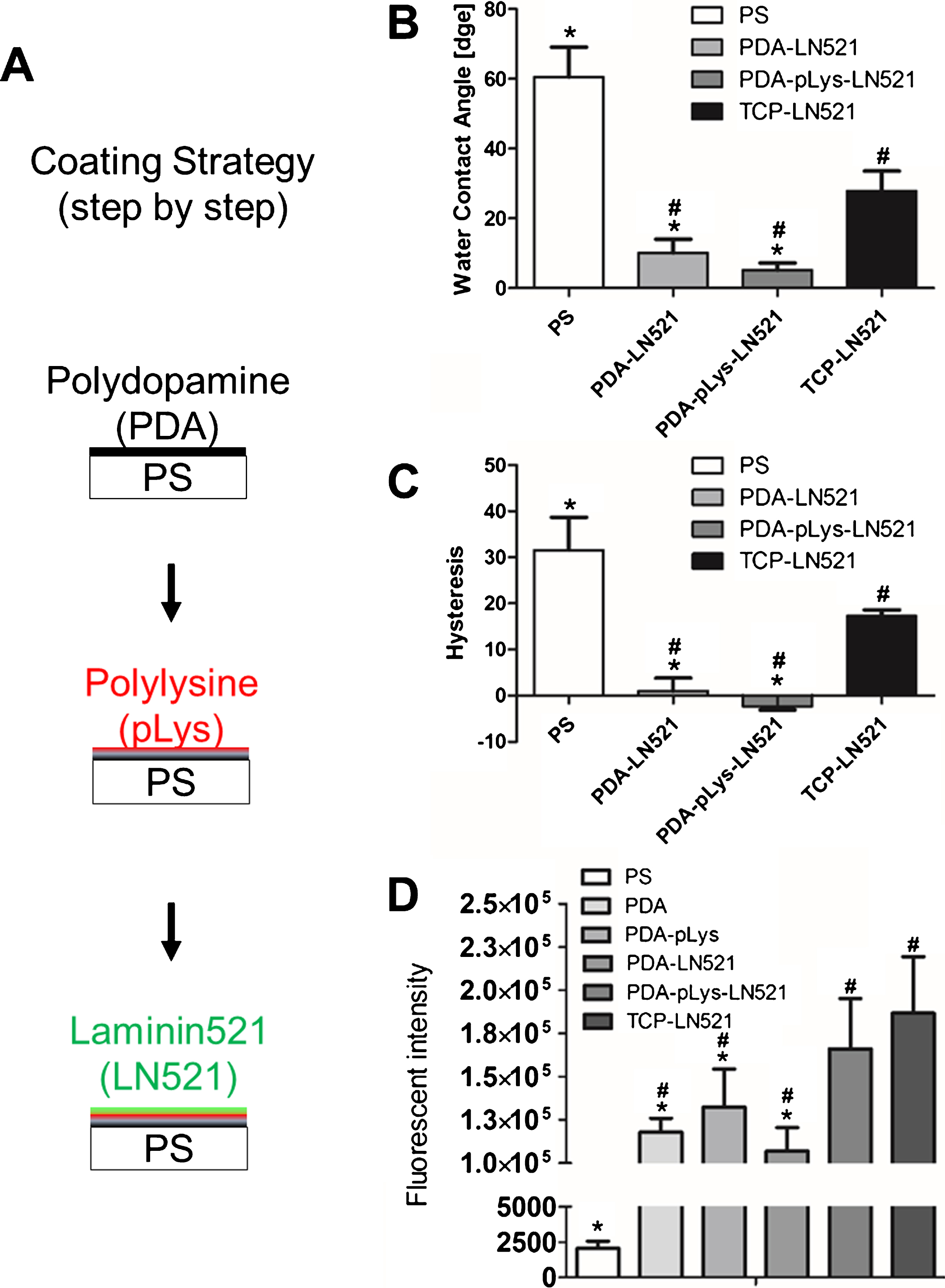
Surface modification strategy and characterization. (A) Scheme of the surface modification protocol. (B) Measurement of water contact angle after surface coating. (C) Analysis of contact angle hysteresis after surface coating. (D) Quantification of primary and secondary amines present on the surface. (n = 3, *p < 0.05 versus TCP-LN521, # p < 0.05 versus PS).
To investigate the effect of surface modification on cell adhesion, the initial adhesion of hiPSCs was evaluated 6 h after seeding (Fig. 2A). Relative fluorescent intensity was used to represent the amount of cells as there was a positive correlation between these two values. The amount of hiPSCs that attached to the surface was used to compare the cell attachment among different coating materials. Very low amount of hiPSCs attached on the surface coating without LN521. There was a strong increasement on cell attachment at 12 h on PDA-pLys-LN521 substrate (Fig. 2B). In addition, the morphology of the cells growing on the substrates was examined at 24 h. As shown in Fig. 2C, hiPSCs on the PDA-pLys-LN521 surface showed a more flatten/atypical morphology with a more homogenous distribution of F-actin.

hiPSC adhesion and morphology. (A) Relative fluorescent intensity of DNA isolated from attached cells at 6 h after seeding (n = 3, *p < 0.05 versus TCP-LN521, #p < 0.05 versus PS). (B) Cell adhesion on differently coated surfaces. Nuclei were counterstained with DAPI (blue), scale bar = 100 μm. (C) Representative staining image of hiPSC morphology (red: F-actin, blue: DAPI, scale bar = 200 μm). The reader is referred to the web version of this article, where the coloured version of the figure is provided.
Cell proliferation was measured to evaluate the growth of hiPSCs on functionalized surfaces (Fig. 3A). hiPSCs proliferated robustly on the PDA-pLys-LN521 surface. There was a significant increase in hiPSC number on PDA-pLys-LN521 coated substrates compared to PDA-LN521 coated surfaces after 48 h. This result was further confirmed by evaluating the expression of the proliferation marker Ki67. Cells on PDA-pLys-LN521 expressed a higher level of Ki67 than on PDA-LN521 (Fig. 3B).
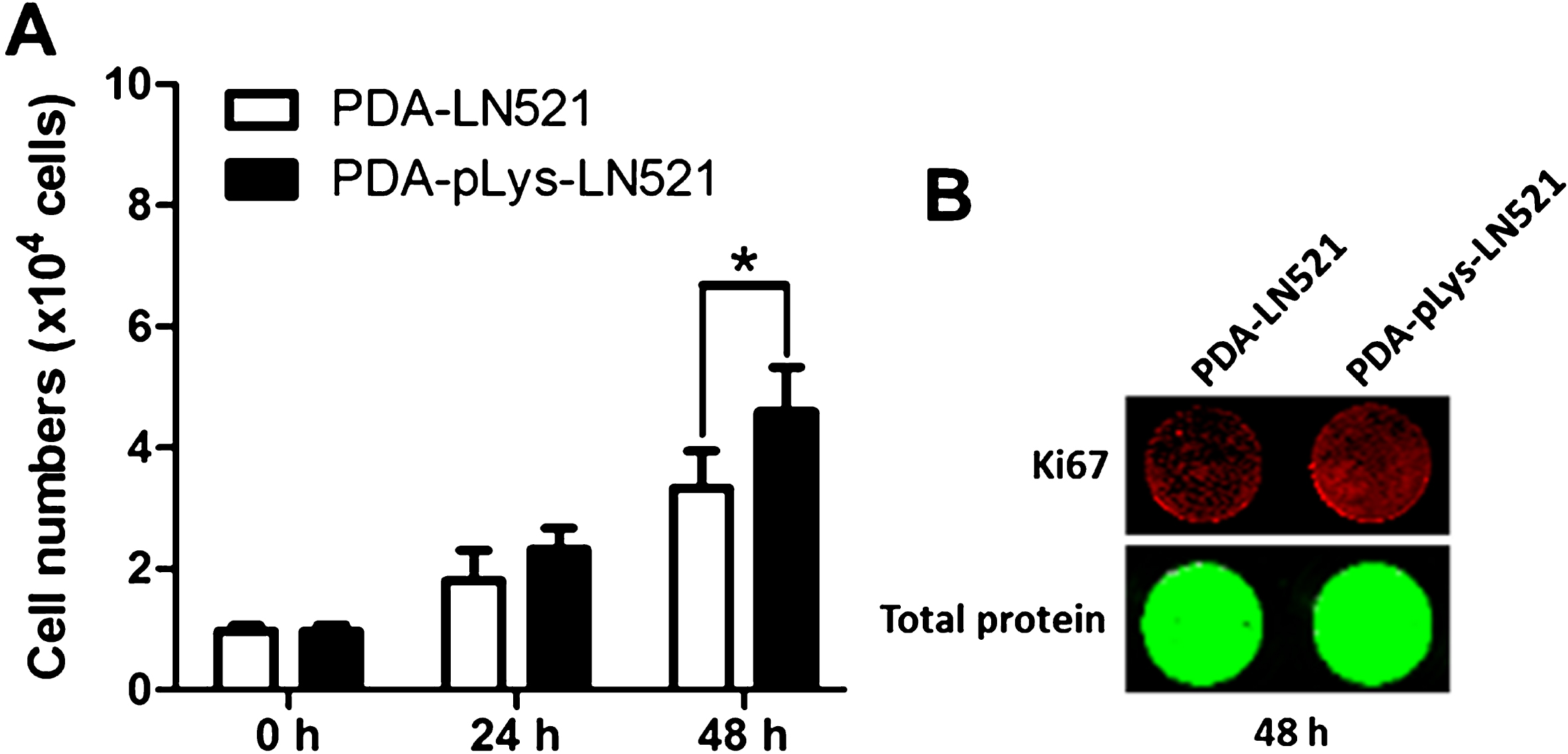
Cell proliferation. (A) Cell proliferation on surfaces with different coatings. Cell numbers were quantified at indicated time points after seeding. (n = 3, *p < 0.05). (B) The representative images of Ki67 expression in cells attached to surfaces with different coatings. Total protein was quantified with NHS ester.
In order to study the influence of surface coating on the pluripotency of hiPSCs, we examined the expression of pluripotent marker Nanog and the differentiation of cells on PDA-LN521 and PDA-pLys-LN521 surfaces. The pluripotency of hiPSCs on the multilayer-coated substrates was fully preserved. hiPSCs cultured on both multilayer-coated surfaces were highly positive for Nanog at 48 h, suggesting the coating materials did not affect the maintenance of pluripotency in a short period (Fig. 4A). Further, hiPSCs cultured on both surfaces were able to differentiate into all three germ layers. Notably, a 10% increase in the mesoderm differentiation was observed in the cells cultured on the PDA-pLys-LN521 surface as compared to those on PDA-LN521 surface (Fig. 4B).
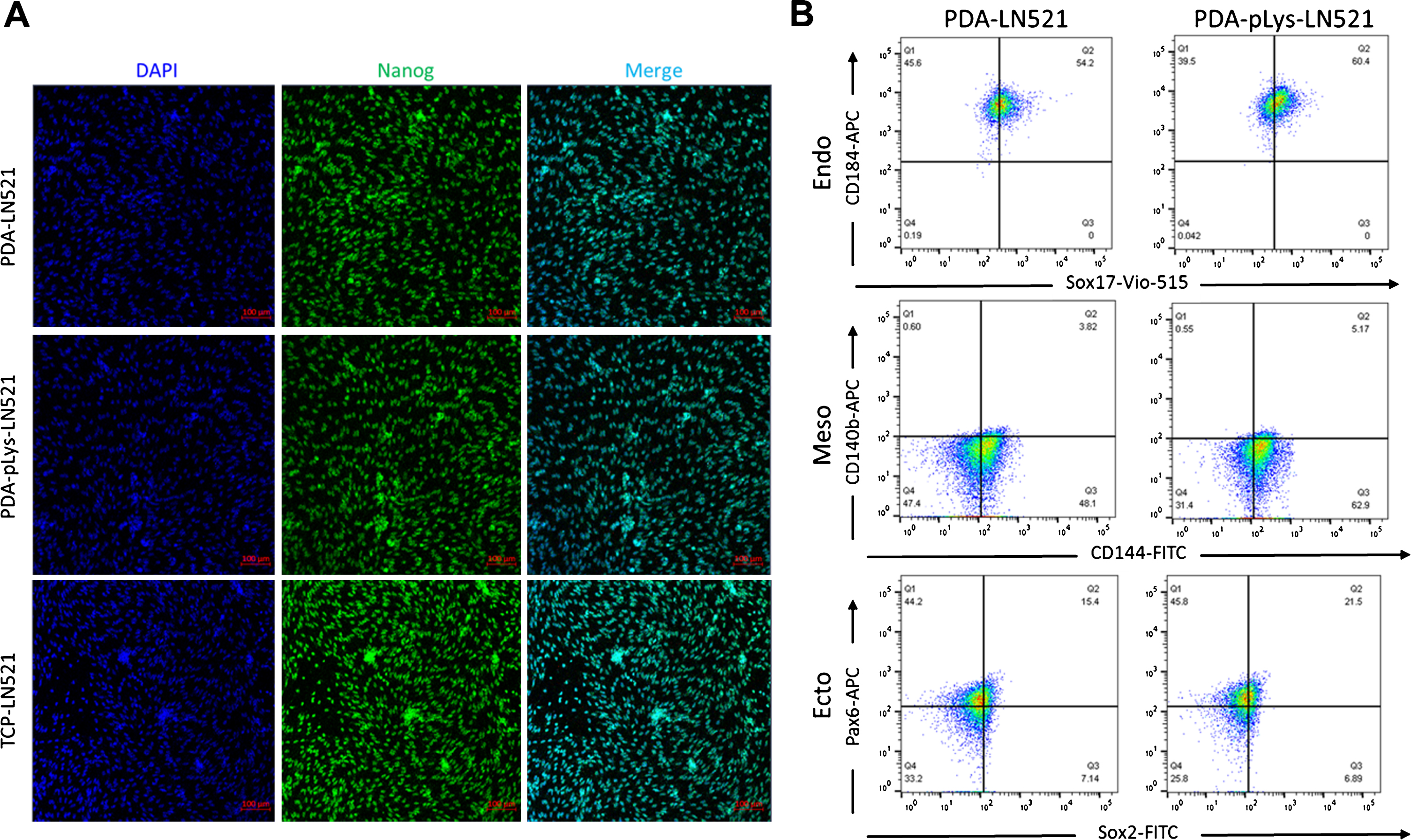
Pluripotency of hiPSCs on multi-layer coated surfaces. (A) Representative staining image of hiPSCs. Pluripotent marker Nanog was immunostained (green) and cell nuclei were counterstained with DAPI (blue) (scale bar = 100 μm). (B) Differentiation of hiPSCs cultured on different surfaces was assessed via flow cytometry analysis. CD184 and Sox17 were used as endodermal lineage markers, CD144 and CD140b were used as mesodermal lineage markers, and Pax6 and Sox2 were used as ectodermal lineage markers.
In this study, we assessed the possibility of dopamine-coating strategy as a universal method to modify the polymeric substrates as hiPSC culture materials. Unmodified hydrophobic polystyrene (PS) was chosen as the starting material and functionalized with PDA. After PDA modification, the substrates were further coated with LN521 with or without the assistance of pLys. The adhesion of hiPSCs was significantly improved on the PDA-pLys-LN521 coated substrates. More important, the pluripotency of hiPSCs on the PDA-pLys-LN521 surface was maintained.
Cells can sense and respond to the physical and chemical features of culture surface, to which they adhere [7, 10]. Recent studies showed that hydrophilic context favored the cell adhesion and further influence cell behaviors including morphology, proliferation, and eventually cell fate choice [9, 26]. In our study, the increase in hydrophilicity of the PS surface was achieved via PDA modification, which introduced hydrophilic groups. The wettability of PDA modified surfaces was further increased after LN521 or pLys-LN521 deposition. The PDA-pLys-LN521 surface showed the highest hydrophilicity as well as the greatest homogeneity. This observation was in accordance with the reported result that modification of PDA coated surface with positively charged molecules and/or ECM proteins could further increase the hydrophilicity as well as the coating homogeneity [27]. The amount of primary and secondary amines on PDA, PDA-pLys, and PDA-LN521 surfaces were significant lower than on TCP-LN521 surfaces. However, no significant difference in the amine amount was observed between the PDA-pLys-LN521 and TCP-LN521 group. This might indicate that the PDA-pLys-LN521 surface was comparable to the TCP-LN521 surface for hiPSC adhesion, as the functional groups present on the culture surface such as carboxylic groups and amino groups can influence the hiPSC adhesion [28].
Engineered substrates with ECM components that provide adhesive signals could enhance the initial cell adhesion to the culture substrates, as the cellular adhesion is dependent on the interaction between the cell membrane receptors and the ligands on the culture substrates [29, 30]. Adhesion molecules, including fibronectin (FN), vitronectin (VN) and LN have been used to modify TCP, which present a well-defined and effective platform for in vitro hiPSC culture [1, 31]. However, based on our result, modification of PS via physical adsorption of LN did not dramatically promote the adhesion of hiPSCs, which complies with the enhanced hiPSC attachment on VN coated PS [27]. Therefore, PDA modification that allows the immobilization of biomolecules to a wide variety of substrate surfaces with batch-to-batch consistence and high conjugation efficiency could be a solution to address this problem [21, 28].
Both PDA-LN521 and PDA-pLys-LN521 surfaces supported the initial adhesion and the survival of hiPSCs as small colonies or single cells during continuous subculture, which is consistent with previous studies, that increased hydrophilicity, favoring the cell adhesion and growth [26]. Recent studies showed that though directly grafted ECM proteins such as VN to the PDA-modified surface enhance the cell adhesion, the long-term survival of hiPSCs was not promoted, which might be attributed to the grafting process affecting the orientation and three-dimensional structure of VN [27, 32]. Decorating the PDA surface with a linker could control the conjugation of biomolecules and preserve their bioactivity [32]. Here, pLys was used to link PDA to LN521. The hiPSCs on PDA-pLys-LN521 showed a spread morphology and the distribution of F-actin was more homogenous as compared to those on PDA-LN521. Taken together, our result might indicate the bioactivity of LN521 was better preserved in the PDA-pLys system [33].
The hiPSC adhesion to substrates was mediated by the interaction between LN and integrins, which also plays a crucial role in cell proliferation [34]. It has been reported that the binding of LN521 to integrin α6β1 could improve the proliferation of hiPSCs [35, 36]. Consistent with the previous studies [18, 37], our data showed that the PDA-pLys-LN521 modified culture system improved cell proliferation as cells generally need to attach to a supporting surface and spread out in order to grow. Thus, PDA-pLys-LN521 coating offered a suitable and applicable approach for the in vitro expansion of hiPSCs, which will stimulate fundamental research as well as future applications [38].
The ability of hiPSC differentiation into cell-type representatives of all three germ layers offers an attractive approach for the bioengineering and in the far future might generate options for cell therapy [15, 38]. Considerable progress has been made in improvement of the efficiency and reliability of hiPSC differentiation [7, 9]. A recent study showed that monolayer-based methods could increase the differentiation efficiency of hiPSCs [39]. The reason might be that the cells are exposed equally to the factors controlling differentiation processes during expansion in monolayer culture systems [40]. The PDA-pLys-LN521 surface supported the maintenance of hiPSCs in monolayer cultures. The pluripotency of hiPSCs on the PDA-pLys-LN521 coated surface was well preserved, as the hiPSCs on the PDA-pLys-LN521 coated substrates were able to differentiate into all three germ layers. Interestingly, there was an increase of approximately 10% in the expression of CD144 (vascular endothelial cadherin), indicating the mesodermal differentiation was promoted under this condition. Our results demonstrate that surface modification of cell culture substrate might be potentially applied to regulate hiPSC differentiation lineage, which might contribute to the development of hiPSC-based cell therapies.
Conclusion
The present study offers a general route to functionalize polymeric substrates to enable hiPSC cultures. This technology effectively extends the applicability of synthetic biomaterials for cell culture system supporting pluripotent stem cells. However, in our study, there is no structural similarity between PDA aggregates and PDA film. Therefore, further research is needed to elucidate the structural difference at the interface and their influence on hiPSC behaviors. Moreover, the uncontrollable self-polymerization of dopamine and the undefined mechanism of PDA formation remain the main handicaps for its application. Conclusively, surface modification using dopamine allowing cell adhesion provides tremendous potential but is still in its initial stages.
Footnotes
Acknowledgments
We thank the Berlin Institute of Health (BIH) Stem Cell Core Facility for kindly providing us with the hiPSC line BIHi001-A. This work was financially supported by the Helmholtz Graduate School of Macromolecular Bioscience (MacroBio), Helmholtz-Portfolio Topic “Technology and Medicine” and Helmholtz Virtual Institute “Multifunctional Biomaterials for Medicine (VH-VI-423), and programme-oriented funding”. Yan Nie is grateful for support from the China Scholarship Council (CSC) (No. 201706205049).