
Abstract
OBJECTIVE:
Here we examined the influence of methane-rich saline treatment (MS) on the whole transcriptome of the skin flaps during the ischemia/reperfusion (I/R) injuryusing RNA-sequence (RNA-seq).
METHODS:
The rats were divided into three groups: the sham surgery group (SH),the I/R surgery group treated with physiological saline (I/R-P) or the I/R surgery group treated with the methane-rich saline (I/R-M) respectively. On the 72 hours after operation, the perfusion and the distribution of micro-circulatoryblood flow in skin flaps were observed by laser doppler flowmeters. The whole transcriptome alteration of the skin flaps was examined using RNA-seq. Moreover, the responses of the skin flaps to MRS treatment were examined using bio-informatic and q-PCR approaches after I/R injury.
RESULTS:
The methane-rich saline (MS) treatment could expand survival area and improve the blood perfusion of the skin flaps after l/R injury. Compared to the I/R-P group, 474 genes significantly altered in the I/R-M group. These genes were mainly associated the development, the cell adhesion and migration. In addition, the PI3K-Akt signal pathway was meaningfully related to regulation of MS treatment. Q-PCR results confirmed that MS treatment positively regulated PI3K-Akt signal pathway relative genes and inhibited the cell adhesion relative genes.
CONCLUSION:
These results proved that methane-rich saline may alleviate I/R injury and improve flap survival rate by regulating cell adhesion and PI3K-Akt signal pathway.
Keywords
List of abbreviations
sham surgery group ischemia/reperfusion ischemia/reperfusion surgery groups treated with physiological saline ischemia/reperfusion surgery groups treated with methane-rich saline methane-rich saline RNA-sequence
Introduction
The skin flap transplantation is widely used for wound repair and reconstruction [1, 2]. Although the technologies of flap surgery have been greatly improved with the development of clinical surgery, the flap loss caused by I/R injury after transplantation is still an unsolved problem [3]. The reactive oxygen and nitrogen species produced during I/R injury could activate the adhesion molecules and lead to massive influx of neutrophils. The neutrophil infiltration would bring more free radicals, cause cell apoptosis and tissue injury [4, 5].
Many studies showed that methane had the protective effect on the ischemia/reperfusion injury in hepatic, myocardial and intestinal ischemia/ reperfusion models [6–8]. Liu et al. proved that hydrogen-rich saline could ameliorate ischemia/reperfusion injury and improve flap survival rate by inhibiting the apoptosis factors [9]. In previous study we demonstrated that methane-rich saline treatment could inhibit apoptosis and improve skin flap survival in rats during I/R injury [10]. However, the exact mechanism of methane-rich saline treatment in skin flap I/R injury was currently unknown.
Extensive gene expression profiling analysis was valuable for the exploration of underlying mechanisms of methane protection. Over the past 10 years, the high throughput next-generation sequencing technologies have revolutionized transcriptomics by providing opportunities for multidimensional studies of cellular transcriptomes [11, 12]. As a main quantitative transcriptome profiling platform, RNA-seq has been considered a new experimental method to replace micro-array which can be used to investigate transcript structures in context of transcription start sites, alternative splicing patterns and other post-transcriptional modifications.
Until now, there were few studies to examine the responses of skin flap in I/R injury to methane-rich saline at entire transcriptome level. In this study, we aimed to characterize the transcriptome profile of skin flap during I/R injury after methane-rich saline treatment and investigate functional regulatory networks of methane-rich saline treatment. This study could provide the useful information for understanding MRS-induced gene expression and ultimately help to explore the underlying mechanism of methane-rich saline treatment in I/R injury at the whole genetic level.
Materials and methods
Animals and grouping
This animal experiment was approved by the Experimentation Ethics Committee on Animal Rights Protection of Peking Union Medical College Hospital. All procedures strictly followed the National Institutes of Health guidelines for the care and use of laboratory animals.
Thirty 8-week-old male SD rats, weighing 280–320 g, were raised in comfortable cages at 22–25°C with adequate food and drink. Before the operation, They were randomly divided into 3 groups: the sham surgery group (SH); the I/R-P group, ischemia for 3 h followed by normal saline (physio-logical saline) treatment; the I/R-M group, ischemia for 3 h followed by saturated methane saline (methane-rich saline) treatment.
Establishment of abdominal flap model
The rats were anesthetized with 7% chloral hydrate (5 mL/kg), shaved in the abdominal flap area to prepare skin. A rectangular 6 cm×6 cm skin flap was designed and marked on the abdomen. After disinfection of the operation area, the flap was cut along the design line, and the superficial vessels under the bilateral abdominal wall were exposed. Then the superficial vessels under the left abdominal wall were ligated and cut off to form the abdominal pedicled flap with the superficial vessels under the right abdominal wall. The blood supply of the flap was blocked by micro-vascular clip for 3 hours, and the flap was sutured in situ after restoring the blood supply of the flap (reperfusion injury). The silicone rubber film of the corresponding thickness of 0.3 mm was placed under the flap. The rats in sham group were not subjected to the ischemia induction. The rats in the control group were injected with physiological saline (I/R-P) and the rats in the experimental group were injected with saturated methane saline (I/R-M) intraperitoneally, 15 min before blood perfusion. The injection was performed every 12 h until 72 hours after operation at a dose of 1 mL/300 g respectively.
Detection of skin flap microcirculation
The rats were anesthetized 72 hours after operation. Then the micro-circulatory perfusion and distribution of the flap were observed by laser Doppler perfusion imager (laser Doppler perfusion imager, LDPI, Moor Instrument, Ltd., Axminster, UK). The necrotic flap was distinguished from the survival flap, and the micro-circulatory function of the flap was monitored.
RNA-seq library preparation and sequencing
Total RNA was isolated from the flaps in I/R-M and I/R-P groups using Trizol reagent (Invitrogen Life Technologies, Paisley, UK). RNA quality was assessed by agarose gel electrophoresis using spectro-photometry. Then the integrity of total RNA was examined by an Agilent Technologies 2100 Bioanalyzer. The mRNA was purified and fragmented from total RNA (2 mg) using poly-T oligo-attached magnetic beads with two rounds of purification. The RNA template was removed and synthesized a replacement strand to generate double-strand cDNA. The libraries were constructed following manufacturer’s instructions.Genome expression and transcriptomes analysis was performed at the Beijing Qian zhao xing ye Biological Technology Co., Ltd. (Beijing, China).
Quantitative RT-PCR
Total RNA isolation, reverse transcription, and the quantification of target gene expression were performed as previously described [13]; β-Actin was used as an internal control. Real-time PCR was performed using a Power SYBR Green PCR Master Mix (Applied Biosystems, Foster City, CA) according to the manufacturer’s instructions. Threshold cycles (Ct) were automatically calculated by the Mastercycler eprealplex (Eppendorf, Hamburg, Germany). All PCR reactions were performed as follows: 95°C for 5 min; 94°C for 40 s; annealing at various temperatures for 40 s, 72°C for 40 s (25 cycles); 72°C for 10 min, 4°C for 5 min. The forward and reverse primers were described in Table 1.
The forward and reverse primers
The forward and reverse primers
All data are reported as the mean±standard error of the mean (SEM). Significant differences were determined via one-way analysis of variance (ANOVA). Statistical significance was set at p < 0.05. All analyses were conducted using SPSS 17.0.
Results
Flap survival
The survived flap tissues were soft, elastic and pink 72 hours after operation. While the necrotic flap tissues were dry, inelastic and dark brown (Fig. 1). The flap tissues in the SH group and the I/R-M group survived well, but the flap tissues in the I/R-P group showed full layer necrosis. The survival rate of the flaps were 38.12±9.40 % in the I/R-M group and 70.31±5.22 % in the sham group, whereas only 21.32±5.21% of the area in the I/R-P group was living which was significantly lower than that in the I/R-M group.
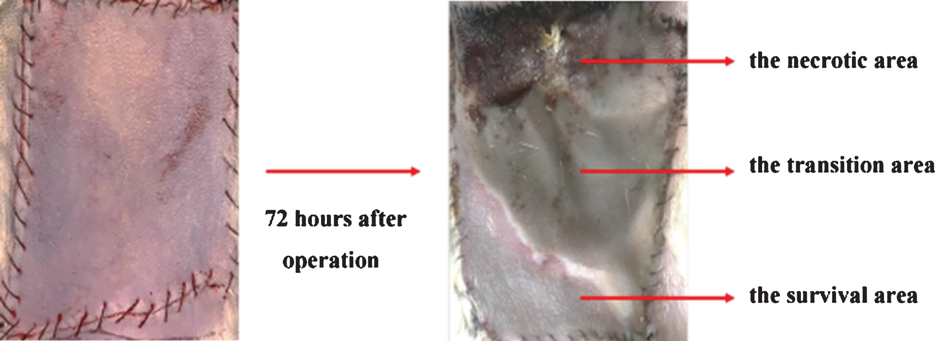
Schematic diagram of different division of skin flap 72 hours after operation. 72 hours after operation, the survived flap tissues were soft, elastic and pink. While the necrotic flap tissues were dry, inelastic and dark brown.
The average blood flow perfusion in the survival area of rats from each group was detected by LDPI. The necrotic area was dark blue, the survival area was red, and the transition area between the dead and survival area showed a progressive color in LDPI scanning image (Fig. 2). The results from LDPI proved that the blood flow of the flap recovered slowly after ischemia treatment but the average blood perfusion of total flap in the l/R-M group(768.1±49.68PU) was much better than that in the l/R-P group (548.0±29.29PU). In addition, the average blood perfusion of survival flap in the SH group (874.8±32.63 PU) had no significant alteration when compared with l/R-M group (Fig. 2, Table 2).

The blood flow perfusion in different groups was detected by LDPI.The average blood flow perfusion in the survival area of rats from the SH group, the l/R-M group or the l/R-P group was detected by LDPI.
The average blood perfusion of survival flap in the different groups
Data=mean±SEM<IF01>. a, SH vs I/R-P, t = 7.454, P<0.0001; b, I/R-P vs I/R-M, t = 3.817, P = 0.0013.
The total RNA was acquired from the skin flaps in the l/R-M group or in the l/R-P group. Ultimately, we identified that 474 genes in skin flap tissues significantly changed between the l/R-M group and the l/R-P group. Among them, 263 genes were up-regulated, and 211 genes were down-regu-lated (Figs. 3A, B).

Genes profile of skin flap from different groups. A. Genes differentially expressed in skin flap tissues from the l/R-M group or from the l/R-P group. B. Heat-map showing gene expression profiles.
GO-based functional analysis could describe gene products and demonstrate their relationships on basis of three ontology categories: biological process, molecular function and cellular component. Here we used GOEAST to analyze the RNA-seq data basing on groups of functionally related genes instead of individual genes. Then we identified significantly enriched GO terms and characterized the responses of skin flap after I /R to MS treatment. In total, the changed genes were enriched in 4379 subcategories under biological process, 483 subcategories under cellular component, and 809 subcategories under molecular function. In biological process ontology, the gens about cell adhesion (Actn3, Dmp1, Stab2, Enpp2, Ibsp), development (ygo2, Foxa2, Atic, Kcna1), cell migration (Cspg4, Tek, Fezf1, Nov, Ace), anatomical structure development (Bcar1, Tgfbr, Isl1, Apcdd1), response to organic substance (Foxa2, Slc34a1, Get4, Aqp3), cell differentiation (Eif2b5, Enam, Abca15, Wdr5) were mainly associated with responses of the skin flap to the MS treatment after I /R (Fig. 4A). According to cellular functional categorization, most of the changed genes were categorized as extracellular region part, extracellular vesicle, extracellular membrane-bounded organelle as shown in Fig. 4B.

Identification of genes function in skin flaps from different groups. A,B. Genes which significantly altered in skin flap tissues from the l/R-M group or from the l/R-P group were analyzed by GO-based functional analysis according to biological process and cellular component.
Then, the changed genes between the l/R-M group and the l/R-P group were analyzed by pathway analysis. The bio-functions and canonical pathways (only p-value less than 0.01) which were most significantly altered were shown in Fig. 5. The top 10 pathways were: PI3K-Akt signaling pathway (Angpt2, Rps6), regulation of actin cytoskeleton, lysosome, staphylococcus aureus infection, focal adhesion, arrhythmogenic right ventricular cardiomyopathy (ARVC), leukocyte transendothelial migration,complement and coagulation cascades, MAPK signaling pathway, groteoglycans in cancer.
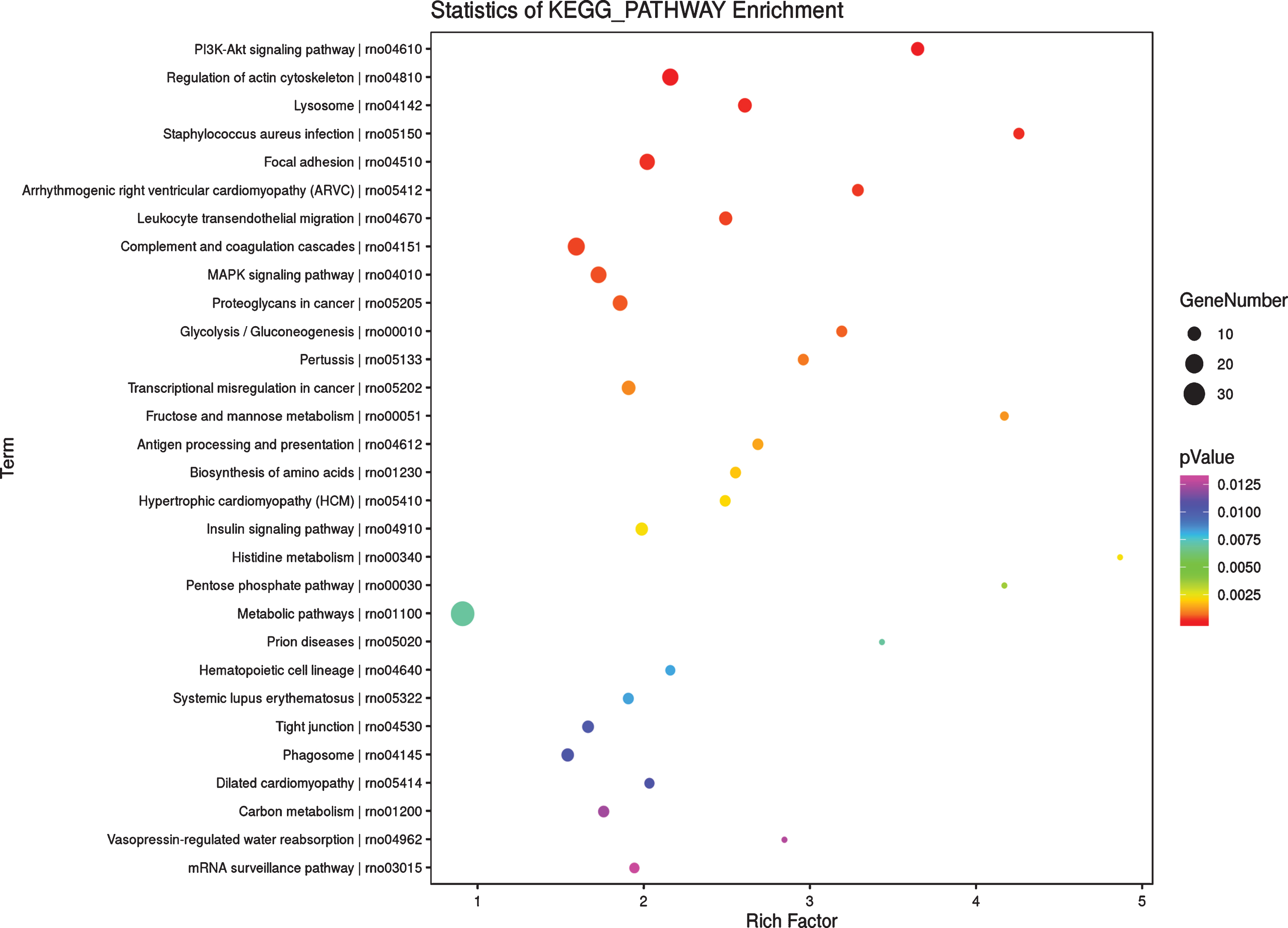
Pathway analysis of genes profile in skin flap from different groups. The differentially expressed genes in skin flap tissues from the l/R-M group and the l/R-P group were analyzed by pathway analysis. The top 10 pathways were shown.
Through GO and pathway analysis, we found that cell adhesion-associated process and PI3K-Akt signaling pathway were closely associated with the responses of skin flap tissues to the l/R-M group treatment. Basing on the results, we selected 7 possible candidate genes (Angpt2, Rps6, Actn3, Dmp1, Stab2, Enpp2, Ibsp) to validate the precision of our RNA-seq data. The mRNA expression of these genes in skin flap tissues from the l/R-M group or from l/R-P group was examined by real-time RT-PCR. As shown in Fig. 6, we verified that the real-time PCR expression profiles were in almost complete agreement with the RNA-seq data (Table 3).
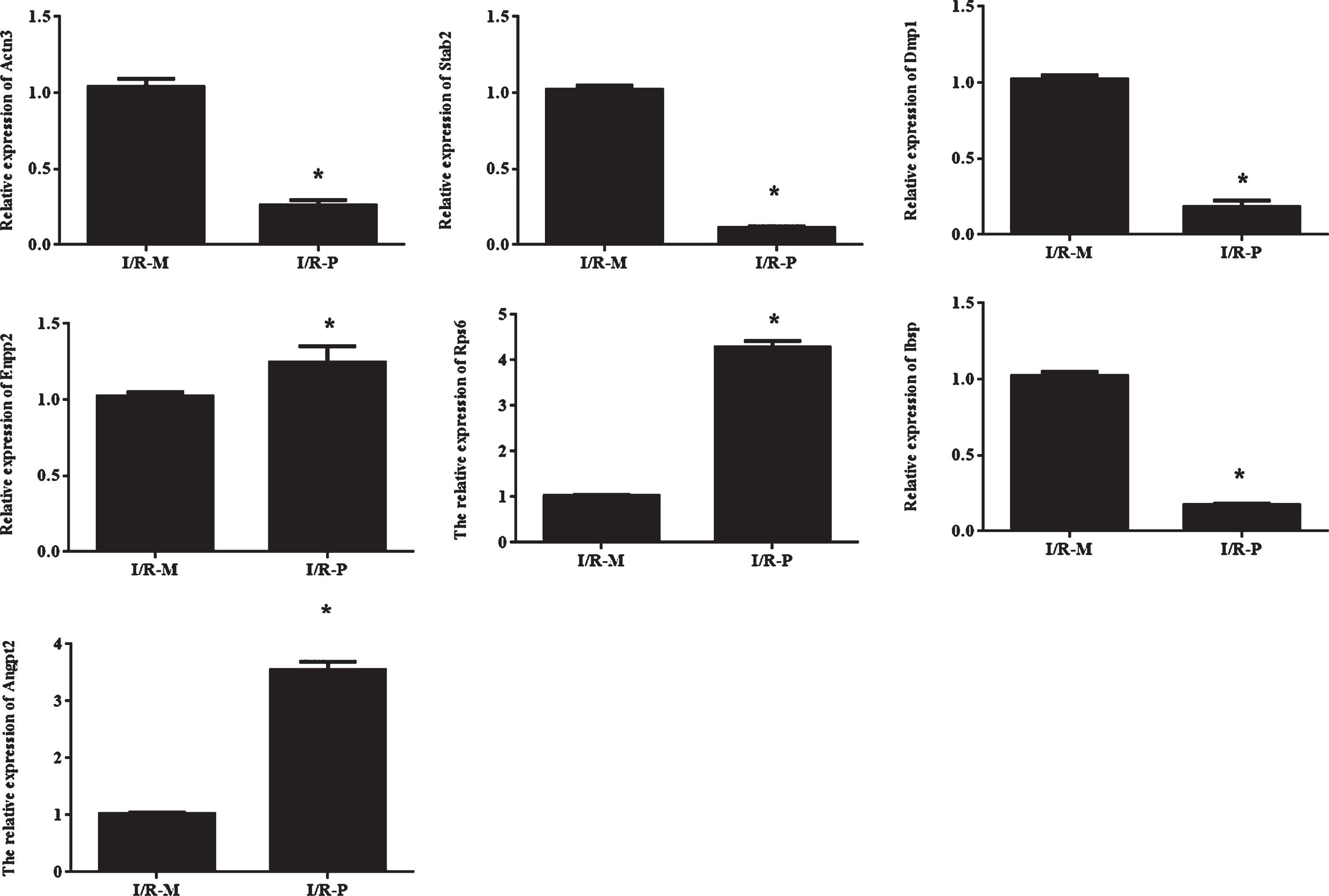
The RNA-seq results were validated by real-time RT-PCR.A. The mRNA expression of the selected genes in the l/R-M group and the l/R-P group was examined by real-time RT-PCR. β-Actin served as an internal control. *P < 0.05 relative to the l/R-M group.
The different genes between the I/R-M group and I/R-P group (M group/P group)
Many studies proved that antioxidants had a protecting effect on the organs by suppressing oxidative stress and inflammation during I/R process [14, 15]. While a lot of antioxidants were not suitable for clinic therapy owing to the impermeability of their membranes [16]. Methane was a nontoxic gas with small molecules [17]. Previous studies pointed out that methane had the proper distribution characteristics to penetrate membranes and diffuse into organs, so it could act as an useful therapeutic agent for I/R injury [18, 19]. In previous study we proved methane-rich saline (MS) could reduce of skin flap necrosis induced by the ischemia/reperfusion injury [10]. However, the mechanism of MS treatment after ischemia/reperfusion injury were still unclear.
In this study, we aimed to examine the extensive responses of skin flap to MS treatment after ischaemia/reperfusion at entire transcriptome level and get a comprehensive view of the mechanism for MS treatment. An abdominal-island skin-flap ischemia and reperfusion model induced by ligating the left superficial epigastric artery was used to investigate transcriptome changes after MS treatment.We found that I/R injury could induce the large necrotic areas and the low levels of flap blood perfusion 72 h after operation, and intraperitoneal injection of methane-rich saline after reperfusion could improve flap blood perfusion and increase the flap survival rate which were in accordance with our previous studies.
Then we explored the whole transcriptome alteration in skin flap tissue with or without MS treatment after ischaemia/reperfusion using transcriptome sequencing. Transcriptome comprised all RNA molecules including mRNAs, non-coding RNAs and other small RNAs in cells. So the quantification of transcript expression levels of genes was needed to further explore the cellular responses under specific biology process such as development and disease [20]. RNA-seq was a well established method to quantitatively measure transcriptome in a high-throughput manner which possess some advantages: First, RNA-seq could detect the transcripts beyond existing genomic sequences. Second, RNA-seq had a low experimental background signal. Third, RNA-seq could acquire the more sequences than micro-array did. Therefore, RNA-seq could detect a higher dynamic range of expression levels over the other methods.
Here, we identified 474 genes with significant changes between the I/R-P group and the I/R-M group by RNA-seq and analyzed these genes by bioinformatic approaches which permitted us to identify the function of them. The results from biological process ontology indicated that cell adhesion, development, cell migration, anatomical structure development were associated with the responses of skin flap to MS treatment. It was well known that adhesion factors could be activated during I/R injury and play the important roles in the inflammation of ischemia/reperfusion injury [21]. Our results confirmed that cell adhesion process could be greatly affected by MS treatment. In addition,the results from cellular functional categorization indicated the significantly changed genes were located in extracellular region part, extracellular vesicle, extracellular membrane-bounded organelle which implied MS treatment may regulate inflammation process by affecting the extracellular micro-environment.
Also, enriched GO molecular function terms showed that transforming growth factor beta–activated receptor activity, transmembrane receptor protein kinase activity, transmembrane receptor protein serine/threonine kinase activity, extracellular matrix binding, scavenger receptor activity, polyol transmembrane transporter activity and glycerol transmembrane transporter activity were obviously affected by MS treatment (Fig. 7). Most of them were associated with extracellular micro-environment which were in accordance with the results from cellular functional categorization, At last, the changed genes in skin flap tissues from the l/R-M group or the l/R-P group were analyzed by pathway analysis.The results showed PI3K-Akt signaling pathway was greatly affected by MS treatment. Previous studies suggested PI3K-Akt signaling pathway was involved in cell survival, metabolism, proliferation, growth and angiogenesis in response to extracellular signals [22–25]. And we firstly prove that MS treatment for skin flap tissues after I/R injury could regulate PI3K-Akt signaling pathway.
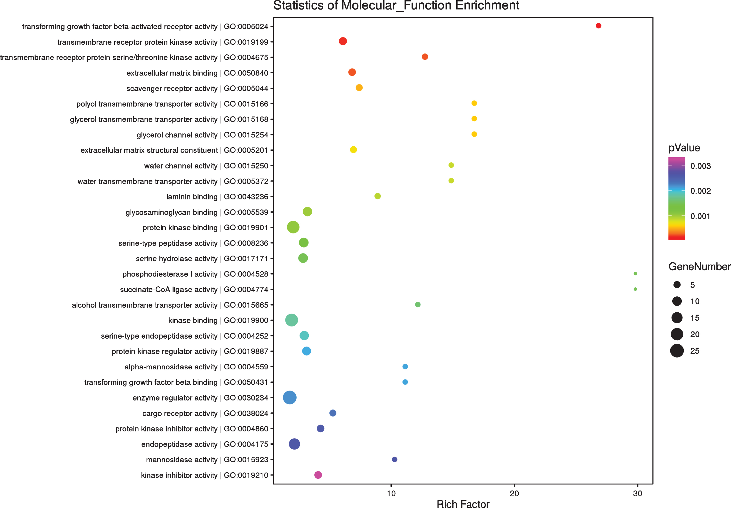
Enriched GO molecular function terms in skin flap from different groups. Genes which significantly differentially expressed in skin flap tissues from the l/R-M group or the l/R-P group were analyzed by GO-based functional analysis according to the molecular function.
On basis of bioinformatic analysis, 7 significant changed genes in PI3K-Akt signaling pathway and cell adhesion pathway (Angpt2, Rps6, Actn3, Dmp1, Stab2, Enpp2, Ibsp) were further explored by real-time RT-PCR. As shown in Fig. 5A, PI3K-Akt signal pathway relative genes (Angpt2, Rps6) were up-regulated and the cell adhesion relative genes (Actn3, Dmp1, Stab2, Enpp2, Ibsp) were down- regulated by MS treatment which verified that correlation between real-time RT-PCR and RNA-seq. These results proved RNA-seq had a high accuracy for quantifying expression levels. However, the function of PI3K-Akt signaling pathway and cell adhesion pathway in skin flap tissues repair after ischemia/reperfusion injury were needed to be further explored.
In this study, we demonstrated that MS treatment could alleviate skin ischemia/reperfusion injury and promote the survival rate of skin flap. Furthermore we firstly proved that methane-rich saline treatment could affect biology processes such as cell adhesion, development and migration and it also significantly affected some pathways including PI3K-Akt signal pathway, regulation of actin cytoskeleton, lysosome pathway and so on. These results may be useful for understanding the therapeutic mechanism of methane.
Perspectives
The methane-rich saline may alleviate ischemia/reperfusion injury.
The methane-rich saline could improve flap survival rate.
The methane-rich saline treatment could regulate cell adhesion and PI3K-Akt signal pathway.
Declarations
Ethics approval and consent to participate
This experiment was approved by the Experimentation Ethics Committee on Animal Rights Protection of Peking Union Medical College Hospital.
Consent for publication
The authors were consent for publication of this article.
Availability of data and materials
Data are available.
Competing interests
The authors declare that they have no competing interests.
Footnotes
Acknowledgments
None.