
Abstract
BACKGROUND:
When performing large volume liposuction, perioperative management of lipedema patients with coagulation disorders remains challenging due to a lack of clinical experience. With a prevalence of 1% of von Willebrand disease (VWD) in the general population, basic knowledge on diagnostic and adapted surgical strategies are essential for patients’ safety.
OBJECTIVE:
Based on a selective literature review, the purpose of this article is to present a standardized algorithm for diagnosis and perioperative treatment of VWD patients undergoing large volume liposuction.
METHODS:
The databases MEDLINE (via PubMed) and Web of Science were selectively searched with the term “(((liposuction) OR (surgery)) OR (lipectomy)) AND (((VWD) OR (hemostaseology)) OR (von Willebrand disease))”. Included were articles published in English or German until November 2020.
RESULTS:
The evidence for large volume liposuctions in patients with VWD is limited. Experience is largely based on operations with similar bleeding risks. A safe performance requires an adjustment of the surgical technique and a customized perioperative drug substitution plan. According to the current literature, perioperative thromboembolic events appear to be rare with adequate drug treatment.
CONCLUSION:
The implementation of the developed diagnostic and treatment algorithm may help further reducing bleeding complications and improve the safety for treated patients.
Introduction
Lipedema is a disproportional and symmetrical accumulation of subcutaneous fat tissue in upper and lower extremities and results in a permanent sensation of tension, pain upon pressure or easy bruising. The disease was first described by Allen and Hines in 1940 and woman are particularly affected in phases of hormonal change like puberty, after child birth or menopause [3]. Lipedema is often misdiagnosed as lymphedema or obesity and its diagnosis is based on the clinical examination and anamnesis of the patient [4, 5]. The pathogenesis of lipedema still remains unclear [6–10]. In the clinical appearance, fat deposits begin abruptly above the wrists and ankles and create the “cuff sign”. The Stemmer’s sign is typically negative. Based on the physical examination of the skin and the subcutaneous tissue, lipedema is classified in three clinical stages (see Fig. 1) [11].
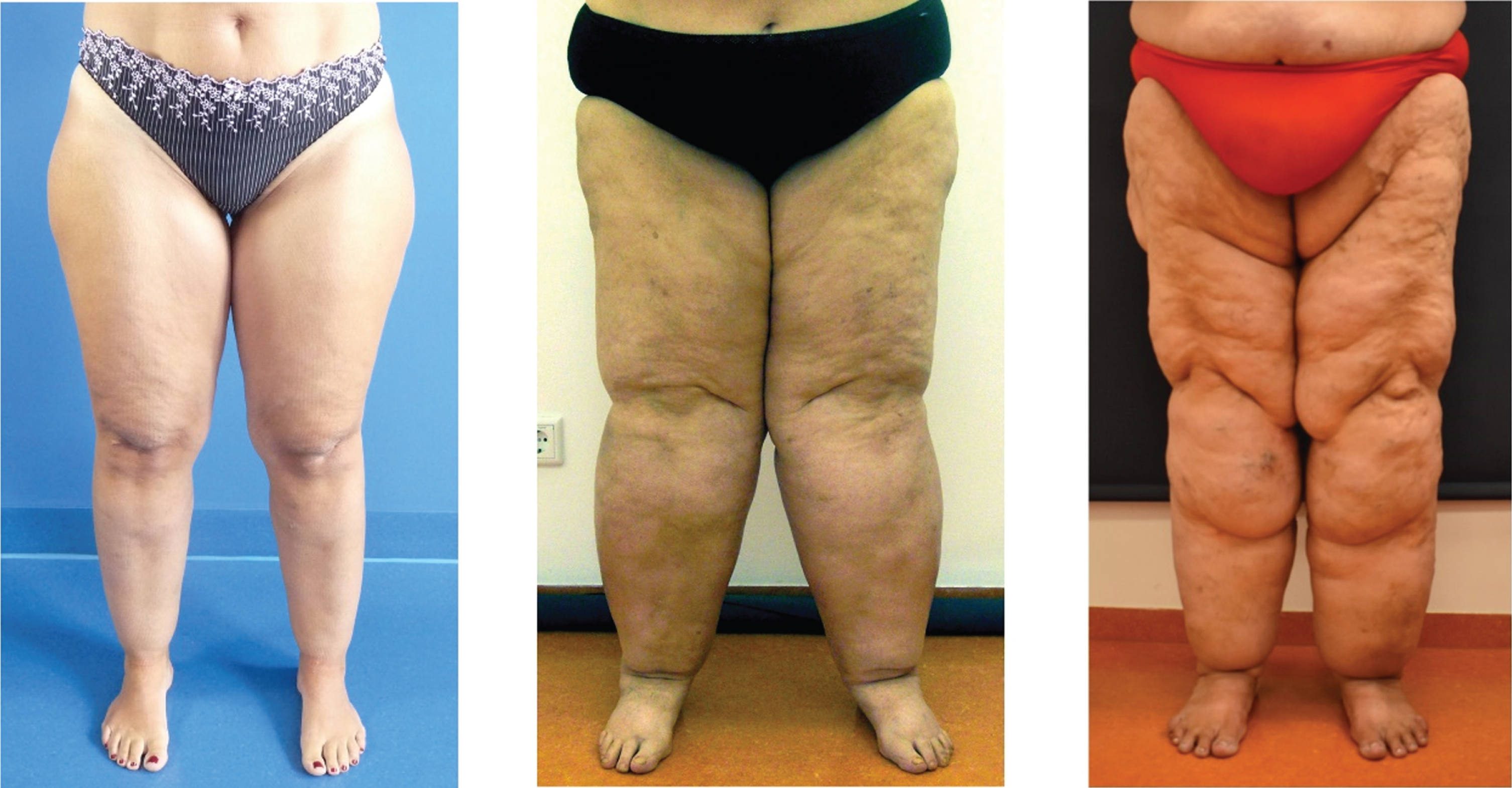
Lipedema classification by clinical stages 1–3.
The complex decongestive therapy (CDT) is still considered as the first-line treatment of lipedema [12, 13]. However, lymph sparing, large volume liposuction has been shown to be a reliable treatment option for lipedema patients in which conservative treatment has failed to sufficiently reduce symptoms [1, 15]. According to the American International Society of Aesthetic Plastic Surgery (ASAPS), liposuction is with 270.670 procedures the second most frequently performed plastic reconstructive surgery in the US in 2019 [16] and with 1.7 million procedures the second most frequently aesthetic operation worldwide [17]. An increasing number of operations are performed with a reconstructive approach for lipedema patients [18]. Sufficient symptom reduction after surgical treatment of lipedema requires in many cases large volume liposuction and the risk of bleeding complications often dictates the limitation of the aspiration volume [1, 19]. Considering the elective nature of this procedure, there is an increased need for risk minimization. From the authors clinical experience in performing lipedema surgery, occasional cases with severe hematoma formation occurred despite inconspicuous laboratory results. A retrospective study on elective plastic and reconstructive surgical procedures describes similar complications on operated patients [20].
In order to prevent bleeding complications, such as severe hematoma or even blood loss requiring transfusion, and therefore to minimize the perioperative risk, a thorough bleeding anamnesis is essential to identify existing coagulation disorders. In the bleeding anamnesis, particular attention should be given to von Willebrand disease (VWD), as this is the most common congenital coagulation disorder with a prevalence of 1%. [21, 22]. Unfortunately, basic laboratory chemistry diagnostics are not capable of detecting VWD in many cases [23]. Therefore, the surgeon’s awareness for coagulation disorders in general and VWD in particular is of special importance.
Clinical guidelines for perioperative management of patients with VWD are complex. Currently, there is a lack of experience for lipedema patients with VWD undergoing large volume liposuction. When considering the large subcutaneous wound areas and potential bleeding complications, there is a need for a systematic and structured approach in the management of these patients.
Based on a selective literature review, the present article aims to present a standardized algorithm in diagnosis and treatment process for VWD patients who are planning to undergo a large volume liposuction.
The databases MEDLINE (via PubMed) and Web of Science were selectively searched with the term “(((liposuction) OR (surgery)) OR (lipectomy)) AND (((VWD) OR (hemostaseology)) OR (von Willebrand disease))” and manually checked for relevance. Additionally, a supplementary search among the references of all publications included was performed. Articles were included that were published in English or German up to November 2020. The developed algorithm was illustratively presented in two lipedema patients with VWD.
Results
Diagnosis
Diagnosis of VWD can be challenging and should be made by a hemostaseologist (24, 25). Due to its high prevalence in the general population, basic knowledge of the disease is essential in all surgical fields (20). VWD is caused by a quantitative or qualitative defect of von Willebrand factor (VWF), a plasma protein that triggers the initial adhesion of platelets at sites of vascular injury (primary hemostasis) and stabilizes coagulation factor VIII (FVIII) in the blood (secondary hemostasis) [26].
VWD can be classified into 3 types: type 1: quantitative defect type 2: qualitative defect type 3: complete absence of VWF
Concerning phenotypic details, type 2 is divided in four variants (2A, 2B, 2M and 2 N) [27]. VWF is needed to stabilize FVIII in the plasma. The key role of VWF stabilizing FVIII in the plasma, is the reason why plasma levels of FVIII in patients with type 1 or 2 VWD are lowered and in type 3 seriously decreased. In consequence, VWF deficiency leads to decreased secreted FVIII and therefore a dual hemostatic defect [28].
In the context of preoperative preparation for large volume liposuctions, coagulation disorders must be investigated in a patient’s medical history [26]. In case of any abnormalities, special focus for VWD should be given. Before each liposuction, routine blood testing, a physical examination and a thorough medical history interview with the question of abnormal bleeding events should be done [26]. If there is a suspicion of a coagulation disorder in the patient’s own history or an abnormal laboratory test, a hemostaseologist should be consulted for further diagnosis and treatment before liposuction [29]. After referral, a detailed questionnaire with screening questions on the patient’s bleeding history helps to identify VWD (Table 1) [30–32].
Questionnaire on bleeding history
Questionnaire on bleeding history
It is important to carry out a detailed medical history interview regarding bleeding (previous spontaneous bleeding, bleeding complications, medication) [33–36]. According to Sramek et al. the most informative questions in identifying patients with possible bleeding disorders are [34]: Prolonged bleeding after surgical procedures Positive family history for bleeding disorders.
Apart from the detailed bleeding anamnesis, a precise physical examination is necessary to confirm the evidence of a bleeding disorder (Table 2) [31, 37].
Extensive physical examination to detect bleeding disorders
When clinically examining the patient for signs of a bleeding diathesis, the first indicators of VWD may be found in the presence of petechiae or mucosal bleeding. In most cases, bleeding is of mild to moderate severity due to the predominance of type 1 VWD. Mild bleeding symptoms are common in healthy population and makes the clinical evaluation difficult [26]. Although bruising easily may be a sign of a bleeding diathesis, it is also regularly seen in lipedema patients due to increased capillary permeability. Therefore, it is of less diagnostic value for the detection of a bleeding disorder in this disease group [3, 39].
Besides the assessment of clinical manifestations, a basic laboratory evaluation of hemostasis should be carried out (Table 3) [5].
Basic Laboratory Evaluation of Hemostasis
All patients suspected of having VWD should have a laboratory-confirmed diagnosis of type and severity of VWD [40, 41]. Unfortunately, a single laboratory test to detect VWD does not exist. Initial hemostasis laboratory is not suitable in identifying VWD, but may differ between a coagulation factor deficiency or a thrombocytopenia [26]. Therefore, bleeding anamnesis is still the most important diagnostic tool, since VWD can be present despite a normal pTT and platelet count. Interestingly, an abnormal pTT is only observed when FVIII is sufficiently reduced as a result of lacking VWF, its carrier protein [26]. When the bleeding history is suspicious or an isolated prolonged pTT is observed, initial VWD assays are performed (see Table 4) [42].
Initial tests for VWD
These three initial tests measure the amount of VWF (VWF:Ag), the function of VWF and its ability to interact with platelets (VWF:RCo) as well as the ability of VWF to sustain normal FVIII levels in plasma [26].
If abnormalities in these initial laboratory tests for VWD are detected, further specialized VWD assays are needed to measure VWF (VWF multimer analysis, Collagen and FVIII binding, etc.) and to classify subtypes [43–45].
It is critical that due to environmental factors (stress, anxiety, systemic inflammation) the levels of VWF can increase and thereby veil low baseline values of VWF. Consequently, in unclear cases repeated VWD testing is obligatory [46].
Using clinical and laboratory criteria, the diagnosis VWD and its subclassification can be made. In general, if VWF level is below 30 IU/dL, patients are likely to have VWD [26].
In order to avoid dangerous intra- and postoperative bleeding complications in VWD patients, a correct diagnosis and pre-operative preparation of a treatment type, substitution plan and adjunctive therapy with antifibrinolytics or antithrombotic prophylaxis are essential. Perioperative drug management in VWD patients with the goal of preventing bleeding during surgery is based on three strategies [20, 48]: Minirin (DDAVP): To increase the plasma concentration of von Willebrand Factor (VWF) by inducing release of endogenous cellular compartments through stimulation of endothelial cells VWF substitution: Replacement of VWF by using human plasma-derived concentrates Cyclocapron (tranexamic acid): Use of substances to promote hemostasis (Cyclocapron serves as an antifibrinolytic by reversibly binding to lysin receptor sites on plasminogen, which reduces the conversion of plasminogen to plasmin)
A patient may be administered one or all three treatment options at the same time depending on the physician’s preference [49]. Perioperative drug treatment of VWD depends on the disease’s subtype and severity of bleeding. Due to the heterogeneity of manifested bleeding in relation to laboratory findings, only few evidence-based recommendations exist on perioperative treatment of VWD [50, 51]. Patients with a mild form and a short immobilization period can be treated with DDAVP (Minirin®), whereas patients with a severe form or type 3 VWD require substitution therapy with concentrates containing VWF (e.g. Haemate HS®) according to the preoperative plan (Table 5).
Product types dependent on VWD subtype in minor and major surgery (84)
Product types dependent on VWD subtype in minor and major surgery (84)
In some types of VWD, the response to DDAVP can be significantly reduced. For an optimal preoperative evaluation, the guidelines for the management of VWD from the National Heart, Lung, and Blood Institute (NHLBI) recommend the measurement of VWF:RCo after the administration of DDAVP in all VWD patients [26].
Antifibrinolytics stabilize clots by inhibiting the conversion of plasminogen to plasmin. In mild to moderate forms of VWD, antifibrinolytics can be applied additionally [52]. When a substitution of VWF/ FVIII or the application of DDAVP is indicated prior of surgical procedure in VWD patients, antifibrinolytics are given additionally to control bleeding [26]. Tranexamic acid is administered intravenously at a dose of 10 mg/ kg every 8 hours [53].
Depending on the subclassification of VWD, a patient-specific individual treatment and substitution plan should be created [50, 54]. Additionally, an adapted, atraumatic liposuction technique based on the concept described by the authors is advisable [1]. Aiming a subtotal resection of the pathological fat tissue in different subcutaneous layers, large wound areas are generated when performing large volume liposuction. Therefore, surgical management should involve an adaption of established procedures in order to reduce bleeding complications. Before the operation, scheduling the procedure on the beginning of the week is advised in order to prevent postoperative problems during on-call duty in weekends. To achieve a maximal reduction of lipedema fatty tissue, the lipectomy is regularly performed with power assisted liposuction (PAL) [1]. The tumescent solution consists of 1000 ml NaCl+1 ml Adrenalin 1:1000 (“wet technique”). Due to increased possible cardiotoxic or CNS-toxic side effects when combined with general anesthesia, local anesthetics and bicarbonate buffer are not added to the tumescence solution [55]. As a result of bulging distribution of the tumescent solution into the tissue and the vasoconstrictive effect of the adrenaline, the danger of bleeding is minimized.
Because of a high bleeding risk in VWD a process changeover to water assisted liposuction (WAL) is advisable, which has shown to have less tissue trauma and reduce bleeding complications [56–58]. In contrast to the “normal” routine liposuction, the surgery should be carried out using only blunt aspiration cannulas with a maximum thickness of 4 mm or less in order to keep the risk of possible bleeding complications low. Strict longitudinal liposuction along the lymphatic vessels is crucial to protect the lymphatic vascular system and prevent injuries. A criss-cross direction, i.e. aspiration in the transverse axis to the lymphatic vessels, must be avoided [58, 59]. A blood gas analysis should be performed intraoperatively to check the patient’s Hb and detect bleeding as early as possible. Another difference in the surgical performance is the continuous, visual control of the bloody phase of the aspirate in the collection bag during aspiration, which is kept below 20% [1].
In the group’s 10-year experience in lipedema surgery, stopping the liposuction in VWD patients before the bloody phase of the aspirate reaches the 20% mark has proven valuable in reducing complications [1]. Following the recommendations of the German National Committee on liposuction in lipedema, it is advisable to keep the lipoaspirate volume significantly lower than 8% of the body weight in patients with VWD [2].
For the patients safety, a multistage procedures with decreased aspirate volumes should be performed [1].
To reduce the wound area by mechanical compression, the compression garment is put on at the end of the operation while the patient is still lying on the operating Table [1]. Post-operatively flat knitted compression class 2 should be continuously worn at least for 6 weeks day and night [1]. Postoperative monitoring is recommended for a minimum of 48 hours under inpatient conditions and the availability of an intensive medical care [26]. In the immediate postoperative period, a two-hourly vital sign checkup should be carried out. Standard thrombosis prophylaxis with LMWH may be initiated 6 h after surgery in clinically stable patients [47, 60]. Hb, electrolytes and basic coagulation parameters should be checked on the first post-operative day. Patient mobilization should be initiated as soon as possible. In addition, daily manual lymphatic drainage is recommended to assist in decreasing the postoperative swelling. After discharge, an additional laboratory control (Hb value, basic coagulation parameters) is repeated on the 5th to 7th post-operative day [61]. The patients are instructed to wear the compression garment continuously for at least 6 weeks and receive manual lymphatic drainage of the operated areas at an increased frequency with at least 3 appointments per week [1] (Fig. 2).

Algorithm for diagnosis and treatment: large volume liposuction and VWD.
The presented algorithm (Fig. 2) was successfully carried out in the surgical treatment of two lipedema patients with concurrent VWD in a specialized clinic for lipedema surgery. Both patients were female. The diagnosis of lipedema (patient 1 stage 2, 45 years; patient 2 stage 3, 66 years) was confirmed by an independent lymphologist. Because of a high suspicion for bleeding disorder in the anamnesis, diagnosis and treatment for both patients were performed according to the presented algorithm. After VWD was diagnosed and subclassified in type 1 VWD, an individualized substitution plan was drawn up. Patients received Minirin (DDAVP) 0.3μg/kg body weight and 1000 mg tranexamic acid (cyclocapron) i.v. directly before the operation. In both patients, the liposuction was performed on the front side of the lower extremities. The aspiration volumes were 7.6 and 12.0 liters, which correlate with 7% and 8% of the patients’ body weight. The percentage of blood in the aspirate was constantly below 20% (Fig. 3). No complications requiring additional treatment (e.g. bleeding, thrombosis, hematoma) were recorded intra- or postoperatively. The patients’ Hb values dropped from 7.9 mmol/L to 6.3 mmol/L and from 8.8 mmol/L to 7.1 mmol/L on the first day after the interventions. These changes corresponded to the normal shifts for patients after large volume liposuctions. The patients did not show any clinical signs of anemia (such as dizziness, orthostatic hypotension, breathlessness etc.) during the two-day inpatient stay. As a minor complication, the formation of a superficial hematoma as well as a moderate post-operative swelling in the lower extremities were recorded in one patient. The symptoms improved with cooling, elevation of the extremity and external application of an ointment containing heparin.
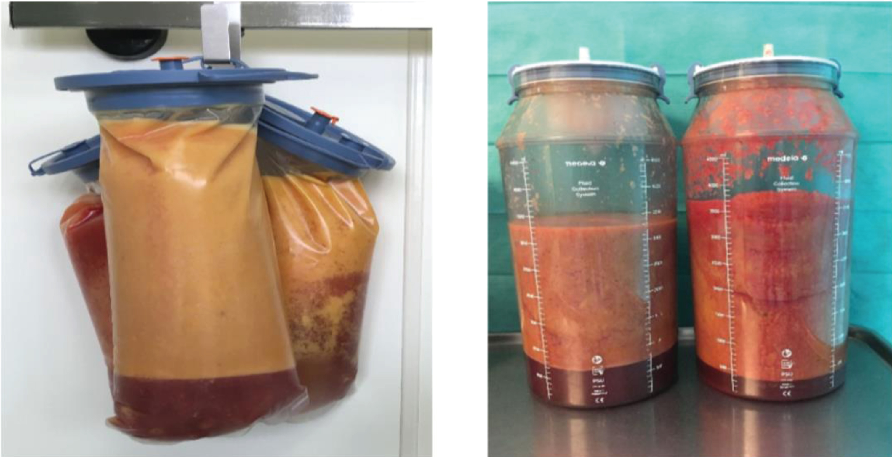
Control of the percentage of blood in the aspirate (< 20%).
Based on a selective literature search, a standard for perioperative management in performing large volume liposuctions in lipedema patients with VWD was successfully performed. The authors were able to practice the therapy regimen without any major complications in 2 patients. Based on limited evidence, general recommendations for elective large volume liposuctions with aspiration volumes > 5,000 ml in patients with VWD can only be made with restrictions. Currently, data on clinical experience of large volume liposuctions in patients with VWD are lacking. Nevertheless, in studies of elective surgical procedures with comparable bleeding risks, there are evidence-based instructions that can be applied to large volume liposuctions [20, 62]. Operations in patients with VWD can be carried out safely if a hemostaseological treatment plan has been drawn up preoperatively, an atraumatic procedure and an intra- and post-operative monitoring under inpatient conditions has been ensured [62]. Treating the diathesis before surgical intervention was crucial for a good outcome [63]. For a safe and successful performance of the procedure, an interdisciplinary collaboration between surgeons, hemostaseologists and anesthesiologists is necessary [24, 25].
As not all diatheses are detected in routine laboratory diagnostics, a detailed bleeding history and physical examination pre-operatively is essential [20, 65]. With VWD Type 1, DDAVP is the method of choice for small to medium-sized operations [26]. When administering Minirin, attention should be paid to achieve stable maintenance levels by limiting fluid intake to 1.5 L, as well as the possibility of decreased serum sodium levels, a possible side-effect of Minirin [66, 67]. The use of DDAVP should be avoided in patients with convulsions and severe cardiovascular diseases [68]. Patients who do not respond or respond insufficiently to DDAVP, as well as patients with a planned major intervention (such as in the case of > 5,000 mL lipoaspirate liposuctions), should receive VWF / FVIII concentrates preoperatively (severe VWD type 1, type 2, type 3) [47, 69]. It is important to notice that different brands of concentrates used for replacement therapy vary strongly in their VWF/ FVIII levels [47]. Since the level of VWF can fluctuate greatly depending on the patient, a substitution and maintenance therapy of VWF / FVIII concentrates tailored to the patient’s needs by the hemostaseologist is required [70]. The target value of surgical prophylaxis is a therapeutic level of 100 IU/dL of VWF:RCo and FVIII activity for severe bleeding or major surgery and > 30–50 IU/dL for minor surgery at least for the first three days of the treatment [71]. An antifibrinolytic treatment with tranexamic acid is an important supplementary therapy for medium to large surgical interventions and its use is therefore indicated for large volume liposuction [72].
It was reported that patients with hemophilic coagulation disorders tend to have a lower risk of developing venous thromboembolism postoperatively [73]. However, preoperative substitution therapy in VWD can lead to an increased postoperative venous thrombosis risk [74, 75]. Therefore, studies recommend precise planning of FVIII/ VWF administration and maintaining a level of FVIII:C between 50 IU/dL and 150 IU/dL [76].
Furthermore, after large volume liposuction and resulting postoperative immobility, all patients should be given thrombosis prophylaxis with low molecular weight heparins and mechanical thrombosis prophylaxis in the form of a compression garment of class II, which additionally reduces the risk of bleeding in a hemophilic diathesis [77]. It is therefore advisable to mobilize patients on the first day after surgery and to put on compression garments immediately after the operation. Subsequently, the medication of LWMH can be discontinued earlier, which may help to reduce bleeding complications.
Intraoperative measures used to lower the risk of perioperative complications involve using smaller diameter cannulas, terminating the liposuction when the bloody phase is over 20% and keeping the amount of lipoaspirate < 8% of the patients’ body weight. Although the authors’ standard created for this selected patient group was successfully realized in two patients, on the whole the positive influence on the outcome seems to be limited and needs to be proofed on large scale studies. Drawing up an optimal substitution plan may be difficult due to the fact that VWF levels can vary strongly in the same patient as a response to several environmental factors. Furthermore, different brand concentrates that are used for replacement therapy can fluctuate strongly in their VWF/ FVIII levels, as well as the overall heterogeneity of appearance of VWD, makes the peri-operative therapy of VWD challenging. Therefore, it is recommended that treating physicians should be familiar with 1-2 brands of VWF/ FVIII concentrates to assure appropriate dosing [47]. Regarding the duration of drug substitution, DDAVP can be administered 0,3μg/kg body weight every 12 hours and should not be administered for more than 3–5 days. 1000 mg Cyclokapron i.v. can be administered up to three times a day over a period of maximum 5 days.
The authors emphasize that the key role in reducing bleeding complications is the initial notice in patient’s anamnesis because a switch in the patient’s therapy with a treatment of VWD is possible before severe intra- or post-operative bleeding complications occur.
Because large volume liposuction with removal of a big part of the subcutaneous fat tissue in the affected areas has been reported to achieve a significant improvement of clinical symptoms and a reduction in need for CDT, the authors suggest that large volume liposuction is the adequate therapy for lipedema patients even with a concurrent VWD [4, 78–83]. Following the presented standardized algorithm, the risks of liposuction in VWD patients might be reduced. Due to the small number of patients the algorithm was practiced on, the developed therapy regime has significant restrictions. As there exists no studies on liposuction in patients with VWD and we evaluated studies of procedures with a comparable risk level, the method has its limitations. The empiric approach of the presented algorithm should be proved by large-scale clinical studies in the future.
Conclusion
Exclusion of coagulation disorders should be part of patient’s anamnesis in lipedema patients. Large volume liposuctions in lipedema patients with WVD require adjusting the surgical technique as well as adhering to an individualized perioperative drug substitution plan drawn up by an hemostaseologist. Substitution regimes are based on Minirin (DDAVP), VWF concentrate or tranexamic acid. According to the current literature, perioperative thromboembolic events appear to be rare after adequate VWF replacement therapy. The implementation of the developed diagnostic and treatment algorithm may reduce bleeding complications and improve the safety for treated patients.
Footnotes
Acknowledgments
None.